BIOCHEMISTRY FOR TEACHERS - F.F. BOYECHKO - 1985
ENERGETICS OF BIOLOGICAL PROCESSES
LIPID METABOLISM IN CELLS
The most crucial phase of lipid transformation is the METABOLISM of these substances within body tissues, which ensures both their assimilation as structural building blocks and their breakdown with the release of energy. Much like CARBOHYDRATES, Lipids serve as a primary energy source for The Cell. Nearly half of the energy required to maintain vital physiological processes is derived by the Organism from Lipid Metabolism. The oxidation of 1 g of fat releases an average of 38 kJ of energy, whereas the oxidation of 1 g of carbohydrates yields 17 kJ. The dominant role of carbohydrates in energy supply is characteristic primarily of Brain Cells, where lipids are not utilized as an energy source.
Body lipids are classified according to various criteria. Based on their biological features, they are divided into reserve and structural (cytoplasmic) lipids. Based on physicochemical properties, they are categorized as Simple Lipids (triacylglycerols, sterides, Waxes), Complex Lipids (Phospholipids, Glycolipids, sulfolipids), and lipid derivatives (carotenes, Fat-soluble Vitamins).
Lipids are distributed unevenly among various Organs and Tissues of the body. They are found in particularly large quantities in specialized connective and adipose tissues, known as fat depots. In the cells of these tissues, the greater part of the Cytoplasm is filled with lipid droplets, in which lipids are predominantly present in the form of triacylglycerols. These represent the main reserve form of lipids in the body, accounting for 99% of all reserve lipids in adipose tissue.
A characteristic feature of reserve lipids is that they contain a significant amount of Unsaturated Fatty acids, which possess a high chemical energy potential while maintaining the fluid state of the stored fat at body Temperature. The total amount of reserve lipids averages about 10% of all body lipids. In obesity, this figure increases to 25–30%. The accumulation of fat reserves in the form of triacylglycerols is characteristic not only of vertebrate cells, but also of plant and microbial cells. Lipids are mobilized from fat depots as needed to support various metabolic processes. The organism continuously maintains two interrelated processes—the deposition and mobilization of lipids from fat depots, and their utilization to meet the body's metabolic demands.
Complex lipids (phospholipids, glycolipids), in the form of lipoprotein complexes, are integral components of various Biomembranes and predominantly perform a structural function. These are the so-called structural or cytoplasmic lipids. Their quantity in the body is relatively constant and changes very little even during prolonged starvation. Depending on their composition, these lipids are subdivided into Glycerophospholipids and Inositol phospholipids. Glycerophospholipids contain a residue of the trihydric alcohol glycerol, whereas inositol phospholipids contain a residue of the polyhydric alcohol inositol. In addition, both groups incorporate residues of phosphoric acid, nitrogenous compounds, and fatty acids. Depending on the nitrogenous compounds present in their composition, glycerophospholipids are classified into lecithins (phosphatidylcholines), cephalins (colamine phospholipids), and Serine phospholipids.
The intracellular metabolism of all groups of simple (triacylglycerols, sterides) and complex (phospholipids) lipids exhibits distinct features along with certain general patterns common to this entire Class of substances.
Metabolism of Simple Lipids
Metabolism of triacylglycerols. The initial stage of intracellular triacylglycerol metabolism is their hydrolytic Cleavage under the action of tissue Enzymes—lipoprotein lipases of Blood Plasma or adipose tissue—into their constituent parts: glycerol and fatty acids. Following subsequent transformations, each of these components is integrated into Various metabolic pathways.
The conversion of glycerol proceeds under the action of the enzyme glycerol kinase and ATP, resulting in The formation of glycerophosphate, which is then oxidized to dihydroxyacetone monophosphate by the enzyme glycerol phosphate dehydrogenase. The latter undergoes isomerization to phosphoglyceraldehyde, which is subsequently incorporated into various metabolic pathways or oxidized to end products.
The intermediates of glycerol metabolism (glycerol phosphate, dihydroxyacetone monophosphate, and glyceraldehyde 3-monophosphate) can also be utilized in the synthesis of simple fats, phosphoglycerides, carbohydrates, and Other Compounds. The Oxidation of Fatty acids resulting from the Hydrolysis of triacylglycerols has specific characteristics that warrant a closer examination.
MOLECULAR MECHANISMS OF Fatty acid oxidation. This process occurs quite intensively within the cell's powerhouses—the Mitochondria—and is directly coupled with dissimilation processes in the Electron Transport Chain and The Tricarboxylic Acid Cycle, which is also characteristic of carbohydrate and amino acid derivative oxidation.
Because fatty acids contain more carbon and hydrogen and less oxygen than carbohydrates, their oxidation requires significantly more oxygen and, consequently, yields a greater amount of ATP via Oxidative Phosphorylation. The oxidation of 1 mol of palmitic acid yields 130 mol of ATP, whereas the oxidation of 1 mol of glucose yields 38 mol of ATP.
In 1904, the German biochemist Knoop experimentally proved that the metabolism of fatty acids in the body proceeds through the cleavage or addition of two-carbon fragments to their molecules. During fatty acid oxidation, acetic acid molecules are progressively cleaved off in the form of acetyl-CoA, thereby shortening the carbon chain of the fatty acid by 2 carbon atoms in each oxidation cycle.
Because hydrogen atom abstraction, followed by the Formation of Acetyl-CoA, occurs predominantly at the β-position of the higher fatty acid molecule, the entire process is termed β-oxidation.
The oxidation process consists of three successive stages: fatty acid activation, β-oxidation, and the oxidation of the resulting acetyl-CoA molecules to CO2 and H2O within the tricarboxylic acid cycle.
The First stage—fatty acid activation—takes place on the outer mitochondrial membrane of various organs and tissues (Heart, Kidneys, mucous membrane of the Small Intestine, and adipose tissue) and is an energy-dependent process. With the participation of ATP, Fatty acids are converted into acyl adenylates:

Subsequently, acyl adenylates react with CoA—SH in the presence of acyl-CoA synthetases, yielding acyl-CoA.

The resulting compounds (acyl-CoA derivatives) cannot penetrate the membranes into the mitochondrial matrix where their subsequent oxidation takes place; therefore, their translocation is mediated by carriers, with carnitine (a trimethyl derivative of β-hydroxy-γ-aminobutyric acid) acting as the carrier. Catalyzed by the enzyme carnitine acyltransferase, an acylcarnitine complex is formed, which easily traverses The inner mitochondrial membrane into the matrix. Within the matrix, the acyl-carnitine complex dissociates under the action of intramitochondrial carnitine acyltransferase. The acyl residue is transferred to intramitochondrial CoA—SH to form acetyl-CoA, while the released carnitine returns to the cytoplasm.
The Second Stage, β-oxidation proper, comprises four sequential steps. In the first step, acyl-CoA undergoes dehydrogenation mediated by a flavoprotein, yielding an α,β-trans-unsaturated derivative. Hydrogen electrons and protons are transferred via FAD to ubiquinone (coenzyme Q), after which electrons are transferred through the cytochrome system to the terminal acceptor, molecular oxygen. These processes drive the synthesis of two ATP molecules. The next step involves the Hydration of the double bond to form β-hydroxy compounds, a reaction catalyzed by the enzyme enolase.
The Third Stage of β-oxidation involves the dehydrogenation of the resulting β-hydroxy compounds by dehydrogenase enzymes, yielding β-keto derivatives. The oxidized form of NAD serves as the hydrogen acceptor. As protons and electrons are transferred along the Respiratory Chain, three ATP molecules are synthesized. In The final stage of β-oxidation, the product of the third step (β-ketoacyl-CoA) interacts with free CoA, producing acetyl-CoA and an acyl-CoA (a CoA ester of a fatty acid) shortened by a two-carbon unit.
The resulting acyl-CoA molecule undergoes the next four-step process of β-oxidation again, in which the fatty acid is shortened by two carbon atoms, and so on until the end. The oxidation of palmitic acid, which contains 16 carbon atoms, yields eight molecules of acetyl-CoA that enter the tricarboxylic acid cycle.
Overall, the reaction for shortening the acyl-CoA chain by two carbon atoms can be written as follows:

The energy released during The breakdown of fatty acids becomes available to the organism through the subsequent transformations of NADH+H+, FADH2, and acetyl-CoA. For example, each oxidation event of palmitoyl-CoA yields one molecule of FADH2 and one molecule of NADH+H+. During The transfer of electrons and protons from reduced NAD via oxidative phosphorylation, three ATP molecules are formed, and during the transfer of protons and electrons from FADH2, two ATP molecules are formed, meaning a total of five ATP molecules are produced per 1 cycle of β-oxidation. Given The Structure of palmitic acid CH3—(CH2)14—COOH, there are 7 such oxidation events. Thus, 7·5 = 35 ATP molecules. In addition, each event of β-oxidation produces eight molecules of acetyl-CoA, which will yield 12·8 = 96 ATP molecules in the Krebs cycle. Therefore, the total energy yield of β-oxidation of a single palmitoyl-CoA molecule is 131 ATP molecules. Of these, one ATP molecule is used for the formation of palmitoyl-CoA from palmitate, resulting in a net yield of 130 ATP molecules.
Considering that 46 kJ of Energy is stored in the high-energy bonds of ATP, a total of 65,100 kJ of energy is generated in the form of ATP. This energy yield represents 60% of the 10,080 kJ released upon the oxidation of 1 mol (256 g) of palmitic acid to CO2 and H2O in a bomb calorimeter. Taking this into account, we can calculate the Energy balance for the breakdown of tripalmitin, which contains three palmitic acid residues and one glycerol molecule (129·3 + 19 = 406 ATP molecules; 406·46 = 18,676 kJ).
Biosynthesis of triacylglycerols. This process requires Fatty Acids and glycerol, the active forms of which participate in the formation of fat molecules.
Glycerol is produced in the organism primarily from dihydroxyacetone monophosphate, an intermediate product of carbohydrate oxidation, under the action of the enzyme glycerol-3-phosphate dehydrogenase.

The resulting α-glycerophosphate is the active form of glycerol and can be used for fat synthesis. Part of the glycerol phosphate is hydrolyzed to glycerol and phosphoric acid under the action of the enzyme glycerol-1-phosphatase. In addition, glycerol is also formed As a result of triglyceride breakdown:

Under The Influence of tissue lipolytic enzymes in the Liver, glycerol can be converted back into glycerophosphate. This reaction proceeds with the participation of ATP and the catalytic action of the enzyme glycerol kinase; however, it cannot occur in adipose tissue and the intestinal mucosa because they lack glycerol kinase. Therefore, the source of glycerol phosphate in these tissues is dihydroxyacetone monophosphate.
In humans and animals, the synthesis of most fatty acids necessary for GROWTH AND DEVELOPMENT takes place in various organs and tissues (liver, kidneys, intestinal mucosa, mammary gland). Their prolonged absence leads to various Metabolic Disorders, which is why unsaturated fatty acids are classified as a group of vitamins (vitamin F).
Unlike β-oxidation, which occurs exclusively in mitochondria, FATTY ACID BIOSYNTHESIS can take place On the surface of The Endoplasmic reticulum, in the Cytosol, and in mitochondria. This process is catalyzed by a complex multienzyme system, fatty acid synthase, the functional activity of which requires reduced nicotinamide Coenzymes, Mn2+ ions, and bicarbonate. The main metabolic pathway of fatty acid biosynthesis—Lipogenesis—takes place in the cytosol.
Medium-chain fatty acids are elongated in mitochondria, whereas the enzymatic process of building up CoA derivatives of polyunsaturated fatty acids occurs in the membranes of the endoplasmic reticulum.
Saturated and monounsaturated fatty acids are synthesized in the organism with the participation of acetyl-CoA. Therefore, any substances whose breakdown yields this compound are potential sources of carbon atoms in fatty acid synthesis reactions. Such sources of acetyl-CoA are predominantly excess dietary carbohydrates that cannot be utilized in Energy Metabolism, Reserve Polysaccharides, and Certain Amino Acids.
Since the capacity of higher organisms to store carbohydrates is limited, their excess in the diet is utilized for the synthesis of fatty acids. This is evidenced by the fact that fat can accumulate quite intensively in the bodies of humans and animals on a fat-free diet containing excess carbohydrates. As a result of oxidation, excess carbohydrates are converted into Pyruvate, which, following oxidative decarboxylation, yields acetyl-CoA—the primary metabolite of fatty acid β-oxidation. Although this compound is also utilized in The process of lipogenesis, these two processes are entirely opposite.
Fatty acid biosynthesis can be divided into three consecutive stages:
a) transport of acetyl-CoA from the mitochondria to the cytosol, where the biosynthetic process takes place;
b) carboxylation of acetyl-CoA to form malonyl-CoA, the true substrate for lipogenesis;
c) Condensation of acetyl-CoA and malonyl-CoA followed by their reduction to form Higher Fatty Acids.
The first stage is accomplished through the interaction of acetyl-CoA with oxaloacetate within the mitochondrial matrix. Unlike acetyl-CoA, the citrate formed in this reaction crosses the mitochondrial membrane quite readily because the membrane contains a specific translocase system for transporting tricarboxylic acids. In the cytoplasm, with the participation of specific enzymes, citrate is cleaved into acetyl-CoA and oxaloacetate, the latter of which is translocated back into the mitochondrial matrix. For each cycle of acetyl-CoA Transport from the mitochondrial matrix to the cytosol, one ATP molecule is consumed.
The carboxylation of acetyl-CoA in the cytosol is carried out by the enzyme acetyl-CoA carboxylase, which contains biotin in its active center. In the presence of ATP, a CO2 molecule binds to the prosthetic group of the enzyme to form a carboxybiotin compound (CO2~biotin-enzyme)—an active form of carbonic acid. From this compound, the carboxyl group is readily transferred to acetyl-CoA, yielding the active form of malonic acid, malonyl-CoA: CO2 + Biotin-enzyme + ATP → Carboxybiotin-enzyme + ADP + Pi
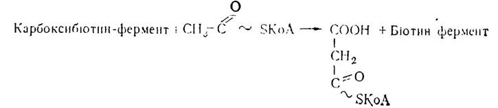
During fatty acid biosynthesis, malonyl-CoA undergoes decarboxylation to yield acetyl and CO2. Thus, malonyl-CoA is not incorporated directly into the fatty acid molecules, but serves merely as an intermediate that facilitates the formation of fatty acids from active acetate. Therefore, the carboxylation process is a critically important regulatory step in fatty acid synthesis, as it controls the incorporation of two-carbon units into higher fatty acid molecules.
At the next stage, acetyl-CoA and malonyl-CoA are enzymatically transferred to a low-molecular-weight protein capable of carrying acyl groups—the acyl carrier protein (ACP). The prosthetic group of this protein is a derivative of vitamin B3 (pantothenic acid) containing a thiol group (ACP-SH), which acts as a specific carrier of acyl residues, thereby determining the catalytic activity of this enzyme.
The acyl carrier protein is a component of the six-component fatty acid synthase enzyme system. Each component of this multienzyme complex catalyzes a specific stage of higher fatty acid biosynthesis. Initially, an acetyl-CoA residue is enzymatically transferred to the thiol group of ACP—SH, yielding acetyl~S—ACP and releasing HS—CoA.

Next, a malonyl residue is transferred to the thiol group of ACP—SH:

The first actual step in fatty acid biosynthesis is the condensation of acetyl- and malonyl-CoA residues, mediated by the condensing enzyme acyl-ACP: malonyl-ACP ligase, accompanied by the decarboxylation of malonyl to form acetoacetyl-CoA.

At this stage, the same $\text{CO}_2$ molecule is released that was previously attached to acetyl-CoA via the biotin-dependent enzyme during malonyl-CoA synthesis. It is believed that the decarboxylation of the malonyl residue shifts the reaction equilibrium toward fatty acid synthesis.
Subsequently, acetoacetyl-S—ACP is reduced by $\beta$-ketoacyl-ACP reductase in the presence of $\text{NADPH} + \text{H}^+$ to yield $\beta$-hydroxyacyl-S—ACP;

Hydroxybutyryl-S—ACP loses a Water molecule under the action of enoyl-ACP dehydratase from the synthetase complex, converting into the $\alpha, \beta$-unsaturated acyl derivative of ACP, crotonyl-S—ACP.

Crotonyl-S—ACP is then reduced to butyryl-S—ACP by crotonyl-S—ACP reductase of the synthetase complex.

The formation of this four-carbon compound completes the first cycle of fatty acid biosynthesis. Chain elongation in fatty acid synthesis proceeds through subsequent reactions occurring in the same sequence. Once again, malonyl-CoA is transferred by malonyl transacylase to the thiol group of SH—ACP to form malonyl-S—ACP, which then interacts with butyryl-S—ACP via the condensing enzyme of the synthetase complex. As a result of this reaction and the decarboxylation of the malonyl residue, a $\beta$-ketoacyl is formed that contains two more carbon units than the preceding acyl group.

Through subsequent reduction and dehydration reactions, the fatty acid chain is lengthened by another two carbon atoms. Repeating this cycle six times yields a palmitic acid molecule.
Thus, chain elongation during fatty acid synthesis begins at the carboxyl group of acetyl-CoA and proceeds via the sequential addition of acetyl residues to the carboxyl end of the growing chain. Each successive acetyl residue is derived from malonyl-CoA, representing the two-carbon fragment of the malonyl-CoA molecule closest to the CoA moiety, while the third carbon atom of malonyl-CoA, located furthest from the CoA group, is lost as $\text{CO}_2$.
The overall process of fatty acid (palmitic acid) biosynthesis can be represented by the following equation:

In other words, the synthesis of a single palmitic acid molecule requires one molecule of acetyl-CoA and seven molecules of malonyl-CoA. Upon completion of biosynthesis, the resulting fatty acid-ACP derivative reacts with coenzyme A to form the active fatty acid form—palmitoyl-CoA, stearoyl-CoA, etc. This process regenerates the ACP—SH of the multienzyme synthetase complex, allowing it to re-enter fatty acid biosynthesis reactions.
Consequently, fatty acid synthesis differs from $\beta$-oxidation in several key aspects. First, the enzyme systems responsible for these processes are localized in different cellular compartments. Second, $\beta$-oxidation of fatty acids proceeds with the release of a large amount of energy; catabolic processes drive the synthesis of ATP and reduced pyridine NUCLEOTIDES, whereas fatty acid assimilation (synthesis) consumes ATP and reduced pyridine coenzymes. Third, different forms of pyridine nucleotides participate in the Synthesis and degradation of fatty acids: $\text{NAD}^+$ is used in $\beta$-oxidation, whereas $\text{NADP}^+$ is used in synthesis.
In the first stage of triacylglycerol biosynthesis, glycerophosphate reacts with two molecules of acyl-CoA to yield phosphatidic acid. This reaction is catalyzed by the enzyme glycerophosphate acyltransferase.
Phosphatidic acid loses a phosphate group through the action of $\text{L}$-$\alpha$-phosphatidate phosphohydrolase, converting into an $\alpha, \beta$-diglyceride. Upon interacting with diacylglycerol acyltransferase, it incorporates another acyl-CoA residue to become a triacylglycerol:

Phosphatidic acid and $\alpha, \beta$-diacylglycerol serve as common intermediates in The biosynthesis of both triacylglycerols and phosphoglycerides.
In addition to the aforementioned phosphatide pathway of fatty acid biosynthesis, there is also a monoglyceride pathway of triacylglycerol biosynthesis. This process occurs predominantly in the mucosa of the small intestine during the absorption of fat breakdown products—mono- and diglycerides. These processes are catalyzed by mono- and diglyceride transacylases.
Metabolism of Complex Lipids
Phosphatides (phospholipids), much like Cholesterol, are essential components of cell membranes, where they perform vital structural Functions and regulate a wide range of critical physiological processes. Furthermore, this group of complex lipids facilitates the absorption, transport, and oxidation of simple fats (triacylglycerols) in tissues; consequently, the metabolism of these two lipid classes is closely intertwined. A deficiency in phospholipids leads to a significant accumulation of fat in the liver, which may result in hepatic steatosis (fatty liver infiltration). Given the stimulating effect of nitrogenous components such as ethanolamine, Choline, and colamine, phospholipids are classified as lipotropic factors. Their lipotropic action stems from their requirement for phospholipid synthesis, which in turn stimulates lipid metabolism.
Breakdown of Phospholipids (Phosphatides). Dietary phospholipids are hydrolyzed in the duodenum through the action of hydrolytic enzymes known as phospholipases, which are present in intestinal and pancreatic juices. The degradation of phospholipids can be illustrated using lecithin (choline phosphatide) as an example; its molecule consists of glycerol, phosphoric acid, choline, and two fatty acid residues.
In the first stage, under the action of phospholipase A, a fatty acid residue is cleaved from the lecithin molecule at the β-position, yielding lysolecithin, which exhibits hemolytic activity and causes erythrocyte hemolysis. Subsequently, phospholipase B removes the second fatty acid residue from lysolecithin, resulting in the formation of α-glycerophosphocholine:

With the participation of phospholipase D, α-glycerophosphocholine splits off choline to form α-glycerophosphate, which is further hydrolyzed by phospholipase C to yield glycerol.

Thus, the ultimate End products of lecithin degradation by phospholipases are glycerol, fatty acids, choline, and phosphoric acid. Other groups of phospholipids—such as cephalins and serine phosphatides—undergo degradation in a similar manner.
The breakdown products of phosphatides are absorbed by the epithelial Cells of the intestinal mucosa, where they undergo organism-specific resynthesis. A fraction of phosphatides can be absorbed without prior hydrolysis in the form of a finely dispersed emulsion. Glycerol is absorbed both in its free state and as glycerol phosphate or glycerophosphocholine. Unlike triglycerides, the majority of resynthesized phospholipids pass directly into capillary vessels and are transported to various organs, predominantly the liver.
While a portion of phospholipids is utilized as structural (plastic) material, the remainder is degraded to end products.
Choline, generated via phospholipid degradation, can undergo various metabolic transformations. In Nervous Tissue, the enzyme choline acetyltransferase converts choline into acetylcholine, a crucial neurotransmitter. Acetylcholine plays a direct role in transmitting nerve impulses across neural centers and from nerve fibers to various target organs.

A particularly important role of choline lies in the metabolism of single-carbon units, specifically methyl groups. Upon oxidation mediated by choline oxidase, choline is converted into betaine aldehyde and subsequently into betaine, which serves as a direct methyl group donor in the biosynthesis of Methionine and homocysteine. Thus, choline helps maintain the cellular pool of methyl groups.
The biosynthesis of choline from Glycine and serine, alongside the involvement of methionine-derived methyl groups, highlights choline as a vital metabolic link between Proteins and lipids.
Biosynthesis of Phosphatides. The biosynthesis of phospholipids, particularly lecithin, is highly active in the walls of the small intestine and the liver, where necessary substrates—glycerol, fatty acids, phosphoric acid, and nitrogenous compounds—are readily available.
The Initial Stages of phosphatide biosynthesis (up to the formation of α,β-diacylglycerols) mirror that of triacylglycerols, whereas subsequent reactions involve the activation of nitrogenous bases and their interaction with α,β-diacylglycerols. Lecithin biosynthesis is preceded by the activation of choline. This activation process involves the reaction of choline with ATP, catalyzed by choline kinase, to produce phosphocholine:

Next, phosphocholine reacts with cytidine triphosphate (CTP) in a reaction catalyzed by cholinephosphate cytidylyltransferase, yielding cytidine diphosphate choline (CDP-choline) with the release of pyrophosphate. CDP-choline then reacts with a diglyceride to form lecithin and cytidine monophosphate (CMP).

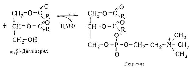
The synthesis of colamine phosphatides, serine phosphatides, and other phospholipids proceeds similarly to that of lecithins. The concurrent synthesis of triacylglycerols and phosphatides in the organism demonstrates a close metabolic relationship between them, further substantiated by the formation of shared intermediates (phosphatidic acids and α,β-diglycerides).
Last update: 06/08/2026
Editorial and Educational Adaptation: This material has been compiled based on the primary/original source text. The project team performed an editorial review, corrected technical inaccuracies, structured sections, and adapted the content for an educational format.
What was processed:
- elimination of formatting defects (OCR errors, structural breaks, corrupted characters);
- editorial organization of content;
- standardization of terminology in accordance with academic sources;
- verification of factual statements against the original source text.
All mentions of the author, publication year, and origin of the primary text have been preserved in accordance with the source.