LEHNINGER PRINCIPLES OF BIOCHEMISTRY - VOL. 2. BIOENERGETICS AND METABOLISM - 2014
PART II. BIOENERGETICS AND METABOLISM
21. LIPID BIOSYNTHESIS
21.2. Biosynthesis of Triacylglycerols
The bulk of the Fatty acids synthesized or ingested by an Organism have two primary possible fates: incorporation into triacylglycerols for the storage of metabolic energy, or incorporation into the phospholipid components of membranes. The choice between these alternative pathways depends on the organism's current physiological needs. During periods of rapid growth, the synthesis of new membranes requires The production of membrane Phospholipids; when an organism is well-fed but not actively growing, it channels the majority of its fatty acids into fat depots (stored fats). Both pathways begin in the same way: with The formation of glycerol and fatty acid esters. In the following section, we examine The pathway of triacylglycerol formation and its regulation, along with the production of glycerol 3-phosphate via glyceroneogenesis.
Triacylglycerols and Glycerophospholipids share common precursors
Animals can synthesize and store enormous amounts of triacylglycerols for subsequent use as fuel (see Box 17-1). The human Liver and Muscles can store only a few hundred grams of Glycogen, which is barely enough to meet the body's energy demands for about 12 hours. In contrast, an average man weighing ~70 kg carries approximately 15 kg of triacylglycerols, which is sufficient to meet the body's basal energy needs for about 12 weeks (see Table 23-5). Triacylglycerols store more than 38 kJ/g of energy—significantly more than other nutrients. Whenever CARBOHYDRATES are ingested in quantities exceeding the body's capacity to store glycogen, the excess is converted into triacylglycerols and stored in adipose tissue. Plants also produce triacylglycerols, energy-rich fuel molecules, accumulating them primarily in juicy fruits, nuts, and seeds.
In animal Tissues, triacylglycerols and glycerophospholipids (such as phosphatidylethanolamine) share two common precursors (fatty acyl-CoA and L-glycerol 3-phosphate), which are generated through several biosynthetic reactions. The bulk of glycerol 3-phosphate is derived from the glycolytic intermediate dihydroxyacetone phosphate via the action of NAD-linked glycerol 3-phosphate dehydrogenase; smaller amounts are also produced in The Liver and Kidneys from glycerol through the action of glycerol kinase (Fig. 21-17). The other precursors of triacylglycerols are acyl-CoAs, formed from fatty acids by acyl-CoA synthetases—the same Enzymes responsible for activating fatty acids during β-oxidation (see Fig. 17-5).
The first step in triacylglycerol Biosynthesis is the acylation of the two free hydroxyl groups of L-glycerol 3-phosphate by two molecules of acyl-CoA, yielding diacylglycerol 3-phosphate, more commonly referred to as phosphatidic acid or phosphatidate (Fig. 21-17). Phosphatidic acid is present in Cells in only trace amounts, yet it serves as a central intermediate in Lipid Biosynthesis, as it can be converted into either triacylglycerols or glycerophospholipids. Along the pathway to triacylglycerols, phosphatidic acid is hydrolyzed by phosphatidic acid phosphatase to form 1,2-diacylglycerol (Fig. 21-18). Subsequent transesterification of the diacylglycerols with a third acyl-CoA molecule yields triacylglycerols.
Class="center">Figure 21-17. Biosynthesis of phosphatidic acid. A fatty acid is activated by conversion to acyl-CoA, then transferred to the ester linkage in L-glycerol 3-phosphate formed via one of the two pathways shown here. Phosphatidic acid is depicted in the correct configuration at C-2 of the glycerol moiety.

Figure 21-18. Phosphatidic acid in lipid biosynthesis. Phosphatidic acid is a precursor to both triacylglycerols and glycerophospholipids. The mechanisms for attaching the polar "HEAD group" during phospholipid synthesis are discussed later.

The biosynthesis of triacylglycerols in animals is hormonally regulated
In humans, total body fat remains relatively constant over extended periods, despite minor short-term fluctuations driven by the caloric content of the diet. Hydrocarbons, fats, or Proteins ingested in excess of Energy Requirements are stored as triacylglycerols. This energy reserve helps the organism withstand periods of fasting.
The pathways of triacylglycerol BIOSYNTHESIS AND DEGRADATION depend on the metabolic resources and current energy demands of the organism. The rate of triacylglycerol biosynthesis is heavily influenced by certain Hormones. For instance, Insulin facilitates The conversion of carbohydrates into triacylglycerols (Fig. 21-19). Patients with severe Diabetes Mellitus, owing to deficient insulin secretion or action, are not only unable to properly utilize dietary glucose but also fail to synthesize fatty acids from carbohydrates or Amino Acids. Without Treatment, diabetic patients exhibit an elevated rate of fat oxidation and ketone body formation (Chap. 17), and consequently, weight loss. ■
Figure 21-19. Regulation of triacylglycerol synthesis by insulin. Insulin stimulates the conversion of dietary carbohydrates and proteins into fat. Individuals suffering from diabetes mellitus lack sufficient insulin; if untreated, fatty acid synthesis declines, and instead, acetyl-CoA derived from carbohydrate and Protein Catabolism is diverted into Ketone Body Synthesis. Individuals with severe Ketosis smell of acetone, which can lead to them being mistaken for intoxicated persons (p. 605).

The balance between triacylglycerol biosynthesis and degradation is underscored by the fact that approximately 75% of all fatty acids released during lipolysis are re-esterified to form triacylglycerols rather than being used as fuel. This ratio persists even under starvation conditions, when METABOLISM/26.html">Energy Metabolism shifts from carbohydrate utilization to Fatty acid oxidation. This recycling of fatty acids occurs partly within adipose tissue, with transesterification taking place prior to their release into the bloodstream. It also occurs partially through an organism-wide cycle: free Fatty acids are transported to the liver, converted there into triacylglycerols, re-exported into the Blood (lipid transport in the blood is discussed in Section 21.4), and—after being released from triacylglycerols by extracellular lipoprotein lipase—are incorporated back into adipose tissue (Fig. 21-20; see also Fig. 17-1). The flux of the triacylglycerol cycle between adipose tissue and the liver may be relatively minor when alternative fuels are available and the release of fatty acids from Adipose tissue is restricted. However, as noted above, the relative fraction of released fatty acids that undergo transesterification remains constant at approximately 75% across all metabolic conditions. Consequently, the concentration of free fatty acids in the blood reflects both the rate of fatty acid release and the balance between triacylglycerol Synthesis and degradation in adipose tissue and the liver.
Figure 21-20. The triacylglycerol cycle. In mammals during starvation, triacylglycerol molecules are degraded and resynthesized within the triacylglycerol cycle. Some fatty acids released via the lipolysis of adipose tissue triacylglycerols enter the bloodstream, and their remnants are utilized in triacylglycerol resynthesis. Blood fatty acids serve as an energy source (e.g., in muscles) or are delivered to the liver for triacylglycerol synthesis. Triacylglycerols produced in the liver are transported via the blood back to adipose tissue, where extracellular lipoprotein lipase releases fatty acids that are re-esterified within adipocytes to form triacylglycerols.

When the mobilization of fatty acids for Energy is required, the hormones Glucagon and epinephrine stimulate their release from adipose tissue (see Figs. 17-3, 17-12). Concurrently, these hormonal signals decrease the rate of Glycolysis and increase the rate of Gluconeogenesis in the liver (supplying glucose to the Brain; for details, see Chap. 23). The released fatty acids are taken up by various tissues, including muscles, where they undergo oxidation to generate energy. The majority of fatty acids absorbed by the liver are not oxidized; instead, they are recycled into triacylglycerols and returned to adipose tissue.
The physiological rationale for this seemingly futile triacylglycerol cycle (futile cycles are discussed in Chapter 15) remains somewhat enigmatic. However, as we gain deeper insight into the metabolic provisioning of the triacylglycerol cycle and its coordinated regulation across two distinct Organs, new avenues for its investigation emerge. For instance, the triacylglycerol cycle (in which fatty acids are rapidly converted back into triacylglycerols rather than being oxidized as fuel) may serve during starvation to maintain an additional energy reserve in the bloodstream—one that can be mobilized much more rapidly in an emergency than stored triacylglycerol.
The continuous turnover of triacylglycerols in adipose tissue during starvation raises another question: What serves as the source of the glycerol 3-phosphate required for this process? As noted earlier, glycolysis is suppressed under these conditions by the action of glucagon or epinephrine, leaving dihydroxyacetone phosphate in short supply. Furthermore, glycerol released during lipolysis cannot be directly converted into glycerol 3-phosphate within adipose tissue due to insufficient levels of glycerol kinase (Fig. 21-17). How, then, is an adequate supply of glycerol 3-phosphate generated? The answer lies in a metabolic pathway discovered more than three decades ago, which until recently received little attention. This pathway is closely linked to the triacylglycerol cycle and, more broadly, to the interplay between Fatty acid and Carbohydrate Metabolism.
Adipose tissue generates glycerol 3-phosphate via glyceroneogenesis
Glyceroneogenesis — a shortened pathway of gluconeogenesis ranging from Pyruvate to dihydroxyacetone phosphate (see Fig. 14-16), after which dihydroxyacetone phosphate is converted into glycerol-3-phosphate via cytosolic NAD-linked glycerol-3-phosphate dehydrogenase (Fig. 21-21). Glycerol-3-phosphate is subsequently utilized in the synthesis of triacylglycerols. Glyceroneogenesis was discovered in the 1960s by Lea Reshef, Richard Hanson, and John Ballard, as well as independently by Eleazar Shafrir and his colleagues, who were intrigued by the presence of two gluconeogenic enzymes—pyruvate carboxylase and phosphoenolpyruvate (PEP) carboxykinase—in adipose tissue, where glucose is not synthesized. Following a long period of inattention to this peculiar fact, interest in this metabolic pathway has been revived, uncovering a link between glyceroneogenesis and late-onset type 2 diabetes.
Fig. 21-21. Glyceroneogenesis. This metabolic pathway is essentially a shortened version of gluconeogenesis from pyruvate to dihydroxyacetone phosphate, followed by the Conversion of the latter into glycerol-3-phosphate, which is used for triacylglycerol synthesis.

Glyceroneogenesis serves multiple Functions. In adipose tissue, glyceroneogenesis coupled with fatty acid re-Esterification controls the rate of fatty acid release into the bloodstream. In brown adipose tissue, this metabolic pathway may regulate the rate at which fatty acids are delivered to Cell/35.html">Mitochondria for use in thermogenesis (see Fig. 19-34). Furthermore, in starving individuals, glyceroneogenesis in the liver alone provides a sufficient supply of glycerol-3-phosphate for up to 65% of fatty acids to be re-esterified into triacylglycerols.
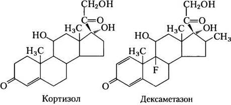
Flux through the triacylglycerol cycle between the liver and adipose tissue is largely controlled by PEP carboxykinase, which acts as the rate-limiting step for both gluconeogenesis and glyceroneogenesis. Glucocorticoid hormones, such as cortisol (a natural steroid derived from Cholesterol; see Fig. 21-45) and dexamethasone (a synthetic glucocorticoid), regulate the levels of PEP carboxykinase in the liver and, consequently, in adipose tissue. Acting through the glucocorticoid receptor, these Steroid Hormones upregulate the expression of the Gene encoding hepatic PEP carboxykinase, thereby enhancing both gluconeogenesis and glyceroneogenesis (Fig. 21-22).
Stimulation of glyceroneogenesis leads to increased triacylglycerol synthesis in the liver and their release into the blood. At the same time, glucocorticoids suppress the expression of the gene encoding PEP carboxykinase in adipose tissue. This results in downregulated glyceroneogenesis in adipose tissue; consequently, fatty acid recycling decreases, and a larger amount of free fatty acids is released into the bloodstream. Thus, glyceroneogenesis is coordinately regulated in the liver and adipose tissue, affecting Lipid Metabolism IN opposite ways: a lower rate of glyceroneogenesis in adipose tissue leads to greater fatty acid release (preventing their recycling), whereas an increased rate of this process in the liver promotes more efficient synthesis and export of triacylglycerols. The net result is an increased flux through the triacylglycerol cycle. When glucocorticoid action ceases, the flux through the cycle diminishes as PEP carboxykinase expression increases in adipose tissue and decreases in the liver.
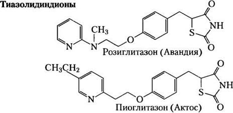
Fig. 21-22. Regulation of glyceroneogenesis. (a) Glucocorticoid hormones stimulate glyceroneogenesis and gluconeogenesis in the liver while simultaneously suppressing glyceroneogenesis in adipose tissue (via Reciprocal Regulation of PEP carboxykinase (PEPCK) Gene Expression IN the two tissues); As a result, the flux through the triacylglycerol cycle increases. Glycerol released from The breakdown of triacylglycerols in adipose tissue is secreted into the blood and transported to the liver, where it is converted primarily into glucose via glycerol kinase, though a fraction is converted into glycerol-3-phosphate. (b) A class of drugs known as thiazolidinediones is currently used to treat type 2 diabetes. In this condition, high blood levels of fatty acids impair glucose uptake in muscles and cause insulin resistance. Thiazolidinediones activate nuclear receptors (peroxisome proliferator-activated receptor y (PPARy)), which induce PEP carboxykinase activity. The therapeutic effect stems from the fact that thiazolidinediones increase the rate of glyceroneogenesis, thereby enhancing triacylglycerol resynthesis in adipose tissue and reducing the concentration of free fatty acids in the blood.

Thiazolidinediones are used in type 2 diabetes to upregulate glyceroneogenesis
The renewed attention paid to glyceroneogenesis is partly due to The Link Between this metabolic pathway and diabetes. High blood levels of free fatty acids impair glucose uptake in muscles and induce insulin resistance, leading to type 2 diabetes. It has been shown that a new generation of drugs, thiazolidinediones, lowers blood fatty acid levels and increases insulin sensitivity. Thiazolidinediones bind to and activate nuclear Hormone Receptors (peroxisome proliferator-activated receptor y (PPARy)), inducing PEP carboxykinase in adipose tissue (Fig. 21-22); the increased PEP carboxykinase activity subsequently leads to enhanced synthesis of glyceroneogenesis precursors. Thus, the therapeutic effect of thiazolidinediones is, at least in part, mediated by an increase in glyceroneogenesis, which in turn promotes triacylglycerol resynthesis in adipose tissue and reduces the release of free fatty acids from adipose tissue into the blood. Unfortunately, the benefits of taking one such drug, rosiglitazone (Avandia), are somewhat diminished by an increased risk of myocardial infarction. The causes of this side effect remain unclear, and research into the drug's MECHANISM OF ACTION is ongoing. ■
Summary of Section 21.2 Biosynthesis of Triacylglycerols
■ Triacylglycerols are formed by the Condensation of two molecules of fatty acyl-CoA with glycerol-3-phosphate to yield phosphatidic acid; this product is dephosphorylated to produce diacylglycerol, which is then acylated by a third fatty acyl-CoA molecule to form triacylglycerols.
■ The synthesis and degradation of triacylglycerols are regulated by hormones.
■ The mobilization and recycling of triacylglycerols establish the triacylglycerol cycle. Triacylglycerols are resynthesized from free Fatty Acids and glycerol-3-phosphate even under conditions of caloric restriction. Dihydroxyacetone phosphate—the precursor of glycerol-3-phosphate—is produced from pyruvate via glyceroneogenesis.
Last update: 06/08/2026
Editorial and Educational Adaptation: This material has been compiled based on the primary/original source text. The project team performed an editorial review, corrected technical inaccuracies, structured sections, and adapted the content for an educational format.
What was processed:
- elimination of formatting defects (OCR errors, structural breaks, corrupted characters);
- editorial organization of content;
- standardization of terminology in accordance with academic sources;
- verification of factual statements against the original source text.
All mentions of the author, publication year, and origin of the primary text have been preserved in accordance with the source.