LEHNINGER PRINCIPLES OF BIOCHEMISTRY - VOL. 2. BIOENERGETICS AND METABOLISM - 2014
PART II. BIOENERGETICS AND METABOLISM
Class="center">I don't remember how the division of labor came about. Perhaps we drew straws. At any rate, David Shemin "drew" Amino acid METABOLISM, which led to his classic work on heme Biosynthesis. David Ritenberg had to continue gratifying his interest in Protein Synthesis AND turnover, and Lipids fell to my lot.
Konrad Bloch, on how fate led him to problems of Lipid Metabolism after the death of his mentor Rudolf Schoenheimer; article in Annual Review of Biochemistry, 1987
21. BIOSYNTHESIS OF LIPIDS
Lipids play a multitude of roles in Cells, some of which have been discovered only recently. In most organisms, lipids serve as the primary form of energy storage and as the predominant component of cellular membranes. Specialized lipids act as pigments (retinal, carotene), Cofactors (vitamin K), detergents (Bile salts), carriers (dolichols), Hormones (vitamin D derivatives, Sex Hormones), intercellular and intracellular messengers (Eicosanoids, phosphatidylinositol derivatives), and anchors for Membrane Proteins (covalently attached Fatty acids, prenyl groups, and phosphatidylinositol). The ability to synthesize A wide variety of lipids is essential to all organisms. This chapter describes the biosynthetic pathways for some of the most common cellular lipids, illustrating the strategies used to assemble these Water-insoluble products from water-soluble precursors such as acetate. Like other biosynthetic pathways, these reaction sequences are endergonic and inherently reductive. They utilize ATP as a source of metabolic energy and reduced electron carriers (usually NADPH) as reducing agents.
We will first describe the Biosynthesis of Fatty acids (the primary components of triacylglycerols) and Phospholipids, and then examine the assembly (involving fatty acids) of triacylglycerols and simpler membrane phospholipids. Finally, we will consider the synthesis of Cholesterol, a component of certain membranes and a precursor to Steroids such as bile acids, sex hormones, and Adrenal hormones.
21.1. Biosynthesis of Fatty Acids and Eicosanoids
Following the discovery that Fatty acid oxidation occurs by the sequential oxidative removal of two-carbon (acetyl-CoA) fragments (see Fig. 17-8), biochemists assumed that FATTY ACID BIOSYNTHESIS probably proceeds simply via the reverse reactions of those same enzymatic steps. However, it was subsequently discovered that the biosynthesis and breakdown of fatty acids proceed via distinct pathways, are catalyzed by different sets of Enzymes, and take place in different cellular compartments. Moreover, biosynthesis requires the participation of the three-carbon intermediate malonyl-CoA, which is not involved in fatty acid degradation.

We will first focus on the reactions of fatty acid synthesis, then shift to The regulation of this metabolic pathway and The biosynthesis of long-chain fatty acids, Unsaturated fatty acids, and their derivatives—the eicosanoids.
Malonyl-CoA Is Formed from Acetyl-CoA and Bicarbonate
The formation of malonyl-CoA from acetyl-CoA is an irreversible process catalyzed by acetyl-CoA carboxylase. The bacterial enzyme consists of three polypeptide subunits (Fig. 21-1); in animal cells, the enzymatic activity is provided by a single multifunctional polypeptide. Plant cells contain both types of acetyl-CoA carboxylase. The enzyme contains biotin as a prosthetic group, which is always covalently linked via an amide bond to the ε-amino group of a Lys residue in one of the three polypeptide subunits or in one of the domains of the multifunctional enzyme. The two-step reaction catalyzed by this enzyme is very similar to other biotin-dependent carboxylation reactions, such as those catalyzed by Pyruvate carboxylase (Fig. 16-16) and propionyl-CoA carboxylase (Fig. 17-11).
Figure 21-1. The reaction catalyzed by acetyl-CoA carboxylase. Acetyl-CoA carboxylase has three functional regions: a biotin-carrier protein (gray); biotin carboxylase, which activates CO2 by attaching it to a nitrogen in the biotin ring via an ATP-dependent reaction (see Fig. 16-16); and transcarboxylase, which transfers the activated CO2 (highlighted in green) from biotin to acetyl-CoA, yielding malonyl-CoA. A long, mobile biotin "arm" carries the activated CO2 from biotin carboxylase to the Active Site of transcarboxylase. The active enzyme in each step is highlighted in blue.

The carboxyl group derived from bicarbonate (HCO-3) is first transferred to biotin in an ATP-dependent reaction. The biotinyl group serves as a temporary carrier of CO2, transferring it In the second step to acetyl-CoA to form malonyl-CoA.
Fatty Acid Synthesis Occurs in Repeating Reaction Cycles
In all organisms, long carbon chains of Fatty acids are assembled via a four-step reaction sequence (Fig. 21-2) catalyzed by an enzyme system called fatty acid synthase (FAS).
Figure 21-2. Addition of two carbons to a fatty acyl chain: the four steps. Any acyl group (malonyl, acetyl, or a longer acyl) is activated by attachment to fatty acid synthase via a thioester linkage. (1) The acyl chain is extended by two carbons through the Condensation of an activated acyl group (acetyl from acetyl-CoA initiates the series of acyls) and two carbons from malonyl-CoA, with the release of CO2 from the malonyl group. To illustrate The Role of decarboxylation in facilitating condensation, The Mechanism of the first step of this reaction is shown. The β-keto product of this condensation is then reduced in three additional steps (nearly identical to the reactions of β-oxidation, but in reverse sequence): (2) the β-keto group is reduced to an alcohol, (3) the removal of H2O creates a double bond, and (4) the double bond is reduced to yield the corresponding saturated fatty acyl group.

The saturated acyl group resulting from these reactions becomes the substrate for subsequent condensation with an activated malonyl group. With each turn of the cycle, the length of the fatty acid chain increases by two carbons.
In this reductive anabolic reaction sequence, both the electron-transfer cofactor and the activating groups differ from those involved in the oxidative catabolic process. Recall that in β-oxidation, the electron acceptors are NAD+ and FAD, and the activating group is the thiol (-SH) group of coenzyme A (see Fig. 17-8). In contrast, the reducing agent in the biosynthetic pathway is NADPH, and the activating groups are, as will be described in the next section, two enzyme-bound -SH groups.
There are two principal types of fatty acid synthase (FAS): type I synthase is found in vertebrates and Fungi, whereas type II synthase is found in plants and Bacteria. The vertebrate enzyme consists of a single multifunctional polypeptide chain (Mr = 240,000). The prototype of such an enzyme is mammalian FAS. The seven active sites of the enzyme, dedicated to carrying out different reactions, are located in separate domains (Fig. 21-3a). The mammalian polypeptide Functions as a homodimer (Mr = 480,000). Apparently, the enzyme subunits operate independently of one another. If mutation inactivates all active sites on one of the subunits, fatty acid synthesis is diminished only slightly. Baker's Yeast and other fungi possess a different type I synthase consisting of two multifunctional polypeptide chains. This enzyme complex differs from the vertebrate enzyme (Fig. 21-3b): of the seven active sites, three are located on the α subunit and the other four on the β subunit.
Figure 21-3. Structure of type I fatty acid synthase. Low-resolution structures of FAS from (a) mammals (pig; based on PDB ID 2CF2) and (b) fungi (based on PDB ID 2UV9, 2UVA, 2UVB, and 2UVC) are shown. (a) In the mammalian enzyme system, all activities reside in distinct domains of a single large polypeptide chain. The following enzymatic activities are present: 3-ketoacyl-[ACP] synthase (KS), [ACP] S-malonyl/acetyltransferase (MAT), 3-hydroxyacyl-[ACP] dehydratase (DH), enoyl-[ACP] reductase (ER), and 3-ketoacyl-[ACP] reductase (KR), where ACP stands for acyl carrier protein. The domain arrangement within this polypeptide is shown below the structure. The seventh domain is thioesterase (TE), which cleaves palmitate from ACP upon completion of synthesis. The ACP and TE domains are disordered in crystals and are therefore not shown here. (b) In the type I fatty acid synthase structure from the fungus Thermomyces lanuginosus, the same active sites are present, but they are distributed between two multifunctional polypeptide chains that act cooperatively. The 12-domain enzyme complex contains six copies of each polypeptide; six α subunits containing ACP along with KS and KR activities are arranged in a ring at the center of the complex: three subunits on one face, three on the other. At the top and bottom, this ring is capped by "domes" formed by trimers of β subunits containing ER and DH activities. The domains of each subunit type are color-coded to match the corresponding active sites in the mammalian enzyme system shown in panel (a).

During fatty acid synthesis via FAS type I, a single product is formed without the release of any intermediates. Once the chain length reaches 16 carbon atoms, the product (palmitate, 16:0, see Table 10-1) leaves the cycle. In the palmitate molecule, the C-16 and C-15 atoms originate from the methyl and carboxyl carbons of acetyl-CoA, which participates in the very first step (Fig. 21-4); the remaining carbon atoms come from malonyl-CoA.
In plants and bacteria, FAS type II operates differently: each stage of synthesis is catalyzed by a separate, freely diffusing enzyme. The biosynthetic intermediates can also diffuse and participate in other biosynthetic pathways (for example, Lipoic Acid synthesis). Unlike FAS type I, FAS type II generates multiple products, including saturated fatty acids of varying chain lengths, as well as unsaturated, Branched-Chain Fatty Acids and hydroxy acids. Type II fatty acid synthase has also been found in vertebrate Cell/35.html">Mitochondria. Below, we will examine FAS type I from mammalian cells in greater detail.
Fig. 21-4. De novo synthesis of palmitate. The acyl chain is elongated by The addition of two-carbon units supplied by activated malonate, with the release of CO2 at each step. The initial acetyl group is highlighted in yellow, C-1 and C-2 from malonate are highlighted in pink, and the carbon atom released as CO2 is shown in green. With each addition of two carbon atoms, the saturated fatty acid chain undergoes reduction and elongation: first to four carbons, then six, eight, and so on. The final product is palmitate.

Mammalian fatty acid synthase contains multiple active sites
The domains of mammalian FAS type I function as separate yet interconnected enzymes. The active sites are located in different domains of the enzyme's polypeptide chain.
Fig. 21-5. Acyl carrier protein (ACP). The prosthetic group is 4'-phosphopantetheine, which is covalently linked to the hydroxyl group of a Ser residue in ACP. Phosphopantetheine contains pantothenic acid (vitamin B5), which is also a component of the coenzyme A molecule. During fatty acid synthesis, malonyl groups are introduced via the -SH group of phosphopantetheine.

The fatty acyl intermediates are covalently bound via a thioester linkage to one or two thiol groups of the synthase complex until The final stage of this enzymatic reaction. Attachment occurs at the -SH group of a Cys residue in one of the seven synthase domains (e.g., β-ketoacyl-ACP synthase; KS) and at the -SH group of the ACP domain in the same polypeptide. Thioester Hydrolysis is exergonic; The energy released renders two stages of fatty acid synthesis thermodynamically favorable ((1) and (5) in Fig. 21-6).
The acyl carrier protein (ACP) is a shuttle that connects individual sections of the enzyme complex. ACP from Escherichia coli is a small protein (Mr = 8860) containing a 4'-phosphopantetheine prosthetic group (Fig. 21-5; compare with pantothenic acid and the β-mercaptoethylamine moiety of coenzyme A in Fig. 8-38, vol. 1). The 4'-phosphopantetheine prosthetic group of ACP is believed to act as a flexible tether that positions the growing fatty acyl chain near The surface of the fatty acid synthase complex as intermediates are transferred from the active site of one enzyme to the next. Mammalian ACP performs a similar function and contains the same prosthetic group. However, as we have seen, it exists as a domain within a much larger multifunctional polypeptide.
Fatty acid synthase attaches acetyl and malonyl groups
Before condensation reactions that extend the fatty acid chain can begin, two thiol groups on the enzyme complex must be primed with ionized acyl groups (Fig. 21-6, top). First, in a reaction catalyzed by malonyl/acetyl-CoA-ACP transferase (designated as MAT in Fig. 21-6 and forming a distinct domain of the multifunctional polypeptide), the acetyl group from acetyl-CoA is transferred to ACP. The acetyl group is then transferred to the -SH group of the Cysteine residue in β-ketoacyl-ACP synthase (KS). The second reaction—The transfer of a malonyl group from malonyl-CoA to the -SH group of ACP—is also catalyzed by malonyl/acetyl-CoA-ACP transferase. Within the primed synthase complex, the acetyl and malonyl groups are held in close proximity, primed for chain elongation. We will now examine the first four steps of this process in detail; the step numbers correspond to those in Fig. 21-6.
Fig. 21-6. Reaction sequence in fatty acid synthesis. Top: The mammalian FAS type I complex; catalytic domains are color-coded as in Fig. 21-3. Each domain of this large polypeptide exhibits one of the six enzymatic activities of the compact, S-shaped complex. Although the acyl carrier protein (ACP) is not shown in Fig. 21-3, it is known to be linked to the KS domain. The phosphopantetheine arm of ACP terminates in an -SH group. Starting from the second diagram, the enzyme active in the next step is highlighted in color. As in Fig. 21-4, the initial acetyl group is yellow, C-1 and C-2 of malonate are pink, and the carbon atom lost as CO2 is green. Steps (1)–(4) are described in the text.

Step (1). Condensation. The Formation of the fatty acid chain formally begins with a Claisen-type condensation involving the activated acetyl and malonyl groups, yielding acetoacetyl-ACP, in which the acetoacetyl group is linked to ACP via the -SH group of phosphopantetheine; a molecule of CO2 is released simultaneously. In this reaction, catalyzed by β-ketoacyl-ACP synthase (KS), the acetyl group is transferred from the enzyme's cysteine -SH group to the malonyl -SH on ACP, becoming the terminal two-carbon unit of the new acetoacetyl group.
The carbon atom of the CO2 released in this reaction is the exact carbon originally introduced into malonyl-CoA from HCO3- via the carboxylation reaction (Fig. 21-1). Thus, during fatty acid biosynthesis, CO2 is bound covalently and only temporarily; it is removed as each two-carbon unit is added.
Why do cells go to the trouble of attaching CO2 to form a malonyl group from an acetate unit, only to lose that CO2 when forming acetoacetate? Recall that during fatty acid β-oxidation (see Fig. 17-8), Cleavage of the bond between two acyl carbons (splitting acetyl off the acyl chain) is highly exergonic, meaning that the direct condensation of two acyl groups (e.g., two molecules of acetyl-CoA) is an extremely endergonic reaction. It is The Use of activated malonate rather than acetate that renders the condensation reactions thermodynamically favorable. The methylene carbon (C-2) of the malonyl group, sandwiched between its carbonyl and carboxyl carbons, serves as an excellent nucleophile. During the condensation stage (Step (1)), decarboxylation of the malonyl group facilitates the nucleophilic attack of the methylene carbon on the thioester linking the acetyl group to β-ketoacyl-ACP synthase, displacing the enzyme's -SH group. Coupling condensation with malonyl decarboxylation makes the overall process highly exergonic. Similar sequential carboxylation-decarboxylation reactions facilitate the formation of phosphoenolpyruvate from pyruvate during Gluconeogenesis (see Fig. 14-17).
By utilizing activated malonates in fatty acid synthesis and activated acetates in their degradation, The Cell makes both opposing pathways energetically favorable. The additional energy required to render fatty acid synthesis energetically favorable is provided by ATP, which participates in the synthesis of malonyl-CoA from acetyl-CoA and HCO3- (Fig. 21-1).
Step (2). Reduction of the carbonyl group. The carbonyl group at C-3 of the acetoacetyl-ACP formed in the condensation step is reduced to D-β-hydroxybutyryl-ACP. This reaction is catalyzed by β-ketoacyl-ACP reductase (KR), with NADPH serving as the electron donor. Note that the Stereochemistry of the D-β-hydroxybutyryl group differs from that of the L-β-hydroxyacyl intermediate formed during fatty acid oxidation (see Fig. 17-8).
Step (3). Dehydration. A molecule of water is removed from C-2 and C-3 of D-β-hydroxybutyryl-ACP, introducing a double bond to yield the product trans-Δ2-butenoyl-ACP. This dehydration is catalyzed by the enzyme β-hydroxyacyl-ACP dehydratase (HD).
Step (4). Reduction of the double bond. Finally, the double bond of trans-Δ2-butenoyl-ACP is reduced by enoyl-ACP reductase (ER) to form butyryl-ACP, with NADPH again acting as the electron donor.
The Reactions Catalyzed by fatty acid synthase repeat until palmitate is formed
The formation of an ACP containing a saturated fatty acyl group completes one full pass through the fatty acid synthase complex. The butyryl group is now transferred from the phosphopantetheine -SH group of ACP to the cysteine -SH group of β-ketoacyl-ACP synthase, which originally carried the acetyl group (Fig. 21-6). To initiate the next four-step cycle that lengthens the chain by another two carbon atoms, another malonyl group is attached to the now vacant phosphopantetheine -SH group of ACP (Fig. 21-7). Condensation occurs—just like the acetyl group in the first cycle, the butyryl group links two carbons of malonyl-ACP with the simultaneous release of CO2. The product of this condensation is a six-carbon acyl group covalently linked to the phosphopantetheine -SH group. In the subsequent Three Reactions of the synthase cycle, the β-keto group is reduced, yielding a six-carbon product, exactly as in The First stage.
Fig. 21-7. Initiation of the second round of the fatty acid synthesis cycle. The butyryl group resides on the Cys -SH group. The incoming malonyl group first attaches to the phosphopantetheine -SH group. Then, during the condensation step, the entire butyryl group on Cys-SH is displaced by the carbanion/carboxyl-derived portion of the malonyl residue, with part of it departing as CO2 (shown in green). This step is analogous to step (1) in Fig. 21-6. The product, a six-carbon β-ketoacyl group, now contains four carbons from malonyl-CoA and two carbons from the acetyl-CoA that initiated the pathway. The β-ketoacyl group then proceeds through steps (2)–(4) (Fig. 21-6).

Seven cycles of condensation and reduction yield a 16-carbon saturated palmitoyl group that remains attached to the ACP. For reasons not yet fully understood, chain elongation by the synthase complex stops at this point, and free palmitate is released from the ACP through the action of the complex's hydrolytic component (a thioesterase, TE).
The overall reaction for the synthesis of palmitate from acetyl-CoA can be considered to consist of two stages. The first stage is the formation of seven molecules of malonyl-CoA:
7 Ацетил-СоА + 7 СO2 + 7 АТР —> 7 малонил-СоА + 7 ADP + 7 Рi (21-1)
This is followed by seven cycles of condensation and reduction:
Ацетил-Со А + 7 малонил-СоА + 14 NАDРН + 14 Н+ —> пальмитат + 7 СO2 + 8 СоА + 14 NАDР+ + 6 Н2O (21-2)
Note that six molecules of water are ultimately produced, because one molecule is consumed to hydrolyze the thioester bond between the palmitate (the reaction product) and the enzyme. The overall reaction (equations 21-1 and 21-2):
8 Ацетил-СоА + 7 АТР + 14 NADPH + 14 Н+ —> пальмитат + 8 СоА + 7 ADP + 7Pi + 14 NADP+ + 6 Н2O (21-3)
Thus, the biosynthesis of fatty acids such as palmitate requires acetyl-CoA and a supply of chemical energy in two forms: group-transfer energy from ATP and reducing power from NADPH. ATP is required to attach CO2 to acetyl-CoA to form malonyl-CoA; NADPH is required for the reduction of double bonds.
In nonphotosynthetic eukaryotes, fatty acid synthesis incurs an additional energy cost because the acetyl-CoA produced in mitochondria must be transported into the Cytosol. As we will see later, this transport of a single acetyl-CoA molecule consumes two ATP molecules, increasing The Energetic Cost of fatty acid synthesis to three ATP molecules per two-carbon unit.
Fatty acid synthesis takes place in the cytosol in many organisms, and in the Chloroplasts in plants
In most higher eukaryotes, the fatty acid synthase complex is located exclusively in the cytosol (Fig. 21-8), as are the enzymes for nucleotide, amino acid, and glucose biosynthesis. This localization ensures the physical Separation of biosynthetic reactions from Catabolic pathways, many of which take place in the mitochondrial matrix. There is a corresponding segregation of the electron-transfer cofactors used in anabolism (primarily in reductive reactions) from those used in Catabolism (primarily in oxidative reactions).
Fig. 21-8. Intracellular Localization of lipid metabolism. Yeast and vertebrate cells differ from those of higher plants in the compartmentalization of lipid metabolism. Fatty acid synthesis occurs in a compartment where NADPH is available for reductive biosynthesis (i.e., where the [NADPH]/[NADP+] ratio is high). Processes described in this chapter are shown in red.
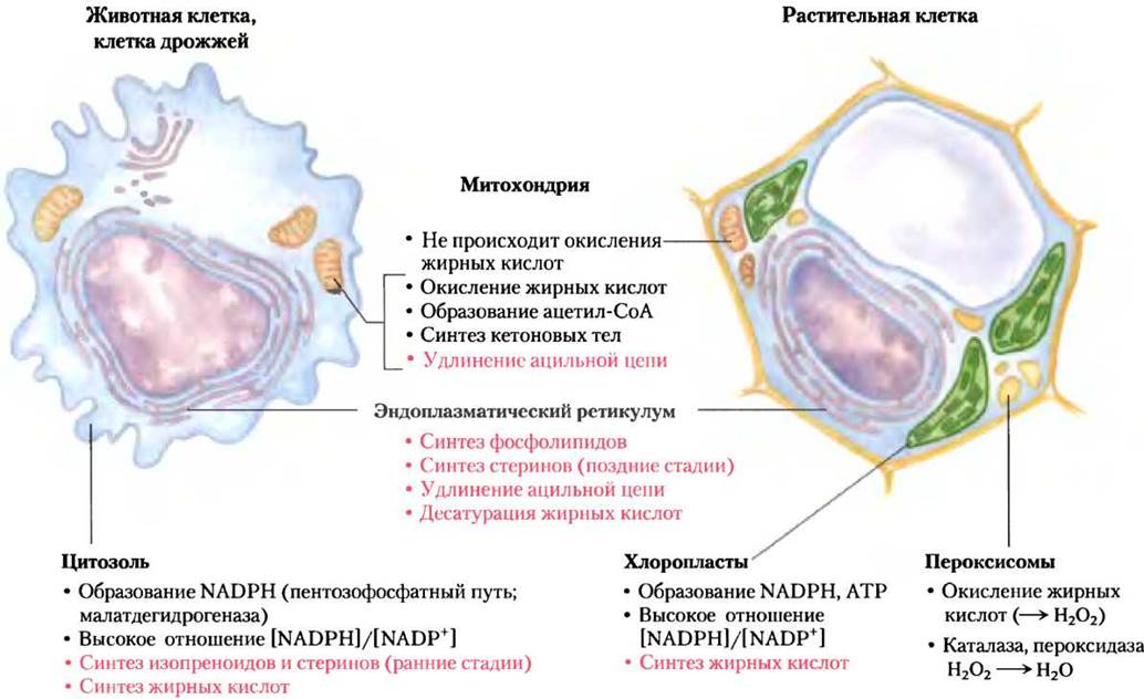
Typically, NADPH serves as the electron carrier in anabolic reactions, whereas NAD+ functions in catabolic pathways. In hepatocytes, the [NADPH]/[NADP+] ratio is exceptionally high (approx. 75), which establishes a strongly reducing environment favorable for the reductive synthesis of Fatty Acids and other Biomolecules. The cytosolic [NADPH]/[NAD+] ratio is much lower, ~8 · 10-4, allowing NAD+-dependent oxidative catabolism of glucose to occur in the same compartment and at the same time as fatty acid synthesis. In mitochondria, the [NADPH]/[NAD] ratio is slightly higher than in the cytosol, driven by the electron flow toward NAD+ during the Oxidation of Fatty acids, Amino Acids, pyruvate, and acetyl-CoA. Within mitochondria, a high [NADPH]/[NAD+] ratio favors oxygen reduction in the Respiratory Chain.
In hepatocytes and adipocytes, cytosolic NADPH is generated primarily via the Pentose Phosphate Pathway (see Fig. 14-21) and through the action of the malic enzyme (malate dehydrogenase, or malic enzyme; Fig. 21-9a). The NADP-producing malate dehydrogenase, which functions in carbon assimilation in C4 plants (see Fig. 20-23), also serves an alternative function. The pyruvate produced in the reaction shown in Fig. 21-9a is returned to the mitochondria. The NADPH required for fatty acid biosynthesis in mammalian hepatocytes and Mammary Glands is supplied by The pentose phosphate pathway (Fig. 21-9b).
Fig. 21-9. Generation of NADPH. Two pathways for NADPH production: (a) catalyzed by the malic enzyme, and (b) the pentose phosphate pathway.

In photosynthetic plant cells, fatty acid synthesis occurs not in the cytosol, but in the chloroplast stroma (Fig. 21-8). This is because NADPH is generated within chloroplasts during the light reactions of Photosynthesis:

Acetate is exported from mitochondria as citrate
In nonphotosynthetic eukaryotes, nearly all the acetyl-CoA used for fatty acid synthesis is produced in mitochondria via pyruvate oxidation and the Catabolism of Amino Acid Carbon Skeletons. Acetyl-CoA derived from fatty acid oxidation does not contribute significantly to animal fatty acid biosynthesis because these two pathways are reciprocally regulated, as described below.
Because The inner mitochondrial membrane is impermeable to acetyl-CoA, acetyl equivalents are transported across this membrane via a specialized shuttle system (Fig. 21-10). Intramitochondrial acetyl-CoA first reacts with oxaloacetate in The Citric Acid Cycle to form citrate, in a reaction catalyzed by citrate synthase (see Fig. 16-7).
Fig. 21-10. Shuttle system for The transport of acetyl groups from mitochondria to the cytosol. The outer mitochondrial membrane is freely permeable to all the components shown. Pyruvate—generated either from Amino Acid Catabolism in the mitochondrial matrix or from glucose via Glycolysis in the cytosol—is converted into acetyl-CoA in the matrix. Acetyl groups leave the mitochondrion in the form of citrate and are delivered to the cytosol for fatty acid synthesis as acetyl-CoA. Oxaloacetate is reduced to malate, which is returned to the mitochondrial matrix and reconverted into oxaloacetate. In the cytosol, malate is oxidized by the malic enzyme to yield NADPH (which remains in the cytosol), while pyruvate returns to the mitochondrial matrix.

Citrate then crosses the inner mitochondrial membrane via the citrate transporter. In the cytosol, citrate is cleaved by citrate lyase in an ATP-dependent reaction that regenerates acetyl-CoA. Oxaloacetate cannot return directly to the mitochondrial matrix because specific transporters are lacking. Instead, cytosolic malate dehydrogenase reduces oxaloacetate to malate, which is transported back into the mitochondrial matrix by the malate-α-ketoglutarate carrier in exchange for citrate. Within the matrix, malate is reoxidized to oxaloacetate, completing the shuttle cycle. However, the bulk of the malate produced in the cytosol is utilized to generate cytosolic NADPH via the malic enzyme (Fig. 21-9a). The resulting pyruvate is imported into the mitochondria via a specialized transporter (Fig. 21-10) and converted back into oxaloacetate in the matrix by pyruvate carboxylase. This cycle consumes two ATP molecules per molecule of acetyl-CoA (one required for the citrate lyase reaction, and another for pyruvate carboxylase). Following the cleavage of citrate to yield acetyl-CoA, the remaining four carbon atoms are converted into pyruvate and CO2 by the malic enzyme, an event that accounts for approximately half of the NADPH required for fatty acid synthesis. The remaining NADPH is provided by the pentose phosphate pathway.
Fatty Acid Biosynthesis Is Strictly Regulated
When a cell or Organism has an Abundance of metabolic energy beyond its immediate needs, this excess is typically converted into fatty acids and stored as lipids (triacylglycerols). The reaction catalyzed by acetyl-CoA carboxylase is the rate-limiting step in fatty acid biosynthesis, and this enzyme plays a central role in Metabolic Regulation. In vertebrates, palmitoyl-CoA—the primary product of fatty acid synthesis—acts as a feedback inhibitor of the enzyme, whereas citrate serves as an allosteric activator (Fig. 21-11a) that increases Vmax. Citrate plays a pivotal role in shifting cellular metabolism from The oxidation of metabolic fuels to their storage as fatty acids. When mitochondrial concentrations of acetyl-CoA and ATP rise, citrate is exported from the mitochondria, where it acts both as a precursor for cytosolic acetyl-CoA and as an allosteric signal that activates acetyl-CoA carboxylase. Simultaneously, citrate inhibits Phosphofructokinase-1 activity (see Fig. 15-14), thereby slowing down carbon flux through glycolysis.
and serves as an allosteric signal to activate acetyl-CoA carboxylase. Furthermore, citrate inhibits phosphofructokinase-1 activity (see Fig. 15-14), reducing the flow of carbon through glycolysis.
Figure 21-11. Regulation of Fatty acid synthesis. (a) In vertebrate cells, both Allosteric Regulation and hormone-dependent covalent modification control The conversion of precursors into malonyl-CoA. In plants, acetyl-CoA carboxylase is activated by changes in Mg2+ concentration and pH triggered by illumination (not shown). (b) Electron micrograph showing filaments of acetyl-CoA carboxylase (the active, dephosphorylated form).

Acetyl-CoA carboxylase is also regulated through covalent modification. Phosphorylation, triggered by the hormones Glucagon and epinephrine, inactivates the enzyme and diminishes its sensitivity to citrate activation, thereby suppressing fatty acid synthesis. In its active (dephosphorylated) form, acetyl-CoA carboxylase polymerizes into long filaments (Fig. 21-11b); phosphorylation induces dissociation into monomeric subunits and a corresponding loss of activity.
The acetyl-CoA carboxylase of plants and bacteria is not regulated by citrate or by phosphorylation-dephosphorylation cycles. Plant enzymes are activated by the increases in stromal pH and Mg2+ concentration that accompany illumination (see Fig. 20-17). Bacteria do not use triacylglycerols for energy storage; in E. coli, the primary role of fatty acid synthesis is to provide precursors for membrane Lipid Biosynthesis. This process is intricately regulated by guanine NUCLEOTIDES (such as ppGpp), which coordinate cell growth with membrane biogenesis (see Fig. 8-39, vol. 1; Fig. 28-24, vol. 3).
Beyond the REGULATION OF ENZYMATIC Activity, these Metabolic pathways are subject to additional control at the level of Gene Expression. For instance, when animals ingest excess amounts of certain polyunsaturated fatty acids, the expression of genes encoding various lipid-producing hepatic enzymes is suppressed. The precise molecular mechanisms governing this gene regulation remain to be fully elucidated.
If fatty acid synthesis and β-oxidation were to occur simultaneously, the two processes would constitute a futile cycle, wasting metabolic energy to no purpose. As noted previously (see Fig. 17-12), β-oxidation is suppressed by malonyl-CoA, which inhibits carnitine acyltransferase I. Thus, the formation of malonyl-CoA—the initial intermediate of fatty acid synthesis—halts β-oxidation at the level of the mitochondrial inner-membrane transport system. This regulatory mechanism exemplifies yet another advantage of compartmentalizing anabolic and catabolic pathways within distinct cellular Organelles.
Long-Chain Saturated Fatty Acids Are Synthesized from Palmitate
In animal cells, palmitate—the principal product of the fatty acid synthase system—serves as the precursor for other long-chain fatty acids (Fig. 21-12). It can be elongated to stearate (18:0) or even longer saturated fatty acids through the sequential addition of acetyl units. This elongation is mediated by the fatty acid elongation system, located in the smooth Endoplasmic reticulum (ER) and mitochondria. The more active ER system extends the 16-carbon chain of palmitoyl-CoA by two carbon atoms to yield stearoyl-CoA. Although the mechanistic steps of ER elongation closely mirror those of palmitate synthesis, different enzyme systems are involved, and coenzyme A rather than ACP typically serves as the acyl carrier. Two-carbon units derived from malonyl-CoA are added in the ER, followed by sequential reduction, dehydration, and reduction steps to yield the saturated 18-carbon chain, stearoyl-CoA.
Figure 21-12. Pathways of fatty acid synthesis. Palmitate is the precursor of stearate and longer-chain saturated fatty acids, as well as the monounsaturated fatty acids palmitoleate and oleate. Mammals cannot convert oleate into linoleate or linolenate (highlighted in pink); consequently, these must be obtained from the diet and are termed Essential Fatty Acids. The schematic outlines the conversion of linoleate into other polyunsaturated fatty acids and eicosanoids. Unsaturated Fatty Acid Nomenclature denotes carbon chain length and double-bond positions, as shown in Table 10-1, vol. 1.

Desaturation of fatty Acids Requires Mixed-Function Oxidases
Palmitate and stearate serve as precursors for the two most abundant monounsaturated fatty acids in animal Tissues: palmitoleate, 16:1(∆9), and oleate, 18:1(∆9). Both contain a single cis double bond situated between C-9 and C-10 (see Table 10-1). The double bond is introduced into the fatty acid chain via an oxidative reaction catalyzed by acyl-CoA desaturase (Fig. 21-13), a mixed-function oxidase (Box 21-1).
Figure 21-13. Electron transfer during fatty acid desaturation in vertebrates. Blue arrows indicate the path of electrons during the oxidation of two substrates, acyl-CoA and NADPH, by molecular oxygen. These reactions take place on the cytosolic surface of the smooth ER. Plants utilize a analogous pathway involving different electron carriers.

Two distinct substrates—the Fatty acid and NADH or NADPH—undergo simultaneous two-electron oxidations. The Electron Transport Chain comprises a cytochrome (cytochrome b5) and a flavoprotein (cytochrome b5 reductase). Both components, alongside acyl-CoA desaturase, reside in the smooth ER. Bacteria possess two distinct cytochrome b5 reductases: an NADH-dependent and an NADPH-dependent form, though the primary physiological electron donor in vivo remains uncertain. In plants, oleate is synthesized within the chloroplast stroma by stearoyl-ACP desaturase, which utilizes reduced ferredoxin as the electron donor.
Mammalian hepatocytes readily introduce double bonds at the ∆9 position of fatty acids but lack the enzymatic machinery to insert additional double bonds between C-10 and the terminal methyl group. Consequently, mammals cannot synthesize linoleate, 18:2(∆9,12), or α-linolenate, 18:3(∆9,12,15). In contrast, plants readily synthesize both compounds; the desaturases that introduce double bonds at the ∆12 and ∆15 positions are localized in the ER and chloroplasts. Plant ER enzymes act not only on free fatty acids but also on the phospholipid phosphatidylcholine containing at least one esterified oleate moiety (Fig. 21-14). Both plants and bacteria must synthesize polyunsaturated fatty acids to maintain essential membrane fluidity at lower temperatures. Figure 21-14. Action of plant desaturases. Plant desaturases oxidize oleate esterified to phosphatidylcholine to yield polyunsaturated fatty acids; certain products are subsequently released from phosphatidylcholine via hydrolysis.
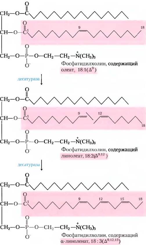
Because linoleate and linolenate are essential precursors for the Synthesis of Other vital metabolites, they are classified as essential fatty acids for mammals, which must acquire them through plant-based diets. Dietary linoleate can be converted into several other polyunsaturated fatty acids, notably γ-linolenate, eicosatrienoate, and arachidonate (eicosatetraenoate)—all of which are derived exclusively from linoleate (Fig. 21-12). Arachidonate, 20:4(∆5,8,11,14), serves as the indispensable precursor for regulatory lipids known as eicosanoids. Through fatty acid elongation reactions analogous to those described on p. 457, linoleate and linolenate are extended to yield 20-carbon fatty acids.
Box 21-1. Mixed-Function Oxidases, Oxygenases, and Cytochrome P-450
Throughout this chapter, we have examined several enzymes that catalyze oxidation-reduction reactions involving molecular oxygen. One such reaction is the formation of a double bond in a fatty acid chain (see Fig. 21-13).
The nomenclature of enzymes catalyzing these reactions often confuses students regarding their catalytic mechanisms. Oxidases are broadly defined as enzymes that catalyze oxidation reactions in which molecular oxygen serves as an electron acceptor, yet the oxygen atoms themselves are not incorporated into the oxidized product (though, as we shall see, exceptions exist!). A classic example of this type of oxidase is the enzyme that introduces a double bond into acyl-CoA during peroxisomal fatty acid oxidation (see Fig. 17-13); another is cytochrome c oxidase of the Mitochondrial Electron Transport chain (see Fig. 19-14). In the former case, the transfer of two electrons to O2 yields hydrogen peroxide; in the latter, four electrons reduce O2 to two molecules of H2O. Many, though not all, oxidases are Flavoproteins.
Oxygenases catalyze oxidative reactions in which oxygen atoms are directly incorporated into the substrate molecule, typically forming a new hydroxyl or carboxyl group. Dioxygenases catalyze reactions in which both atoms of an O2 molecule are incorporated into an organic substrate. A prominent example is Tryptophan 2,3-dioxygenase, which catalyzes the cleavage of the indole ring of tryptophan during the catabolism of this amino acid. When this reaction is carried out in the presence of 18O2, the labeled oxygen atoms are recovered in the two carbonyl groups of the product (highlighted in red).

Monooxygenases are more numerous and exhibit more complex Mechanisms of action. They catalyze reactions in which only one of the two oxygen atoms of an O2 molecule is incorporated into an organic substrate, while the second oxygen atom is reduced to H2O. Monooxygenases require two substrates to effect the reduction of both oxygen atoms of O2: the principal substrate accepts one of the two oxygen atoms, whereas the cosubstrate provides the hydrogen atoms needed to reduce the second oxygen atom to H2O. The overall reaction equation for monooxygenases is as follows:
АН + ВН2 + O-O —> А-ОН + В + Н2O
where AH is the principal substrate and BH2 is the cosubstrate. Because the majority of monooxygenases catalyze Reactions Involving the hydroxylation of the principal substrate, they are also referred to as hydroxylases. Sometimes they are called mixed-function oxidases or mixed-function oxygenases to indicate that they simultaneously oxidize two different substrates. (Note that the use of the term "oxidase" here is a departure from its conventional meaning, as discussed above.)
Various classes of monooxygenases exist, depending on The Nature of the cosubstrate. Some monooxygenases utilize flavin nucleotides (FMNH2 or FADH2), others use NADH or NADPH, and still others employ α-ketoglutarate as a cosubstrate. The enzyme that hydroxylates the phenyl ring of phenylalanine to yield Tyrosine is a monooxygenase that uses tetrahydrobiopterin as its cosubstrate (see Fig. 18-23). This is the enzyme that is defective in humans suffering from the genetic disorder phenylketonuria.
The most numerous and complex monooxygenase reactions involve a hemoprotein known as cytochrome P-450. This cytochrome is typically localized in the smooth ER rather than in mitochondria. Like mitochondrial cytochrome oxidase, cytochrome P-450 can react with O2 and bind carbon monoxide (CO); however, it can be distinguished from cytochrome oxidase because the complex of its reduced form with carbon monoxide exhibits a sharp absorption peak at 450 nm—hence the designation P-450.
Cytochrome P-450 catalyzes hydroxylation reactions in which an organic substrate RH is converted to R-OH, meaning it incorporates one oxygen atom from an O2 molecule, while the other oxygen atom is reduced to H2O using reducing equivalents provided by NADH or NADPH, typically transferred to cytochrome P-450 via an iron-sulfur protein. Figure 1 presents a simplified scheme of cytochrome P-450 action, which includes intermediate stages that are not yet fully understood.
Fig. 1.

Cytochrome P-450 is actually represented by a whole family of similar proteins; several hundred members of this family are known, each possessing distinct substrate Specificity. For example, in the adrenal cortex, a specific cytochrome P-450 is involved in the hydroxylation of steroids to yield hormones (see Fig. 21-46). Cytochrome P-450 plays a crucial role in the hydroxylation of many drugs, such as barbiturates, as well as other xenobiotics (foreign chemical substances), particularly hydrophobic compounds that are practically insoluble in water. The environmental carcinogen benzo[a]pyrene (found in cigarette smoke) undergoes hydroxylation by cytochrome P-450 as part of its detoxification. Hydroxylation of xenobiotics increases their water solubility, thereby facilitating their excretion in the urine. Unfortunately, the hydroxylation of certain compounds converts them into toxic substances that compromise the detoxification system.
The reactions catalyzed by mixed-function oxidases described in this chapter belong to the desaturation pathways of fatty acyl-CoA (Fig. 21-13); these reactions also occur in the synthesis of Leukotrienes (Fig. 21-16), plasmalogens (Fig. 21-30), the conversion of squalene to cholesterol (Fig. 21-37), and the biosynthesis of Steroid Hormones (Fig. 21-46).
Eicosanoids are derived from 20-carbon polyunsaturated fatty acids
Eicosanoids are a family of highly potent signaling biomolecules that function as short-range messengers, affecting tissues in the immediate vicinity of the cells that produce them. In response to hormonal or other stimuli, phospholipase A2—present in most mammalian cells—attacks membrane phospholipids, releasing arachidonate esterified at the middle carbon of glycerol. Enzymes in the smooth ER then convert arachidonate into Prostaglandins, beginning with the formation of prostaglandin H2 (PGH2), the immediate precursor to numerous other Prostaglandins and thromboxanes (Fig. 21-15, a). The two steps in the biosynthesis of PGH2 are catalyzed by a bifunctional enzyme called cyclooxygenase (COX), also known as prostaglandin H2 synthase. In the first step, the cyclooxygenase activity introduces molecular oxygen, converting arachidonate into PGG2. In the second step, the peroxidase activity of COX converts PGG2 into PGH2.
Fig. 21-15. The "cyclic" pathway from arachidonate to prostaglandins and thromboxanes. (a) Following the release of arachidonate from phospholipids by the action of phospholipase A2, the cyclooxygenase and peroxidase activities of COX (prostaglandin H2 synthase) catalyze the formation of PGH2, the precursor of other prostaglandins and thromboxanes. (b) Aspirin inhibits the first reaction by acetylating an essential Ser residue in the enzyme. Ibuprofen and naproxen inhibit the same reaction, likely by mimicking The structure of the substrate or a reaction intermediate. (c) Specific COX-2 inhibitors developed for pain relief (see text).

Mammals express two isoforms of prostaglandin H2 synthase—COX-1 and COX-2. They have distinct physiological functions yet share highly similar Amino acid sequences (nearly 60% identity) and catalytic mechanisms at both active sites. COX-1 is responsible for the synthesis of prostaglandins that regulate gastric mucus secretion, whereas COX-2 mediates prostaglandins involved in inflammation, pain, and fever.
Pain can be alleviated by inhibiting COX-2. The first drug widely used for this purpose was aspirin (acetylsalicylate; Fig. 21-15, b). The trade name "aspirin" (derived from *a* for acetyl and *spir* from *Spiraea*—the German botanical name for meadowsweet, *Spiraea ulmaria*, a source of salicylic acid) was introduced in 1899 when the drug began to be manufactured by Bayer. Aspirin inhibits the cyclooxygenase activity of both COX isoforms to a similar extent by acetylating a Serine residue and blocking the active site of each enzyme, thereby also suppressing the synthesis of prostaglandins and thromboxanes. Ibuprofen and other widely used nonsteroidal anti-inflammatory drugs (NSAIDs; Fig. 21-15, b) also inhibit this pair of enzymes. However, the inhibition of COX-1 can lead to unpleasant side effects, including gastric irritation and more severe complications. In the 1990s, following the Determination of the crystal structures of COX-1 and COX-2, compounds with significantly greater specificity for COX-2 were developed. These substances effectively relieve pain of various origins. Three drugs came into widespread use: rofecoxib (Vioxx), valdecoxib (Bextra), and celecoxib (Celebrex) (Fig. 21-15, c). Developed in the late 1990s, they immediately attracted the attention of pharmaceutical companies. However, as clinical trial reports accumulated, enthusiasm waned because these drugs were shown to increase the risk of myocardial infarction and stroke. The exact causes of these side effects remain unclear, but some researchers suggest that COX-2 inhibitors disrupt the delicate balance between prostacyclin—a hormone that dilates Blood Vessels, inhibits platelet aggregation, and whose production is suppressed by COX-2 inhibitors—and thromboxanes, which are synthesized via COX-1 and promote blood clotting. Vioxx was pulled from production and withdrawn from the market in 2004, soon followed by a ban on Bextra. Celebrex remains in use (as of 2013), but with heightened caution.
Blood Platelets contain thromboxane synthase, which converts PGH2 into thromboxane A2, the parent compound for other thromboxanes (Fig. 21-15, a). Thromboxanes cause blood vessel constriction and platelet aggregation during the Cytology/cytology/16.html">Early stages of blood clotting. Low doses of aspirin taken regularly reduce the likelihood of Heart attacks and strokes by diminishing thromboxane production. ■
Like prostaglandins, thromboxanes contain a five- or six-membered ring; the pathway from arachidonate to these two classes of compounds is sometimes referred to as the "cyclic" pathway to distinguish it from the "linear" pathway leading from arachidonate to leukotrienes, which feature a linear carbon chain (Fig. 21-16). Leukotriene synthesis is initiated by the action of several lipoxygenases, which catalyze the insertion of molecular oxygen into arachidonate. These enzymes, found in leukocytes, heart, Brain, Lungs, and Spleen, belong to the mixed-function oxidase family; the most prominent mixed-function oxidase is cytochrome P-450 (Box 21-1). Different leukotrienes are distinguished by THE POSITION OF the peroxide group introduced by lipoxygenases. Unlike the "cyclic" pathway, the "linear" pathway from arachidonate is not inhibited by aspirin or other NSAIDs.
Fig. 21-16. The "linear" pathway from arachidonate to leukotrienes.

Plants also synthesize vital signaling molecules from fatty acids. As in animals, the key initiation step of the signaling pathway involves the Activation of a specific phospholipase. In plants, the released fatty acid substrate is α-linolenate. In the first step of the pathway, a lipoxygenase catalyzes the conversion of linolenate to jasmonate. Jasmonate (see Fig. 12-32 in Vol. 1) is known to function in defense signaling against insect herbivory, resistance to fungal pathogens, and pollen maturation. Additionally, it regulates seed germination, ROOT growth, and fruit and seed development.
Summary of Section 21.1 Biosynthesis of Fatty Acids and Eicosanoids
■ Long-chain fatty acids are synthesized from acetyl-CoA by a cytosolic enzyme complex (possessing six active sites) and an acyl carrier protein (ACP). Two Types of fatty acid synthase exist. FAS type I is found in vertebrates and fungi; it consists of multifunctional Polypeptides. FAS type II is composed of a complex of separate proteins and is found in bacteria and plants. These two synthases contain distinct -SH groups (provided by the phosphopantetheine of ACP and a Cys residue of the β-ketoacyl-ACP synthase) that function as carriers of the growing fatty acyl intermediates.
■ Malonyl-ACP, formed from acetyl-CoA (mitochondrial shuttle) and CO2, condenses with an acetyl group linked to Cys-SH to yield acetoacetyl-ACP and CO2. This is followed by reduction to the D-β-hydroxy derivative, dehydration to yield trans-Δ2-unsaturated acyl-ACP, and a final reduction to butyryl-ACP. NADPH serves as the electron donor for both reduction steps. Fatty acid synthesis is regulated primarily at the level of malonyl-CoA formation.
• Six more malonyl-ACP molecules successfully enter the reaction, attaching to the carboxyl end of the growing fatty acyl chain to form the final product of fatty acid synthase, palmitoyl-ACP. Palmitate is subsequently released via hydrolysis.
• Palmitate can be elongated to form the 18-carbon stearate. Both palmitate and stearate can undergo desaturation through the action of mixed-function oxidases, yielding palmitoleate and oleate, respectively.
• Mammals are unable to synthesize linoleate and must obtain it from plant sources; they convert exogenous linoleate into arachidonate, the precursor for the biosynthesis of eicosanoids (prostaglandins, thromboxanes, and leukotrienes—a family of highly potent signaling molecules). The synthesis of prostaglandins and thromboxanes is inhibited by NSAIDs, which target The activity of prostaglandin H2 synthase.
Last update: 06/08/2026
Editorial and Educational Adaptation: This material has been compiled based on the primary/original source text. The project team performed an editorial review, corrected technical inaccuracies, structured sections, and adapted the content for an educational format.
What was processed:
- elimination of formatting defects (OCR errors, structural breaks, corrupted characters);
- editorial organization of content;
- standardization of terminology in accordance with academic sources;
- verification of factual statements against the original source text.
All mentions of the author, publication year, and origin of the primary text have been preserved in accordance with the source.