MEDICAL BIOLOGY, ANATOMY, HUMAN PHYSIOLOGY AND PATHOLOGY - Y.I. Fedoniuk 2010
ANATOMY, PHYSIOLOGY, PATHOLOGY
CHAPTER 5. THE PROCESS OF BLOOD AND LYMPH CIRCULATION. ITS PATHOLOGY
VESSELS OF THE SYSTEMIC CIRCULATION
2. VEINS OF THE SYSTEMIC CIRCULATION
2.1. System of the Superior Vena Cava
The superior vena cava (v. cava superior) collects Blood from the HEAD, neck, upper limbs, and walls of the thoracic cavity. It is formed by the junction of the right and left brachiocephalic Veins at the level of the Articulation of the first rib with the Sternum. The azygos vein empties into it.
The brachiocephalic veins (right and left) are each formed by the merger of the subclavian and internal jugular veins. Veins of The Thyroid Gland and internal thoracic veins empty into the brachiocephalic veins.
The VEINS OF THE Head and Neck drain into three main paired trunks: the internal, external, and anterior jugular veins.
The internal jugular vein collects blood from the cranial cavity, as well as from the external surface of the head, Tongue, Pharynx, and thyroid gland. It begins in the region of the jugular foramen and runs in the neck as part of the neurovascular bundle.
The External Jugular Vein collects blood from the occipital region and auricle. It empties into the subclavian vein.
The anterior jugular vein collects blood from the anterior region of the neck above the Hyoid bone and empties into the external jugular or subclavian vein.
Veins of the upper limb. There are superficial and deep veins of the upper limb (Fig. 5.33). The superficial veins are the cephalic vein (lateral) and the basilic vein (medial). In the region of the cubital fossa, they are connected by a short median cubital vein. The cephalic vein empties into the axillary vein, and the basilic vein empties into the brachial vein. These deep veins originate in the hand (predominantly two veins accompanying one artery). The ulnar and radial veins form two brachial veins, which then unite to form a single axillary vein. The axillary vein continues as the subclavian vein.
Class="center">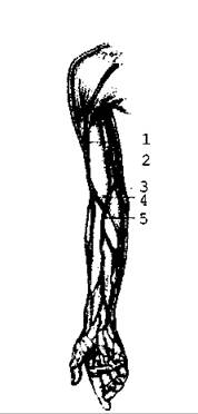
Fig. 5.33. Superficial (subcutaneous) veins
The subclavian vein lies on the upper surface of the first rib, anterior to the artery of the same name. By uniting with the internal jugular vein, it forms the brachiocephalic vein. The left and right brachiocephalic veins merge to form the superior vena cava.
The veins of the thoracic cavity are represented by the azygos and hemiazygos veins, which collect blood from the walls and Organs of the right and left halves of the thoracic cavity. They lie in the posterior Mediastinum; the hemiazygos vein empties into the azygos vein, and the latter empties into the superior vena cava.
The internal thoracic veins are located on the anterior wall of the thoracic cavity. They accompany the corresponding Arteries and empty into the brachiocephalic vein.
Last update: 08/08/2026
Editorial and Educational Adaptation: This material has been compiled based on the primary/original source text. The project team performed an editorial review, corrected technical inaccuracies, structured sections, and adapted the content for an educational format.
What was processed:
- elimination of formatting defects (OCR errors, structural breaks, corrupted characters);
- editorial organization of content;
- standardization of terminology in accordance with academic sources;
- verification of factual statements against the original source text.
All mentions of the author, publication year, and origin of the primary text have been preserved in accordance with the source.