Anatomy of the Head and Neck Vessels and Nerves (Angioneurology) - Chornokulskyi S.T. 2009
Veins of the head and neck
Internal jugular vein
The Veins OF THE HEAD and Neck are tributaries of the SUPERIOR VENA CAVA (v. cava superior) and comprise the following paired vessels:
— Internal jugular vein (v. jugularis interna);
— External Jugular Vein (v. jugularis externa);
— Anterior jugular vein (v. jugularis anterior).
The internal jugular vein (v. jugularis interna) is the largest of the jugular veins, serving as a direct continuation of the sigmoid sinus of the dura mater. It begins at the level of the jugular foramen (foramen jugulare), just below which lies its dilation known as the superior bulb of the internal jugular vein (bulbus superior venae jugularis). Initially, the vein lies posterior and lateral to the Internal Carotid Artery, and subsequently lateral to the common carotid artery. Between them and posteriorly lies the Vagus nerve, completing the neurovascular bundle of the neck.
Before merging with the subclavian vein, the v. jugularis interna forms a second dilation called the inferior bulb of the internal jugular vein (bulbus inferior venae jugularis), above and below which paired Valves are located.
The tributaries of the internal jugular vein are divided into 2 groups:
— intracranial and
— extracranial.
Intracranial tributaries of the internal jugular vein (Figs. 21, 21a).
I. Dural Venous Sinuses of the Brain (sinus durae matris):
1 — superior sagittal sinus (sinus sagittalis superior);
2 — inferior sagittal sinus (sinus sagittalis inferior);
3 — straight sinus (sinus rectus);
4 — transverse sinus (sinus transversus);
5 — confluence of sinuses (confluens sinuum);
6 — marginal sinus (sinus marginalis);
7 — occipital sinus (sinus occipitalis);
8 — superior petrosal sinus (sinus petrosus superior);
9 — inferior petrosal sinus (sinus petrosus inferior);
10 — cavernous sinus (sinus cavernosus);
11 — anterior intercavernous sinus (sinus intercavemosus anterior);
12 — posterior intercavernous sinus (sinus interca vernosus posterior);
13 — sphenoparietal sinus (sinus sphenoparieralis);
14 — sigmoid sinus (sinus sigmoideus).
Class="center">
Fig. 20. Veins of the head and neck
1 - superior sagittal sinus (sinus sagittalis superior);
2 - inferior sagittal sinus (sinus sagittalis inferior);
3 - confluence of sinuses (confluens sinuum);
4 - straight sinus (sinus rectus);
5 - great cerebral vein (v. cerebri magna);
6 - transverse sinus (sinus transversus);
7 - superior petrosal sinus (sinus petrosus superior);
8 - sigmoid sinus (sinus sigmoideus);
9 - superficial temporal veins (venae temporales superficiales);
10 - inferior petrosal sinus (sinus petrosus inferior);
11 - cavernous sinus (sinus cavernosus);
12 - superior ophthalmic vein (vena ophthalmica superior);
13 - angular vein (vena angularis);
14 - pterygoid plexus (plexus pterygoideus);
15 - facial vein (vena facialis);
16 - vagus nerve (n. vagus);
17 - retromandibular vein (vena retromandibularis);
18 - middle scalene Muscle (m. scalenus medius);
19 - anterior scalene muscle (m. scalenus anterior);
20 - external jugular vein (v. jugularis externa);
21 - Subclavian Artery (a. subclavia);
22 - subclavian vein (v. subclavia);
23 - internal thoracic vein (v. thoracica interna);
24 - superior thyroid vein (v. thyroidea superior);
25 - internal jugular vein (v. jugularis interna);
26 - anterior jugular vein (v. jugularis anterior);
27 - Right lymphatic duct (ductus lymphaticus dexter)

Fig. 20a. Blood supply to the Cerebral Cortex (schematic):
1 - Arteries;
2 - capillaries;
3 - veins

Fig. 21. Dural venous sinuses at the Base of the Skull
1 - crista galli (crista galli);
2 - anterior intercavernous sinus (sinus intercavemosus anterior);
3 - superior sagittal sinus (sinus sagittalis superior);
4 - internal carotid artery (a. carotis interna);
5 - superior ophthalmic vein (v. ophthatmica superior);
6 - sphenoparietal sinus (sinus sphenoparietalis);
7 - cavernous sinus (sinus cavernosus);
8 - inferior petrosal sinus (sinus petrosus inferior);
9 - superior petrosal sinus (sinus petrosus superior);
10 - basilar plexus (plexus basilaris);
11 - foramen magnum (foramen magnum);
12 - marginal sinus (sinus marginaUs);
13 - sigmoid sinus (sinus sigmoideus);
14 - occipital sinus (sinus occipitalis);
15 - transverse sinus (sinus transversus);
16 - confluence of sinuses (confluens sinuurn)

Fig. 21 a. Dural venous sinuses: A — frontal section; B — posterolateral view; C — artery of the pia mater of the brain.
A:
1 - superior sagittal sinus (sinus sagittalis superior);
2 - Parietal bone (os parietale);
3 - dura mater of the brain (dura mater encephali);
4 - straight sinus (sinus rectus);
5 - transverse sinus (sinus transversus);
6 - tentorium cerebelli (tentorium cerebelli);
7 - falx cerebri (falx cerebri).
B:
1 - superior sagittal sinus (sinus sagittalis superior);
2 - inferior sagittal sinus (sinus sagittalis inferior);
3 - crista galli (crista gaili);
4 - straight sinus (sinus rectus);
5 - tentorium cerebelli (tentorium cerebelli);
6 - sigmoid sinus (sinus sigmoideus);
7 - transverse sinus (sinus transversus);
8 - occipital sinus (sinus occipitalis);
9 - falx cerebelli (falx cerebelli);
10 - confluence of sinuses (confluens sinuum).
C:
1, 2 - dural venous sinuses (sinus durae matris);
3 - periarterial adventitial space;
4,10 - external glial limiting membrane (membrana limitans gliocytica externa);
5 - neuron (neurocytus);
6 - pericellular space;
7 - gliocyte (gliocytus);
8 - gliocyte process (processus gliocyticus);
9 - perivascular glial limiting membrane (membrana limitans glialis perivascularis);
11 - artery of the pia mater;
12 - subarachnoid space (spatium subarachnoidale);
13 - subdural space (spatium subdurale)
Venous blood from the dural sinuses collects into the sigmoid sinus and further into the internal jugular vein. The sinuses receive blood from the cerebral veins, internal ear, dura mater (except for the middle meningeal vein, v. meningea media), and diploic veins (diploë) (Fig. 21 b).
II. The veins of the cerebrum are classified as superficial and deep.
1. Superficial cerebral veins (vv. superficiales cerebri):
— Superior, middle, and inferior cerebral veins (vv. superiores mediae et inferiores superficiales cerebri).
The superficial cerebral veins collect blood from the venous network of the pia mater, pierce the arachnoid mater, and drain into the dural venous sinuses.
2. Deep cerebral veins (vv. profundae cerebri).
— Basal vein (v. basalis) is paired and formed by the anterior cerebral veins (vv. anteriores cerebri): deep middle cerebral vein (v. profunda media cerebri); inferior thalamostriate veins (vv. thalamostriatae inferiores): inferior ventricular and choroid veins, olfactory gyrus vein (v. gyri olfactorii); and peduncular veins (vv. pedunculares).
The 2 basal veins, anterior communicating vein, and 2 posterior inferior communicating veins curve around the structures of the Hypothalamus and Brainstem, forming the cerebral venous circle. The deep veins collect blood from the basal nuclei, hippocampus, septum pellucidum, hypothalamus, and the choroid plexuses of the brain ventricles, among other structures.
— Internal cerebral veins (vv. internae cerebri) (Fig. 22) are two veins located within the tela choroidea of the Third ventricle which, by uniting, form the great cerebral vein (v. cerebri magna) (of Galen) (Fig. 20, 22).
— The great cerebral vein lies above the tectum of the Midbrain and drains into the straight sinus.
— Hypophysial portal veins (vv. portales hypophysiales) (Fig. 23) drain into the cavernous and intercavernous sinuses of the dura mater.
3. Veins of the brainstem (vv. trunci encephali).
4. Cerebellar veins (vv. cerebelli):
— superior and inferior veins of the vermis and Cerebellum (vv. superiores et inferiores vermis et cerebelli).
III. Ophthalmic veins (vv. orbitales) (Fig. 21).
1. The superior ophthalmic vein (v. ophthalmica superior) is a continuation of the nasofrontal vein (v. nasofrontalis), which in turn is formed by the confluence of the supratrochlear and angular veins. The superior ophthalmic vein passes through the superior orbital fissure and drains into the cavernous sinus. It is of great clinical importance because, lacking valves, it connects the extracranial (v. angularis) and intracranial veins of the head.

Fig. 21 b. Intracranial tributaries of the internal jugular vein on a frontal section of The Skull and brain.
1 - parietal bone (os parietale);
2 - superior sagittal sinus (sinussagittalissuperior);
3 - dura mater of the brain (dura mater encephali);
4 - subdural space (spatium subdurale);
5 - cerebral vein (vena cerebralis);
6 - cerebral artery (cortical branch) (arteria cerebralis (ramus corticalis));
7 - pia mater of the brain (pia mater encephali);
8 - perivascular space (spatium perivasculare);
9 - medullary branch (ramus meduliaris);
10 - cerebral cortex (cortex cerebri);
11 - falx cerebri (falx cerebralis);
12 - subarachnoid space (spatium subarachnoidale);
13 - arachnoid mater of the brain (arachnoidea mater encephali);
14 - vein of the dura mater (vena durae matris s. meningeae);
15 - diploic vein (vena diploica);
16 - venous lacuna of the dura mater (lacuna venosa durae matris);
17 - emissary vein (vena emissaria);
18 - arachnoid granulations (Pacchionian bodies) (granulationes arachnoideae Pacchioni)
2. The inferior ophthalmic vein (v. ophthalmica inferior) is formed by the veins of the lower eyelid, lies on the inferior orbital wall, and drains into the superior ophthalmic vein, or alternatively, into the cavernous sinus or pterygoid plexus.
IV. The veins of the internal ear (vv. labyrinthi) emerge through the internal acoustic meatus and drain into the inferior petrosal sinus.
V. The diploic veins (vv. diploicae) run within the channels of the spongy substance of the cranial vault bones, communicating with the dural venous sinuses and the subcutaneous venous Vessels of the head via emissary veins. These include the frontal, anterior and posterior temporal, and occipital diploic veins (Fig. 22 a).

Fig. 21 c. Veins of the Spinal Cord on its transverse section:
1 - left posterolateral longitudinal vein (vena longirudinalis dorsolateralis sinistra);
2 - radicular veins (venae radiculares);
3 - intervertebral vein (vena intervertebralis);
4 - anterior internal vertebral venous plexus (plexus venosus vertebralis internus anterius);
5 - anterior median longitudinal vein (vena longirudinalis mediana anterior);
6 - White matter OF the spinal cord (substantia alba medullae spinalis);
7 - posterior internal vertebral venous plexus (plexus venosus vertebralis internus posterior);
8 - posterior median longitudinal vein (vena longirudinalis mediana posterior)

Fig. 21 d. Hemomicrocirculatory bed of the spinal cord on a transverse section.
VI. Emissary veins (vv. emissariae) include the parietal, mastoid, condylar, and occipital veins (vv. emissariae parietalis, mastoidea, condylaris et occipitalis), which emerge from the cranial cavity through the correspondingly named apertures and canals of the cranial bones.
VII. Intracranial venous plexuses:
— basilar plexus (plexus basilaris) (Fig. 21);
— plexus of the hypoglossal canal (plexus venosus canalis nervi hypoglossi);
— plexus of the foramen ovale (plexus venosus foraminis ovalis);
— internal carotid plexus (plexus venosus caroticus internus).

Fig. 22. Internal cerebral veins (1) and the great cerebral vein in the tela choroidea of the third ventricle with the choroid plexus (2) of the Lateral ventricles
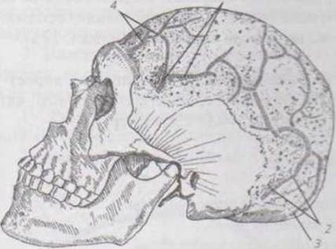
Fig. 22 a. Diploic veins of the BONES OF THE cranial vault
1. Anterior temporal diploic vein (v. diploica temporalis anterior);
2. Posterior temporal diploic vein (v. diploica temporalis posterior);
3. Occipital diploic vein (v. diploica occipitalis);
4. Frontal diploic vein (v. diploica frontalis)
Last update: 11/08/2026
Editorial and Educational Adaptation: This material has been compiled based on the primary/original source text. The project team performed an editorial review, corrected technical inaccuracies, structured sections, and adapted the content for an educational format.
What was processed:
- elimination of formatting defects (OCR errors, structural breaks, corrupted characters);
- editorial organization of content;
- standardization of terminology in accordance with academic sources;
- verification of factual statements against the original source text.
All mentions of the author, publication year, and origin of the primary text have been preserved in accordance with the source.