Human Anatomy and Physiology - I. V. Gayvoronsky 2011
Cardiovascular System
Hemomicrocirculatory bed
From the intraorgan Arteries with the smallest diameter, Blood enters the Vessels of the hemomicrocirculatory bed, which is represented by arterioles, precapillary arterioles, capillaries, postcapillary venules, and venules (Fig. 12.18). These vessels are involved in supporting metabolic processes in Organs and Tissues.
A characteristic feature of arterioles—the afferent vessels of the microcirculatory bed—is the presence of all three tunic layers in their walls. Precapillary arterioles contain circularly arranged myocytes only in their initial segments. They form precapillary sphincters that act as vascular Valves. These microscopic structures regulate blood flow into the capillary bed.
From the precapillary arterioles, blood flows into the capillaries. These are the thinnest-walled vessels, consisting solely of flat endothelial Cells resting on a basement membrane. Their diameter ranges from 3 to 10 µm. Capillaries facilitate Metabolic exchange between the blood and tissue fluid: various substances pass from the capillary lumen into the tissues and vice versa. However, not all capillaries are filled with blood at all times; at rest, only a fraction of them (10 to 30%) function actively, which is why a distinction is made between nutritive and reserve capillaries. The latter dilate and fill with blood only during functional activity, significantly increasing the number of active capillaries and thereby intensifying METABOLISM between the blood and tissues.
Class="center">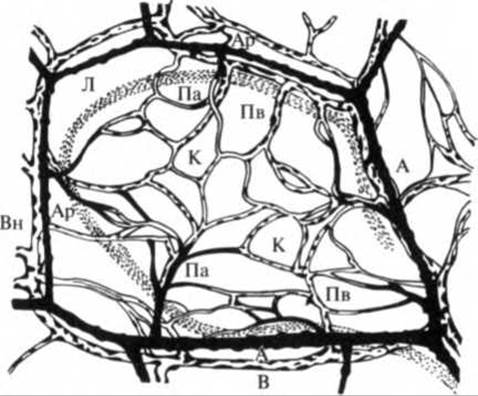
Fig. 12.18. Vessels of the microcirculatory bed (diagram):
А — artery; Ар — arteriole; Вн — venule; Па — precapillary arteriole; Пв — postcapillary venule; К — capillary; Л — lymphomicrocirculatory bed; В — vein
From the capillaries, blood flows into the efferent vessels of the hemomicrocirculatory bed—postcapillary venules and venules. The wall of postcapillary venules is structurally similar to that of capillaries. Venules have wider lumens, and their walls may feature scattered myocytes, while their lumens may occasionally contain isolated valves.
In certain tissues, arteriovenous anastomoses (shunts) are present, designed to regulate blood flow at the microcirculatory level. Through these, blood bypasses the capillaries and flows directly from arterioles into venules. Arteriovenous anastomoses thus regulate the blood volume within organs.
Last update: 08/08/2026
Editorial and Educational Adaptation: This material has been compiled based on the primary/original source text. The project team performed an editorial review, corrected technical inaccuracies, structured sections, and adapted the content for an educational format.
What was processed:
- elimination of formatting defects (OCR errors, structural breaks, corrupted characters);
- editorial organization of content;
- standardization of terminology in accordance with academic sources;
- verification of factual statements against the original source text.
All mentions of the author, publication year, and origin of the primary text have been preserved in accordance with the source.