Human Anatomy - Kotsan I. Y. 2009
Brain
Lateral ventricles
The lateral ventricles (ventriculi laterales) are located within the hemispheres of the cerebrum and represent the remnant of the cavity of the first Brain vesicle (Fig. 246).
There are two lateral ventricles: the left (first), which corresponds to the left hemisphere, and the right (second), located in the right cerebral hemisphere.
Each lateral ventricle is lined with ependyma, filled with CEREBROSPINAL FLUID, and communicates with the Third ventricle via the interventricular foramen. The interventricular foramen (foramen interventriculare) is paired and situated between the Column of the fornix and the thalamus.
Class="center">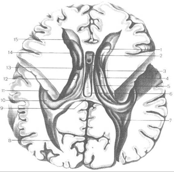
Fig. 246. Lateral ventricles of the cerebrum — horizontal section (adapted from M. R. Sapin)
1 — septum pellucidum; 2 — cavity of the septum pellucidum; 3 — terminal stria; 4 — central part of the lateral ventricle; 5 — crus of the fornix; 6 — commissure of the fornix; 7 — calcar avis; 8 — posterior horn; 9 — splenium of the corpus callosum; 10 — hippocampus; 11 — inferior horn of the lateral ventricle; 12 — thalamus; 13 — interventricular foramen; 14 — HEAD of the caudate Nucleus; 15 — anterior horn of the lateral ventricle
The lateral ventricle comprises a central part, as well as anterior, posterior, and inferior horns. These ventricular regions are situated in all lobes of the hemispheres (with the exception of the insula).
The central part (pars centralis) of the lateral ventricle is located in the parietal lobe of the cerebral hemispheres. Its medial wall is formed by the body of the fornix, the superior wall by the corpus callosum, and the inferior wall by the body ofторы the caudate nucleus, the thalamus, and the stria terminalis. The superior and inferior walls of the lateral ventricle meet at an acute angle, which is why the lateral wall in the central part of the lateral ventricle is virtually absent.
The anterior (frontal) horn (cornu frontale — anterius) is a slit-like space located in the frontal hemisphere anterior to the interventricular foramen. The medial wall of the anterior horn is formed by the septum pellucidum, while the lateral and partially the inferior walls are formed by the head of the caudate nucleus. The anterior, superior, and inferior walls of the anterior horn are bounded by the fibers of the corpus callosum.
The inferior (temporal) horn (cornu temporale — inferius) of the lateral ventricle is the cavity of the temporal lobe. The lateral and superior walls of the inferior horn are formed by the corpus callosum and the tail of the caudate nucleus. The medial wall is formed by the hippocampus, which extends toward the anterior Regions of the inferior horn and terminates in a Swelling. This hippocampal swelling is divided by fine sulci into individual elevations (digitationes hippocampi). Medially, the fimbria hippocampi — a continuation of the crus of the fornix — fuses with the hippocampus, and to it is attached the choroid plexus of the lateral ventricle, which descends here from the central part.
The posterior (occipital) horn (cornu occipitale — posterius) of the lateral ventricle extends into the occipital lobe of the hemisphere. Its superior and lateral walls are formed by the fibers of the corpus callosum, while the inferior and medial walls are formed by the White matter OF the occipital lobe. The medial wall of the posterior horn features two elevations: superior and inferior. The superior elevation, the bulb of the posterior horn (bulbus cornus posterioris), is represented by the fibers of the corpus callosum on their way into the occipital lobe, where they curve around the parieto-occipital sulcus. The inferior elevation, the calcar avis, is formed by the indentation into the cavity of the posterior horn of brain tissue located deep within the calcarine sulcus. On the inferior wall of the posterior horn lies the slightly convex collateral trigone (trigonum collaterale), which is the impression into the ventricular cavity made by the cerebral tissue located deep within the collateral sulcus.
Within the central part and the inferior horn of the lateral ventricle lies the choroid plexus (plexus choroideus ventriculi lateralis), which produces cerebrospinal fluid. This plexus is a derivative of the pia mater of the brain and a branch of the anterior choroidal artery (a branch of the Internal Carotid Artery). Within the ventricular cavity, it forms a web of vessels covered by an epithelial layer — the remnant of the inner wall of the first brain vesicle. These structures of the choroid plexus (Blood vessel wall and epithelium) constitute the morphological substrate of the blood-brain barrier, which prevents most harmful substances from passing from the bloodstream into the cerebrospinal fluid (with the exception of alcohol and certain other neurotropic substances).
The choroid plexus enters the cavity of the lateral ventricle through the choroid fissure (fissura choroidea), which is located in its central part between the thalamus and the fornix, and in the inferior horn — between the stria terminalis and the fimbria of the hippocampus.
The site of attachment of the choroid plexus to the thalamus is called the taenia choroidea. In the anterior regions, the choroid plexus of the lateral ventricle communicates with the choroid plexus of the third ventricle via the interventricular foramen (foramen interventriculare).
Last update: 08/08/2026
Editorial and Educational Adaptation: This material has been compiled based on the primary/original source text. The project team performed an editorial review, corrected technical inaccuracies, structured sections, and adapted the content for an educational format.
What was processed:
- elimination of formatting defects (OCR errors, structural breaks, corrupted characters);
- editorial organization of content;
- standardization of terminology in accordance with academic sources;
- verification of factual statements against the original source text.
All mentions of the author, publication year, and origin of the primary text have been preserved in accordance with the source.