Human Anatomy - Kotsan I. Ya. 2009
Brain
Basal (subcortical) nuclei and white matter of the telencephalon
Besides the cortex, which forms the superficial layers of the Telencephalon, the Gray matter in each cerebral hemisphere also occurs as distinct nuclei or masses embedded within the White matter (closer to the Base of the Brain). Due to their anatomical Location, these accumulations of gray matter are known as the basal nuclei (central or subcortical nuclei) (Fig. 245). The basal nuclei of the cerebral hemispheres include the corpus striatum, the claustrum, and the amygdaloid body.
Class="center">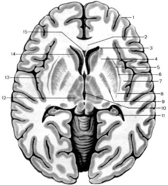
Fig. 245. Horizontal section of the brain. Basal nuclei (after M. R. Sapin)
1 — Cerebral Cortex (Pallium); 2 — genu of the corpus callosum; 3 — anterior horn of the lateral ventricle; 4 — internal capsule; 5 — external capsule; 6 — claustrum; 7 — extreme capsule; 8 — putamen; 9 — globus pallidus; 10 — Third ventricle; 11 — posterior horn of the lateral ventricle; 12 — thalamus; 13 — cortical substance (cortex) of the insula; 14 — HEAD of the caudate Nucleus; 15 — cavity of the septum pellucidum
The corpus striatum received its name because, on horizontal and frontal sections of the brain, it appears as alternating stripes of gray and white matter. It comprises the caudate nucleus and the lentiform nucleus.
The caudate nucleus (nucleus caudatus) is located lateral to the thalamus, from which it is separated by a layer of white matter known as the stria terminalis. The caudate nucleus consists of a head (caput nuclei caudati), which lies in the frontal lobe and bulges into the anterior horn of the lateral ventricle; a body (corpus nuclei caudati), located deep within the parietal lobe at the floor of the central part of the lateral ventricle; and a tail (cauda nuclei caudati), which extends into the temporal lobe and contributes to The formation of the superior wall of the inferior horn of the lateral ventricle. Lateral to the head of the caudate nucleus lies a layer of white matter—the anterior limb of the internal capsule—which separates this nucleus from the lentiform nucleus.
The lentiform nucleus (nucleus lentiformis) is situated lateral to the thalamus and the caudate nucleus, separated from the thalamus by the posterior limb of the internal capsule. On a frontal section of the brain, the lentiform nucleus has a wedge-shaped appearance, with its apex pointing medially and its base laterally. Two parallel vertical medullary laminae in the sagittal plane divide the lentiform nucleus into three parts. The most lateral is the putamen, which has a darker coloration. Medial to the putamen lie two slightly lighter medullary laminae (medial and lateral), collectively known as the globus pallidus. The medial lamina is referred to as the medial medullary lamina (or medial globus pallidus), and the lateral one as the lateral globus pallidus. Comparative anatomical and embryological studies have demonstrated that the globus pallidus is an evolutionarily older Structure, whereas the caudate nucleus and putamen belong to the phylogenetically newer structures known as the neostriatum (striatum).
To denote the functional unity of the Components of the corpus striatum that differ in histological structure and origin yet are topographically closely related—namely, the caudate nucleus and putamen on the one hand, and the globus pallidus on the other—the term striopallidal system is used. The striopallidal system constitutes the core of the extrapyramidal system, which regulates habitual movements, Muscle tone, and certain autonomic (vegetative) reactions, particularly thermoregulation and Carbohydrate METABOLISM.
The claustrum is a thin sheet of gray matter situated lateral to the putamen of the lentiform nucleus. It is separated from the putamen by a layer of white matter called the external capsule (capsula externa), and from the insular cortex by a similar layer called the extreme capsule (capsula extrema).
The amygdaloid body (corpus amygdaloideum) is located within the WHITE MATTER OF the temporal lobe, approximately 1.5–2 cm posterior to the temporal pole. It is almond-shaped and consists of three parts: the anterior amygdaloid area (area amygdaloidea anterior), the basolateral part (pars basolateralis), and the corticomedial (olfactory) part (pars corticomedialis [olfactoria]). The anterior amygdaloid area and the corticomedial (olfactory) part of the amygdaloid body are relatively small, belong to the central part of the rhinencephalon, and are connected to the anterior perforated substance by the stria terminalis of the thalamus.
The white matter of the cerebral hemispheres is represented by various systems of fibers, among which associative, commissural, and projection fiber bundles are distinguished. These are regarded as the conduction PATHWAYS OF THE brain (and Spinal Cord). Associative nerve fibers originating from the cerebral cortex (extracortical) remain within the confines of a single hemisphere, interconnecting various functional centers. Commissural nerve fibers form the corpus callosum, the anterior commissure, and the fornix commissure, linking homologous centers of the right and left cerebral hemispheres. Projection nerve fibers, which run from the cerebral hemispheres to lower divisions (Diencephalon, mesencephalon, etc.) and the spinal cord, as well as those running in the reverse direction from these structures, form the internal capsule and its corona radiata.
The internal capsule (capsula interna) is a thick, bent plate of white matter. Laterally, it is bounded by the lentiform nucleus, and medially by the head of the caudate nucleus (anteriorly) and the thalamus (posteriorly). Three parts are distinguished within the internal capsule: the anterior limb, the posterior limb, and the genu. The anterior limb (crus anterius capsulae internae) lies between the caudate and lentiform nuclei, while the posterior limb (crus posterius capsulae internae) lies between the thalamus and the lentiform nucleus. The junction of these two segments, forming an angle open laterally, is called the genu of the internal capsule (genu capsulae internae).
All projection fibers connecting the cerebral cortex with other PARTS OF THE Central Nervous system pass through the internal capsule. The genu of the internal capsule contains fibers of the corticonuclear pathway, which run from the precentral gyrus cortex to the motor cranial nerve nuclei. The anterior portion of the posterior limb, immediately adjacent to the genu of the internal capsule, contains the corticospinal fibers. Like the preceding pathway, this motor tract originates in the precentral gyrus and extends to the motor nuclei of the anterior horns of the spinal cord. Posterior to these pathways, within the posterior limb, are the thalamocortical fibers, represented by processes of thalamic Cells projecting to the postcentral gyrus cortex. This pathway includes fibers conducting all modalities of general sensitivity (pain, Temperature, Touch, pressure, and Proprioception). Even further posterior, in the central Regions of the posterior limb, lies the temporoparieto-occipitopontine tract. Its fibers originate from cells in various cortical areas of the occipital, parietal, and temporal lobes and project to the pontine nuclei located in the anterior part of the Pons. The auditory and visual pathways occupy the posterior sections of the posterior limb, originating from subcortical auditory and visual centers and terminating in their respective cortical centers. The anterior limb of the internal capsule contains the frontopontine tract.
Fibers of the ascending pathways diverge in various directions toward the cerebral cortex, forming the so-called corona radiata (radiant crown). Fibers of the descending pathways of the internal capsule descend in compact bundles into the cerebral peduncle.
Thus, we have outlined only The most significant pathways whose fibers course through the internal capsule. A more detailed Discussion of the BRAIN AND SPINAL cord pathways will follow below.
Last update: 08/08/2026
Editorial and Educational Adaptation: This material has been compiled based on the primary/original source text. The project team performed an editorial review, corrected technical inaccuracies, structured sections, and adapted the content for an educational format.
What was processed:
- elimination of formatting defects (OCR errors, structural breaks, corrupted characters);
- editorial organization of content;
- standardization of terminology in accordance with academic sources;
- verification of factual statements against the original source text.
All mentions of the author, publication year, and origin of the primary text have been preserved in accordance with the source.