Human Histology - O.D. Lutsyk 2003
General Histology
The Doctrine of Tissues. Epithelial Tissues. Glandular Epithelium
A tissue is a biological system consisting of Cells and their derivatives that formed during phylogeny and performs specific Functions. The cellular elements and their derivatives are the fundamental units of a tissue as a complex heterogeneous system. In turn, tissues serve as the structural basis for building Organs. Cells determine the primary properties of a tissue, and their destruction leads to The breakdown of the system, rendering the tissue non-viable. Besides cells, non-cellular structures are also distinguished within tissues. These include symplasts (Muscle fibers, the outer part of the trophoblast), syncytia (certain Developmental Stages of Male Germ Cells), post-cellular structures (erythrocytes, platelets, cornified scales of the epidermis), and Extracellular matrix (ground substance and fibers - Collagen, elastic, and reticulin). All non-cellular structures are Cell derivatives. Cells within a tissue system interact with each other and with the extracellular matrix. Cell-to-cell interactions, both directly and via the extracellular matrix, ensure the functioning of the tissue as a unified system.
The term "tissue" was first coined by the English scientist Nehemiah Grew in 1671. He used it in a literal sense when describing plant structures, where the intertwining of fibers resembled textile fabric. Thanks to the work of the French anatomist Xavier Bichat (1801), the Concept of Tissues firmly established itself in animal and human anatomy, although his proposed tissue Classification was incorrect as it was not based on microscopic data (Bichat distinguished 21 tissues). It was not until the second half of the 19th century (1857–1859) that the German microscopists Franz Leydig and Albert von Kölliker proposed the tissue classification that we practically use today. They divided all tissues into four groups: epithelial, connective, muscular, and nervous.
A major contribution to The Development of histophysiology, particularly The Theory of tissue evolution, was made by the works of the histologists Alexander Zavarzin and Nikolai Khlopin. In 1934, Alexander Zavarzin proposed dividing all tissues according to their functions into two groups: general and special. Zavarzin classified epithelia and Internal Environment Tissues (the latter including Connective Tissues, Blood, and Lymph) as general tissues, and muscular and nervous tissues as special ones. In modern practice, histologists use the division of tissues into the aforementioned four morphofunctional types (epithelia, Tissues of the Internal Environment, muscular, and nervous) (Table 5).
Table 5. Main characteristics of the four primary tissue types
|
Tissues |
Cells |
Extracellular matrix |
Main functions |
|
Epithelial |
Sheets of polyhedral cells |
Practically absent |
Lining of body surfaces or cavities, glandular secretion |
|
Internal environment |
Various fixed and wandering cells |
Abundant |
Support-mechanical, protective, trophic |
|
Muscular |
Elongated contractile cells or symplasts |
Moderate amount |
Contractile (movement) |
|
Nervous |
Cells with long processes |
Practically absent |
Perception of stimuli, generation, and transmission of nerve impulses |
Tissue development—histogenesis—occurs during the Embryonic period of ontogeny following The formation of germ layers (ectoderm, endoderm, and mesoderm). Tissues arise from the cellular material of the germ layers through differentiation. The foundation of differentiation, i.e., The Emergence of cellular differences (biochemical and morphological), is The process of determination—the genetic specification of a cell's future developmental pathway resulting from the blockage of certain genome components. The restriction of developmental potentials due to determination is termed "commitment." It occurs gradually. For example, a population of cells belonging to a single embryonic primordium can give rise to several tissues; their further determination takes place during histogenesis. This process involves smaller portions of The Genome than the Formation of the primordia did; therefore, differences between tissues belonging to the same type are not as pronounced as those between tissues belonging to different types.
Every tissue possesses or has possessed during Embryogenesis so-called stem cells. These are the least differentiated and least committed cells, which are presumably determined within the germ layers before the end of Gastrulation. Stem cells form a population characterized by self-maintenance, multi-directional differentiation, and the generation of functionally mature cells of that tissue via precursor cells. When a stem cell embarks on a pathway of differentiation, a sequential series of committed mitoses yields first semi-stem cells and subsequently differentiated cells with specific functions. The departure of a stem cell from the population serves as a signal for another stem cell to divide via non-committed mitosis. As a result, the total number of stem cells is restored, remaining relatively constant under normal conditions.
The population of cells developing sequentially from a single type of stem cell to a mature specialized cell is called a differon, or histogenetic Lineage. Tissues typically comprise several differons. Alongside performing specific functions, specialized cells are capable of synthesizing unique substances known as chalones, which inhibit the proliferation of precursor and stem cells. When the number of mature cells decreases for any reason (e.g., following an injury), the inhibitory effect of chalones weakens, the mitotic activity of precursor cells increases, and the number of specialized cells is restored.
The process of structural restoration of a biological object after damage is termed regeneration. Depending on the level of biological Organization, subcellular, cellular, tissue, and organ regeneration are distinguished. General Histology studies regeneration at THE TISSUE LEVEL. There is physiological regeneration, which occurs continuously in a healthy Organism, and reparative regeneration, which takes place as a result of injury. Regenerative capacities vary across different tissues and are associated with the presence of stem cells and precursor cells. In adult humans, some tissues have limited regenerative capacities; however, recent studies have established that even Neurons—long thought to be incapable of regeneration in the adult organism—retain regenerative potential in certain regions of The Nervous system.
Epithelial Tissue (textus epithelialis). Phylogenetically, epithelium is one of the oldest tissues, having emerged first at the dawn of multicellular organism evolution. The term "epithelium" was proposed by Frederik Ruysch and derives from the Greek words *epi* (upon) and *thele* (nipple). This is because the scientist was studying the epidermis—an epithelium located over the papillary layer of the dermis. Epithelium is a component of almost all organs, largely determining the Specific features of their Structure and function. Most glands are also constructed from this tissue.
Epithelial tissue performs a range of vital functions in the Human and Animal body. For instance, epithelium protects underlying tissues from mechanical, chemical, infectious, and radiation damage. This function predominates in the epithelium of the Skin, oral mucosa, and certain other areas. Another function of epithelium is METABOLISM, which involves the absorption of substances and their excretion outwards through this tissue. This function is characteristic of the epithelium of the intestines, Stomach, skin, Lungs, and Kidneys. Furthermore, epithelium performs a secretory function inherent to the so-called Glandular Epithelium, which forms glands.
Morphofunctional Features of epithelium. A key morphological characteristic of epithelial tissue is that it consists exclusively of epithelial cells (epitheliocytes) and contains practically no extracellular matrix (Fig. 3.1). Interconnected by various types of junctions, the cells form a continuous sheet. Epithelium retains its ability to form cellular sheets both in tissue culture and under pathological conditions, such as tumor growth.
An epithelial cell sheet always rests on a basement membrane (Fig. 3.2). Under a Light Microscope, the latter appears as a homogeneous lamina up to 1 µm thick. Under an Electron microscope, it reveals a three-dimensional network of cords 3–4 nm in diameter, composed of five components: type IV collagen, heparan sulfate proteoglycan, entactin, Laminin, and Fibronectin. The basement membrane separates the epithelium from the underlying loose Connective Tissue, prevents the epithelium from growing into the connective tissue, and thus performs a barrier function. It also provides adhesive properties for both tissues and is important for the Nutrition of the avascular epithelium, ensuring its trophic supply via the Blood Vessels of the loose connective tissue through the basement membrane.
Class="center">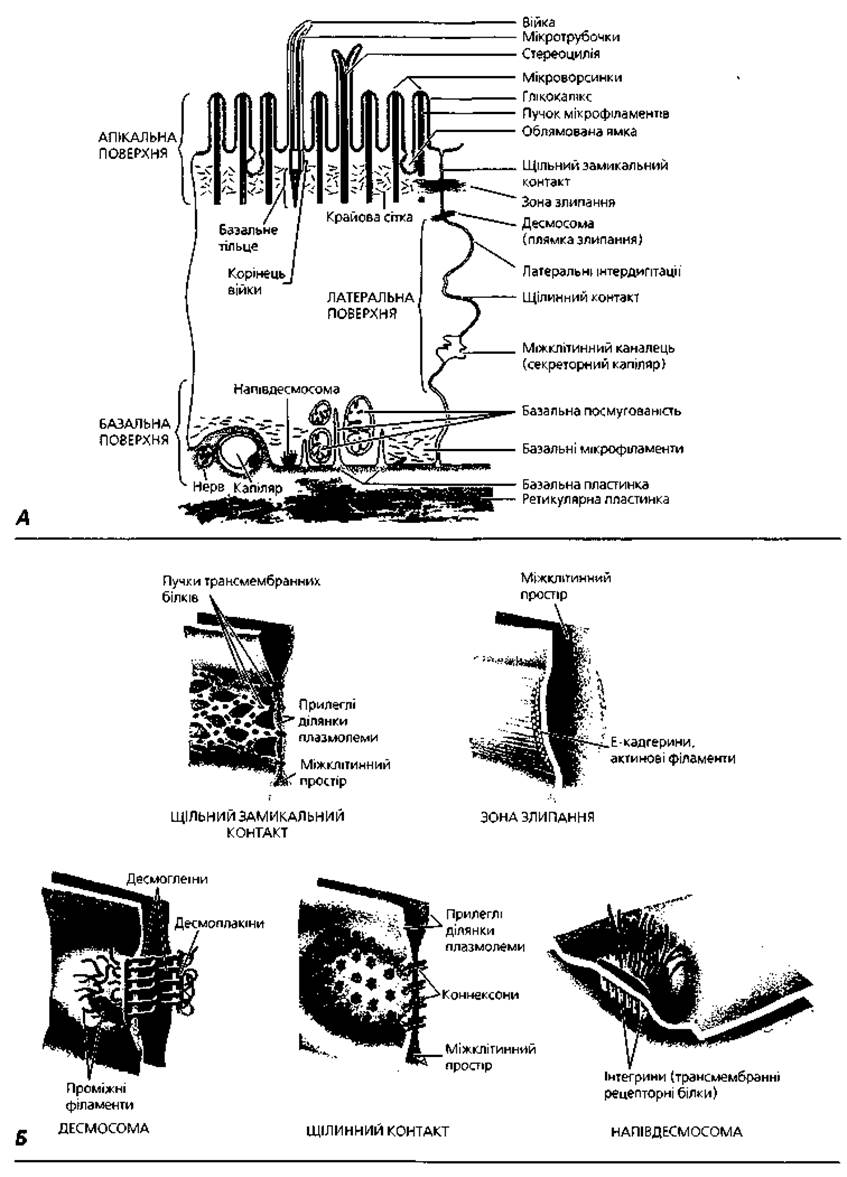
Fig. 3.1. Structure of an epithelial cell: A - schematic representation of a generalized epitheliocyte demonstrating specialized structures of its basal, lateral, and apical surfaces; B - ultrastructural details of Intercellular junctions between epitheliocytes

Fig. 3.2. Two Types of basement membranes: A - bilayered basement membrane separating the epithelium from connective tissue while ensuring their firm attachment; B - trilayered basement membrane separating the endothelium from the epithelium within renal corpuscles and pulmonary alveoli, formed by the fusion of two membranes whose components are produced by both endotheliocytes and epitheliocytes
Due to its position at the boundary between body tissues and the external environment, epithelial cells or the epithelial sheet as a whole exhibit a characteristic structural feature known as polar differentiation. This implies the presence of two cellular poles: an apical pole, facing the external environment, and a basal pole, located on the basement membrane. The apical and basal poles display distinct morphological features (Fig. 3.1). The basal region contains The Nucleus, and Mitochondria may be localized here, forming the so-called basal striation. The basal Plasma Membrane can form deep invaginations. The apical pole of an epitheliocyte is characterized by structures such as microvilli, a brush border, cilia, etc.
Epithelial tissue possesses a high regenerative capacity—both physiological and reparative. This is due to its boundary position and direct contact with the external environment, making the epithelium the body's primary barrier against harmful agents. Regeneration of the epithelium is carried out by stem cells, which vary for each type of epithelium.
Classification of epithelial tissues. There are two classifications of epithelia: phylogenetic (or simply genetic) and morphofunctional. The phylogenetic classification, proposed by Nikolai Khlopin, is based on THE ORIGIN OF various types of epithelium from different germ layers. According to this classification, the following types of epithelia are distinguished:
1) cutaneous - derived from the ectoderm; structurally stratified or pseudostratified; its function is protective; localization includes the skin, Oral Cavity, Esophagus, Cytology/practical/76.html">Cornea of the eye, Vagina, anus, etc.;
2) intestinal - derived from the endoderm; structurally simple columnar; function is absorption; localization includes The Stomach, small and large intestines;
3) renal - derived from the intermediate mesoderm; structurally simple; function is the reabsorption of substances from primary urine into the blood; localization includes renal tubules;
4) coelomic - derived from the ventral mesoderm; structurally simple squamous; function is demarcating; localization includes serous membranes;
5) ependymo-glial - derived from the neural tube; structurally simple; localization includes the lining of Brain cavities.
6) angiodermal - of mesenchymal origin; structurally simple squamous; it lines Blood and Lymphatic vessels as well as The Heart, and is termed "endothelium".
A more common classification of epithelial tissues, which is also widely used by pathologists, is the second, morphofunctional classification. It is based on the structural and Functional Characteristics of various types of epithelium. According to this classification, epithelial tissues are divided into glandular and lining (surface) epithelia, with the latter subdivided into simple and stratified based on their relationship to the basement membrane (Fig. 3.3). In Simple Epithelium, all cells rest on the basement membrane, whereas in Stratified Epithelium, only the Cells of the basal layer are in direct contact with it, while all other cells form layers above and have no direct connection to the basement membrane.
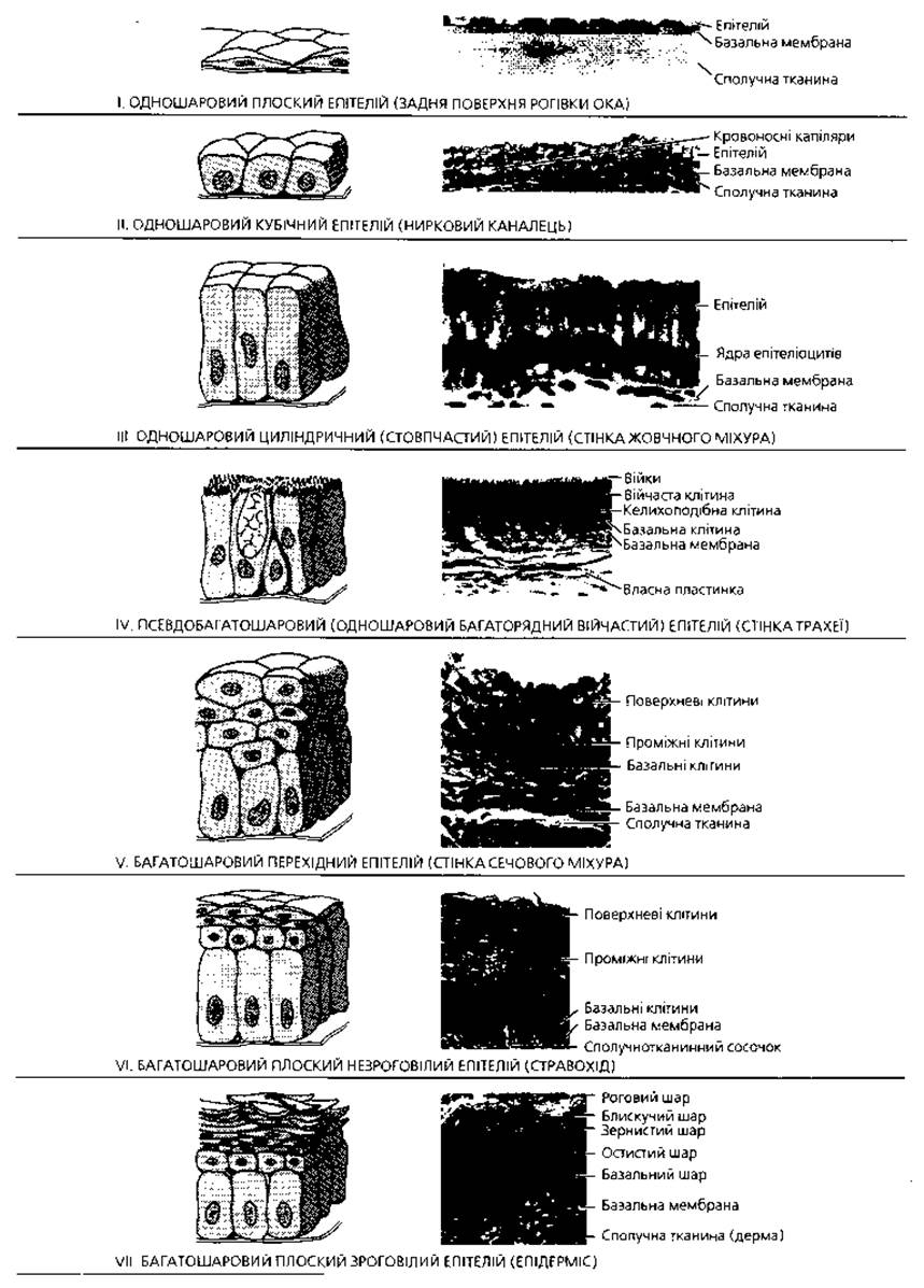
Fig. 3.3. Seven MAIN TYPES OF surface epithelium: left - schematic representation, right - light micrograph view, x300
Simple epithelium is subdivided into single-layered (simple) and pseudostratified. Simple epithelium is defined as having all cells of identical shape (also called isomorphic), with their nuclei located at the same level, forming a single row. Based on cell shape, this epithelium is classified as columnar, cuboidal, or squamous. Pseudostratified epithelium contains cells of varying shapes (hence its alternative name, anisomorphic), with their nuclei located at different levels, forming multiple rows. This epithelium is also referred to as pseudostratified because it resembles stratified epithelium in appearance, yet all its cells actually contact the basement membrane, thus forming a single layer. Stratified squamous epithelium is divided into keratinized and non-keratinized stratified squamous epithelium. In certain organs, stratified cuboidal, stratified columnar, and transitional epithelia are also found (Table 6).
Structure of various types of epithelium. Simple squamous epithelium (mesothelium) lines the peritoneal, pleural, and pericardial cavities. It is a thin sheet of polygonal cells with irregular, wavy borders (cell boundaries become clearly visible after silver nitrate impregnation). Some cells are binucleated or trinucleated, and their surface features microvilli. The mesothelium facilitates exchange between the fluid filling the secondary body cavity and blood (or lymph), a function supported by the pinocytotic activity of its cells.
Table 6. Main types of surface epithelia in humans
|
By number of layers |
By cell shape |
Examples of localization |
Main functions |
|
Simple |
Squamous |
Blood vessels (endothelium); serous membranes (mesothelium) |
Lining, pinocytosis, secretion of BIOLOGICALLY ACTIVE SUBSTANCES, facilitating internal organ movement |
|
Simple |
Cuboidal |
Renal tubules, lung bronchioles |
Lining, secretion, and Transport of substances |
|
Simple |
Columnar |
Stomach, intestine |
Lining, protection, absorption, and secretion of substances |
|
Pseudostratified (simple pseudostratified) |
Varying in shape and height |
Lining, protection, secretion |
|
|
Stratified |
Keratinized squamous |
Skin |
Lining, protection |
|
Stratified |
Non-keratinized squamous |
Esophagus, vagina |
Lining, protection |
|
Stratified |
Cuboidal |
Lining of excretory ducts |
|
|
Stratified |
Columnar |
Conjunctiva |
Lining, protection |
|
Stratified |
Transitional |
Lining, protection |
Simple cuboidal epithelium is found in the renal tubules, excretory ducts of numerous glands, and pulmonary bronchioles. The cells of this epithelium have equal height and width, with structural features varying across different organs.
Simple columnar epithelium forms the inner lining of the stomach, small and large intestines, Gallbladder, excretory ducts of The Liver and Pancreas, certain renal tubules, uterine cavity, and Fallopian tubes. The following types of this epithelium are distinguished:
1) striated (brush-bordered) - in the intestine and gallbladder; its cells possess a striated apical border;
2) ciliated - in the Uterus and Fallopian tubes; the cells bear motile cilia (Fig. 3.4) that facilitate ovum transport;
3) glandular - in the stomach; these cells are capable of producing a mucus-like secretion and are termed glandulocytes.
Pseudostratified columnar epithelium lines the respiratory tract and certain segments of the Reproductive System. It is commonly referred to as pseudostratified ciliated epithelium. Its main cell types include ciliated cells, intercalated (short and long) cells, mucous (goblet) cells, and endocrine cells. Ciliated cells are wedge-shaped, with their wide apex facing the epithelial surface and bearing cilia, while their narrow base attaches to the basement membrane. Intercalated cells are also wedge-shaped, but oriented with their wide base resting on the basement membrane and their narrow ends wedged between ciliated cells without reaching the epithelial surface, leaving the entire surface covered with cilia. Among the intercalated cells are stem cells, which differentiate into ciliated and mucous cells. Ciliary beating and mucus produced by goblet cells help clear dust particles from the respiratory tract. Endocrine cells produce biologically active substances (Hormones) that mediate local Regulation of Respiratory functions.
Stratified non-keratinized squamous epithelium is located in the corneal epithelium, oral cavity, esophagus, vagina, and anal canal. It comprises three distinct cellular layers:
1) basal layer - columnar cells forming a single layer on the basement membrane, actively dividing by mitosis and containing stem cells, thus serving as the germinative (cambial) layer;
2) spinous layer - polygonal cells with cytoplasmic projections (spines) that interdigitate with the apical ends of the basal cells, arranged in multiple layers;
3) squamous cell layer - superficial layers of dying and desquamating cells.

Fig. 3.4. Ultrastructure of microvilli and cilia: A - Longitudinal section of small intestinal microvilli, x40,000; B - cross-section of microvilli, x50,000; C - longitudinal section of cilia and microvilli of the Uterine tube epithelium, x36,000; D - cross-section of cilia, clearly showing the microtubule arrangement described by the 9x2+2 formula, up to x100,000
Stratified keratinized squamous epithelium covers the skin surface and is known as the epidermis. It consists of multiple cell layers, among which several strata can be distinguished. The epidermis of the palms and soles features five layers:
1) basal layer, which, In addition to poorly differentiated stem epitheliocytes, contains dendritic pigment cells (melanocytes), Langerhans cells (dendritic macrophages), and Merkel cells (mechanoreceptors);
2) spinous layer, structurally similar to the non-keratinized epithelium described above; together, the basal and spinous layers form the germinative zone of the epidermis (Malpighian layer);
3) granular layer - composed of flattened cells containing granules of the fibrous protein keratohyalin;
4) clear (lucid) layer - appears as a homogeneous, light-refracting band in histological preparations due to the presence of eleidin within its flattened cells; eleidin represents a complex of keratohyalin with tonofibrils and marks the next stage in the formation of the cornified protein, keratin;
5) cornified (keratinized) — consists of cornified scales filled with keratin and air bubbles; under The Influence of lysosomal Enzymes, the outer scales lose their intercellular connections and constantly desquamate from the epithelial surface.
Transitional epithelium lines the Urinary Tract—the renal calyces and pelvis, Ureters, and urinary bladder. It consists of three layers:
1) basal — composed of small, heavily stained cells;
2) intermediate — contains cells of various shapes, mostly polygonal;
3) superficial — consists of large, light cells that often contain 2–3 nuclei.
Depending on the state of the organ wall, these cells may be flattened or pear-shaped. The apical region of these cells contains specific structures—plasma membrane invaginations and fusiform vesicles, which serve as a reserve of the Plasmalemma and are incorporated into it during organ stretching. When the wall contracts, the thickness of the epithelial layer increases because some cells of the intermediate layer are "squeezed" upward, while the superficial cells acquire a pear-shaped form.
Glands (glandulae). The vast majority of glands are derivatives of glandular epithelium (Fig. 3.5). Glandular cells are called glandulocytes. Glands are divided into two major groups: exocrine (external secretion glands) and endocrine (internal secretion glands). Accordingly, glandulocytes are subdivided into exocrinocytes and endocrinocytes. Exocrine glands always consist of two parts—a terminal (secretory) portion and an excretory duct. They produce secretions that are released onto The surface of the epithelial sheet. Endocrine glands lack excretory ducts; their products—hormones—are secreted directly into the BLOOD AND LYMPH. Glands are classified by their structure, type of secretion, and The Nature of their secretory product.

Fig. 3.5. Formation of various types of glands from the covering epithelium: when contact with the epithelial sheet is maintained, glands discharge their secretion onto its surface (exocrine glands); if contact with the epithelial sheet is lost, the secretion is released into the blood or intercellular fluid (endocrine glands)
Based on their Location relative to the epithelial sheet, glands are classified into intraepithelial and extraepithelial. The former are located entirely within the epithelial sheet and do not extend beyond its boundaries. In humans, intraepithelial glands are unicellular. These are mucous goblet cells (exocrinocytes) located within the pseudostratified ciliated epithelium of the respiratory tract and the simple columnar epithelium of the intestine. Extraepithelial glands in The Human Body are multicellular. They are situated outside the epithelial sheet in the connective tissue and are connected to the epithelium by an excretory duct.
Based on the number of excretory ducts, extraepithelial exocrine glands (Fig. 3.6) are divided into simple, which have a single excretory duct, and compound, in which the excretory duct branches. Depending on the number of terminal secretory portions, simple glands can be branched or unbranched. The former have multiple terminal portions, whereas the latter have only a single terminal secretory portion.
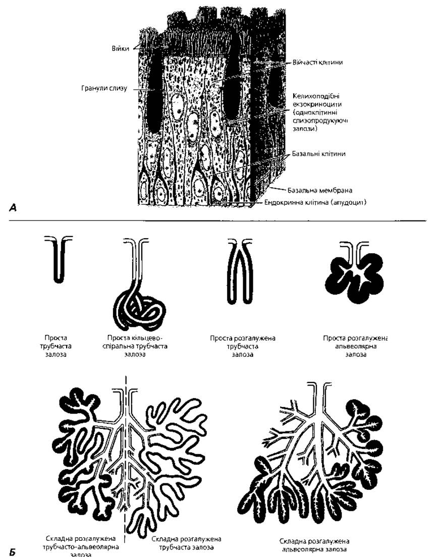
Fig. 3.6. Various types of glands: A — unicellular glands: dissociated exocrinocytes (goblet cells) and endocrinocytes (apudocytes) within the epithelial lining of the trachea; B — classification principles for multicellular exocrine glands: secretory cells are shown in black, duct cells are light; compound glands possess branched ducts
Compound glands are always branched because their numerous excretory ducts terminate in many secretory portions. Based on the shape of their secretory portions, glands are categorized into tubular (the terminal portion is shaped like a tubule), alveolar (the terminal portion is sac-shaped), and tubulo-alveolar (containing both types of terminal portions).
According to the type of secretion (The Mechanism of releasing the secretory product), glands are classified into the following varieties:
1) merocrine — the secretion is released from The Cell without disrupting its integrity; the majority of glands in the human body secrete via the merocrine pathway; the name derives from the Greek words "meros" (part) and "krino" (to secrete);
2) apocrine — the apical portion of the cell is pinched off along with the secretion; in humans, Mammary Glands and specific sweat glands secrete via the apocrine pathway; the name originates from the words "apex" (tip) and "krino" (to secrete); this type of apocrine secretion is currently termed macroapocrine, as distinguished from microapocrine, where only the tips of microvilli detach from the cell;
3) holocrine — following the accumulation of secretion, the cell completely disintegrates and its remnants become part of the secretion; in humans, Sebaceous Glands of the skin secrete via the holocrine pathway; the name derives from the Greek words "holos" (whole) and "krino" (to secrete).
Based on The chemical composition of their secretion (Fig. 3.7), glands are divided into proteinaceous, mucous, mixed (protein-mucous), sebaceous, and sudoriferous (sweat) glands.
Structure of secretory cells. THE CONCEPT OF the secretory cycle. The vast majority of secretory glandular cells (glandulocytes) are distinguished by the presence of secretory inclusions in their Cytoplasm. Cell shape varies and changes depending on the phase of secretion. Nuclei are mostly large with irregular, indented surfaces. In the cytoplasm of glandulocytes that produce protein secretions, the granular Endoplasmic reticulum is well developed. In cells synthesizing non-protein secretions (Lipids, Steroids), the agranular (smooth) endoplasmic reticulum is better developed. Mitochondria are numerous. Glandulocytes are characterized by a well-developed Golgi apparatus, where secretory granules are formed. Glandular cells exhibit polarity caused by a specific directional flow of secretory processes—for instance, in external secretion, from the basal to the apical region of the secretory cell.
Secretion is a complex process comprising four phases:
1. Uptake of raw Materials by glandulocytes from the blood and lymph across the basal surface.
2. Synthesis and accumulation of the secretion, which takes place in the granular or agranular endoplasmic reticulum; secretory products are packaged within the Golgi apparatus.
3. Release of the secretion from glandulocytes—extrusion—which occurs via various pathways depending on the secretion type: merocrine, apocrine, or holocrine.
4. Restoration of the glandular cell's initial state. These phases may occur sequentially in glandulocytes in a cyclic manner, forming the so-called secretory cycle. In other cases, they occur simultaneously, which is characteristic of diffuse or spontaneous secretion.
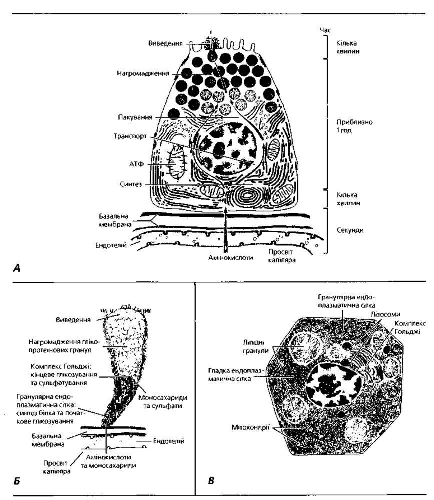
Fig. 3.7. Schematic representation of Three types of glandular cells: A - protein-producing acinar cell of the pancreas; B - mucus-producing goblet cell of the small intestinal epithelial lining; C - steroid-secreting cell (testicular glandulocyte)
Terms to Remember
1. Tissue. 2. General tissues. 3. Special tissues. 4. Epithelial tissues. 5. Connective tissues. 6. Muscle Tissues. 7. Nervous Tissue. 8. Differentiation. 9. Determination. 10. Commitment. 11. Stem cell. 12. Progenitor cell (semistem cell). 13. Differentiated cell. 14. Differon. 15. Chalone. 16. Regeneration. 17. Physiological regeneration. 18. Reparative regeneration. 19. Cell sheet. 20. Basement membrane. 21. Polar differentiation of epithelial cells. 22. Phylogenetic classification of epithelium. 23. Skin-type epithelium. 24. Intestinal-type epithelium. 25. Renal-type epithelium. 26. Coelomic-type epithelium. 27. Ependymal-glial-type epithelium. 28. Angiodermal-type epithelium. 29. Morphofunctional classification of epithelium. 30. Simple epithelium. 31. Uniseriate epithelium (squamous, cuboidal, columnar). 32. Pseudostratified epithelium. 33. Stratified epithelium. 34. Stratified squamous non-keratinized epithelium. 35. Stratified squamous keratinized epithelium. 36. Transitional epithelium. 37. Glands. 38. Glandulocytes. 39. Exocrinocytes. 40. Endocrinocytes. 41. Exocrine glands. 42. Endocrine glands. 43. Intraepithelial glands. 44. Extraepithelial glands. 45. Simple glands. 46. Compound glands. 47. Branched glands. 48. Unbranched glands. 49. Alveolar glands. 50. Tubular glands. 51. Tubulo-alveolar glands. 52. Merocrine glands. 53. Apocrine glands. 54. Holocrine glands. 55. Mucous glands. 56. Serous glands. 57. Seromucous glands. 58. Sebaceous glands. 59. Sweat glands. 60. Secretory cycle.
Last update: 09/08/2026
Editorial and Educational Adaptation: This material has been compiled based on the primary/original source text. The project team performed an editorial review, corrected technical inaccuracies, structured sections, and adapted the content for an educational format.
What was processed:
- elimination of formatting defects (OCR errors, structural breaks, corrupted characters);
- editorial organization of content;
- standardization of terminology in accordance with academic sources;
- verification of factual statements against the original source text.
All mentions of the author, publication year, and origin of the primary text have been preserved in accordance with the source.