Human Anatomy and Physiology (with Age-Related Features of the Child's Body) - M.R. Sapin, V.I. Sivoglazov 2002
Structure of the Human Body
Tissues
Muscle Tissues
Muscle tissue is a group of Tissues (striated, smooth, and cardiac) of different origin and Structure, unified by a functional characteristic—The ability to contract, alter their length, and shorten.
Striated (skeletal) muscle tissue is formed by muscle fibers containing myofibrils, whose relative arrangement creates transverse striation (Fig. 9). Striated muscle tissue forms skeletal Muscles Attached to the BONES OF THE Skeleton. An important property of skeletal muscles is their ability to contract (shorten) under conscious voluntary control. The main tissue element of skeletal striated muscle tissue is muscle fibers, which in individual muscles can reach 10—12 cm in length. Externally, each muscle fiber is covered by a membrane—the sarcolemma, into which thin Collagen fibers, known as the endomysium, are interwoven. Within each muscle fiber, beneath the sarcolemma in the Cytoplasm (sarcoplasm), lie numerous nuclei (up to 100), general-purpose Organelles, as well as specialized organelles and inclusions (Myoglobin, Glycogen). Myoglobin, dissolved in the sarcoplasm, is a pigment-containing protein similar in its properties to erythrocyte Hemoglobin.
The main part of a muscle fiber is typically composed of specialized organelles—myofibrils. Each myofibril consists of regularly alternating regions—dark anisotropic bands (A) and light isotropic bands (J). In the middle of each A band runs the M line, or mesophragma. Through the middle of the J band runs the Z line—the telophragma. The alternation of dark and light bands in adjacent myofibrils aligned at the same level creates the appearance of transverse striation on a histological section of Skeletal Muscle. Each dark band is formed by thick filaments (10 nm), which are primarily composed of the high-molecular-weight protein Myosin. Each light band consists of thin filaments (5 nm) composed of the low-molecular-weight protein Actin, as well as the low-molecular-weight Proteins Tropomyosin and troponin.
Class="center">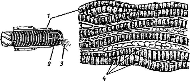
Fig. 9. Striated (skeletal) muscle tissue: 1 — muscle fiber, 2 — sarcolemma, 3 — myofibrils, 4 — nuclei
The region of a myofibril between two Z lines is called a sarcomere, which is the functional unit of the myofibril.
A sarcomere includes a dark band and half of a light band adjacent to it on each side. Both ends of the thick filaments are free, whereas only one end of the thin filaments is free. Thus, thin filaments extend from the Z discs and interdigitate between the thick filaments. During Muscle contraction, Actin and myosin filaments slide past each other; during relaxation, they move in opposite directions. Based on The amount of myofibrils and sarcoplasm, muscle fibers are classified into slow ("red") fibers, which contain few myofibrils and abundant sarcoplasm, and fast ("white") fibers, which contain numerous myofibrils and sparse sarcoplasm. "Red" muscle fibers contract slowly but can sustain activity for a long time. "White" muscle fibers contract rapidly and fatigue quickly. The combination of Slow and fast striated muscle fibers in muscles ensures both rapid response (contraction) and long-term endurance.
The source of development for striated (skeletal) muscle tissue is the Cells of the myotomes of somites. In the Cytology/cytology/16.html">Early stages of embryonic development, mononuclear spindle-shaped cells—myoblasts—migrate from the mesoderm of the myotomes. Proliferating rapidly, myoblasts form the primordia of future muscles in their respective locations. Rapid nuclear division leads to the loss of cellular structure in myoblasts, and they transform into large multinucleated complexes—muscle fibers. In the developing muscle fibers, the number of myofibrils increases, and transverse striation appears. During the second half of fetal development and in postnatal ontogeny, muscle fibers grow in length and thickness by increasing the number of myofibrils they contain. Along with the growth and differentiation of muscle fibers, they fuse with satellite cells. Satellite cells are located beneath the sarcolemma of muscle fibers and serve as a source of new fibers. Satellite cells are capable of dividing and giving rise to myoblasts following muscle injury.
Smooth muscle tissue forms the contractile apparatus in the walls of Internal Organs, gland ducts, Blood Vessels, and Lymphatic vessels. The Structural elements of this tissue are smooth muscle cells (myocytes). Smooth myocytes are spindle-shaped cells 20—100 µm long and 5—8 µm thick. A single rod-shaped Nucleus is located in the center of The Cell. Upon contraction of the myocyte, The Nucleus bends and may even twist spirally. Organelles, including numerous Mitochondria, are located closer to the poles of the cell. The Endoplasmic reticulum and the Golgi complex are poorly developed, indicating a low synthetic function of the myocytes. The cytoplasm of myocytes contains many actin and myosin filaments arranged not parallel but at an angle to each other. The proportion of actin (compared to myosin) in smooth muscle cells is higher than in striated muscle fibers. The interaction of actin and myosin filaments occurs via the sliding mechanism, but it is carried out differently than in skeletal muscle tissue. Smooth myocytes lack transverse striation; they contract involuntarily, and their Functions are controlled by the autonomic (vegetative) division of The Nervous system.
Smooth myocytes are grouped into bundles, whose formation involves thin collagen and elastic fibers.
Cardiac striated muscle tissue is formed by tightly apposed, transversely striated muscle cells—cardiomyocytes. At the same time, cardiac muscle cells contract automatically, obeying the rhythm of the conducting System of the Heart and the Functions of the Autonomic (vegetative) nervous system. Cardiomyocytes are elongated cells (up to 100—150 µm) with a thickness of 10—20 µm, containing one nucleus (sometimes two nuclei) located in the center of the cell. General-purpose organelles are concentrated closer to the ends of the cell. Mitochondria are arranged in chains along the myofibrils. Cardiomyocytes contain inclusions such as glycogen and Lipids. Actin and myosin filaments in cardiomyocytes are arranged in much the same way as in skeletal muscle. Thin actin filaments are attached at one end to the telophragma, which forms the Z line. Thick (myosin) filaments, located between the actin ones, are attached at one end to the mesophragma (M line) and are directed toward the telophragma at the other.
Cardiomyocytes, by contacting one another, form a functionally and structurally integrated contractile system. Intercalated discs are located at the boundaries of adjacent cardiomyocytes. They consist of contacting Regions of the cytolemma of adjacent cells in the area of myofibril localization, resembling expanded desmosomes. Within the intercalated discs, in areas not occupied by myofibrils, there are so-called Gap Junctions, or nexuses. Intercalated discs perform a mechanical function, firmly connecting adjacent cardiomyocytes, while simultaneously ensuring the rapid passage of impulses, which enables all cardiac myocytes to contract synchronously. Intercalated discs provide not only structural but also functional integration of cardiomyocytes into a cohesive cardiac muscle (myocardium).
Last update: 10/08/2026
Editorial and Educational Adaptation: This material has been compiled based on the primary/original source text. The project team performed an editorial review, corrected technical inaccuracies, structured sections, and adapted the content for an educational format.
What was processed:
- elimination of formatting defects (OCR errors, structural breaks, corrupted characters);
- editorial organization of content;
- standardization of terminology in accordance with academic sources;
- verification of factual statements against the original source text.
All mentions of the author, publication year, and origin of the primary text have been preserved in accordance with the source.