Cytology, General Histology and Embryology - V. K. Napkhanyuk 2002
Tissues
Internal Environment Tissues
Blood
Blood (sanguis) is a fluid body tissue of mesenchymal origin that circulates through a closed Vascular System and forms the body's internal environment.
Class="center">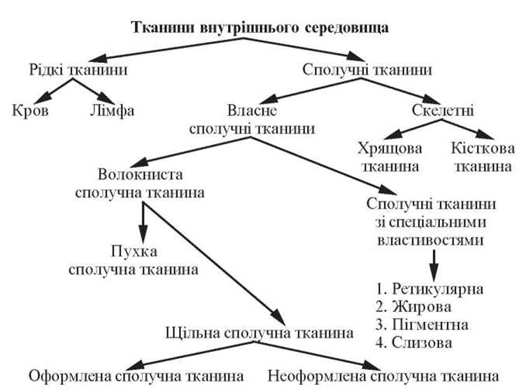
Fig. 43. Classification of internal environment Tissues
Like any other tissue, blood is a system in which all elements are histogenetically and functionally interconnected and subject to the general laws of neurohumoral regulation.
Blood accounts for 5 to 9% of total human body mass (5.0–5.5 L).
All Blood Cells develop from a common pluripotent stem Cell, whose differentiation into various blood cell types is determined by microenvironmental factors (reticular tissue of Hematopoietic Organs) and hemopoietins.
The blood cell population is a continuous, self-replenishing cell system with a short developmental cycle, in which mature forms represent terminal cells.
Under physiological conditions, The formation of new cells and the destruction of old ones are balanced to maintain the constancy of both the quantitative and qualitative composition of blood cells.
Blood consists of two main components: plasma (the intercellular substance) and formed elements suspended in the plasma.
Plasma volume accounts for 55–60%, and formed elements account for 40–45%.
The primary Functions OF BLOOD include:
— transport: carrying oxygen from the Lungs to tissues and carbon dioxide from tissues to the lungs (respiratory function), delivering nutrients from their site of entry to sites of utilization (trophic function), and transporting Hormones and Enzymes from their production sites to target organs;
— homeostatic: blood participates in maintaining the constancy of the body's internal environment;
— protective: blood provides humoral and cellular Immunity, as well as phagocytosis.
Plasma
Blood Plasma is a colloidal system consisting of Water (90–93%), Organic compounds (Proteins: albumins, globulins, fibrinogen — 7%), and other organic and Inorganic Compounds (3%). The total concentration of Mineral Substances in blood plasma is 0.9%, with a pH of 7.36.
— albumins: account for about 4%, binding and transporting a wide range of substances via the bloodstream;
— globulins: account for about 1.1–3.1% (subdivided into α-globulins, β-globulins, and γ-globulins, which contain Antibodies);
— fibrinogen: accounts for 0.2–0.4%, a water-soluble protein that can convert into an insoluble form—fibrin—under certain conditions. This property of fibrinogen enables Blood Coagulation. Plasma from which fibrinogen has been removed is called blood serum.
Formed Elements
The formed elements of blood include erythrocytes, platelets (post-cellular structures), and leukocytes (cells).
Erythrocytes (erythrocytes) are red blood cells in humans and mammals. They are non-motile, highly differentiated post-cellular structures that lose their Nucleus and all cytoplasmic Organelles during maturation.
The total number of erythrocytes in the entire human blood volume is 25∙1012, and their total volume is about 2 L. One liter of blood contains (4.9–5.5)∙1012 erythrocytes in men and (3.7–4.9)∙1012 in women. In newborns, the erythrocyte count is (6.0–9.0) ∙ 1012, and in the elderly, up to 6.0∙1012. In clinically healthy individuals, erythrocyte counts may fluctuate depending on physical exertion, high-altitude exposure, and the Introduction/43.html">Action of Certain hormones.
Most erythrocytes have the shape of biconcave discs (discocytes). The erythrocyte shape is maintained by a complex protein, β-sialoglycoprotein, embedded in The erythrocyte membrane, as well as a specialized membrane Skeleton composed of spectrin—a protein underlying the Plasmalemma from the inside—which is linked to another protein, ankyrin.
Studies of erythrocytes using a Scanning Electron microscope have demonstrated that, In addition to discocytes, which account for 80% of the total number, other erythrocyte forms exist: planocytes (with a flat surface), stomatocytes (with a dome-shaped surface), saddle-shaped (biconcave), spherocytes (sphere-shaped), and echinocytes (spicule-shaped).
Spherocytes and echinocytes represent Aging forms of erythrocytes.
This variety of erythrocyte forms, the proportion of which does not exceed 20%, is referred to as physiological poikilocytosis. Values above this level are considered pathological (pathological poikilocytosis).
The total surface area of an erythrocyte is 125 µm2. The diameter of a human erythrocyte is 7.1–7.9 µm, with a thickness of 2.0–2.5 µm at the periphery and 1 µm in the center. In the center of the erythrocyte, the membrane forms a depression (physiological excavation). This structural feature of the erythrocyte helps increase its surface area and accelerate oxygen saturation.
Under physiological conditions, these dimensions are characteristic of 75% of erythrocytes (normocytes).
The plasmalemma (membrane) of an erythrocyte is about 20 nm thick. Its outer surface contains Phospholipids, malonic acid, antigenic Oligosaccharides, and adsorbed proteins. The inner surface of the membrane contains glycolytic enzymes, Na+, K+, ATPases, Glycoproteins, and Hemoglobin.
The hyaloplasm of an erythrocyte contains numerous hemoglobin granules 4–5 nm in size and consists of water (60%) and dry residue (40%); 95% of the dry residue is hemoglobin. In humans, Two Types of hemoglobin are present: HbA (characteristic of adults) and HbF (characteristic of the fetus).
At birth, HbF accounts for 80% and HbA for 20%. In adults, HbA is 98% and HbF is 2%.
Slides for Study
Slide 14. Frog blood smear (Fig. 44).
Low magnification. Under this magnification, select the area with the highest concentration of erythrocytes.
High magnification. Examine and draw the erythrocytes. Frog erythrocytes are significantly larger than mammalian erythrocytes. They are oval in shape. The rod-like Nucleus of the erythrocytes is intensely stained, and the erythrocyte Cytoplasm is stained bright red due to oxyphilia.
Label the following on the drawing: 1) erythrocyte nucleus; 2) erythrocyte cytoplasm.
Slide 15. Human blood smear (Fig. 45).
Low magnification. Select an area on the slide with well-fixed erythrocytes.
High magnification. Examine and draw the slide. Erythrocytes are stained pink with eosin. The oxyphilia of erythrocytes is due to hemoglobin, which saturates mature erythrocytes. They have the shape of a biconcave disk. The smear clearly shows a central indentation in the erythrocyte—the site formerly occupied by The Nucleus. This area appears lighter on the slide.
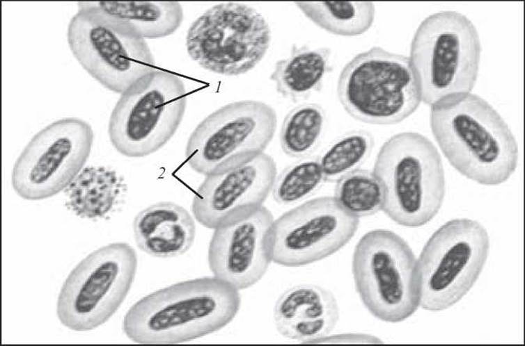
Fig. 44. Frog blood smear. Hematoxylin and eosin staining. x 400:
1 — erythrocyte nucleus; 2 — erythrocyte cytoplasm
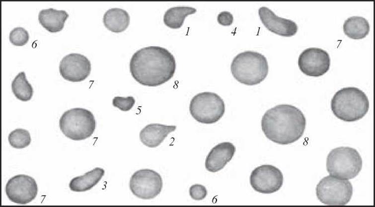
Fig. 45. Human blood erythrocytes. Romanowsky–Giemsa staining. x 900:
1, 2, 3 — poikilocytes (atypical erythrocytes differing from the round shape); 4–6 — microcytes (erythrocytes of regular round shape, but of atypical size); 7 — normocytes (erythrocytes of normal shape and size); 8 — megalocytes (large-sized erythrocytes)
Leukocytes (leucocytes) are white spherical blood cells that contain a nucleus and all cytoplasmic organelles, and are capable of exiting Blood Vessels and actively moving by forming pseudopodia.
In an adult human, 1 L of blood contains (4.0–9.0)∙109 leukocytes. An increase in the leukocyte count in the blood is called leukocytosis, and a decrease is called leukemia.
Depending on the presence or absence of specific granulation in their cytoplasm, all leukocytes are divided into granulocytes and agranulocytes. Based on the staining Properties of the granules, granulocytes are subdivided into neutrophilic, acidophilic, and basophilic (Fig. 46).
Neutrophilic granulocytes (granulocytus neutrophilicus) are round cells with a diameter of 7–9 µm in fresh blood and 10–12 µm in smears. In adult human blood, neutrophilic granulocytes outnumber other leukocytes, accounting for 65–75% of the total leukocyte count.
Among neutrophils, segmented-nuclear (mature, 60–65%), band-nuclear (3–5%), and juvenile (0–0.5%) forms are distinguished.
The nuclei of mature cells consist of 2–5 segments connected by Chromatin strands and containing heterochromatin. Female neutrophils are characterized by the presence of sex chromatin (the X chromosome) in the form of a drumstick known as a Barr body. Other neutrophils feature an S-shaped, horseshoe-shaped (band-nuclear), or Kidney-shaped nucleus (juvenile forms).
The Superficial layer of the cytoplasm contains only Glycogen granules, Actin filaments, and microtubules, which facilitate pseudopodia formation for cell motility.
The inner part of the cytoplasm houses organelles (Golgi apparatus, granular ER, scattered Mitochondria) and visible granularity. Depending on Structure and Chemical composition, primary non-specific (azurophilic — Lysosomes) and secondary specific (neutrophilic, 80–90%) granulations are distinguished.
The primary function of neutrophils is the phagocytosis of microorganisms, which is why they are referred to as microphages.
The ratio of the Three types of neutrophils holds definite diagnostic value and is clinically relevant. For instance, an increase in juvenile and band-nuclear forms accompanied by leukocytosis indicates an ongoing inflammatory process.
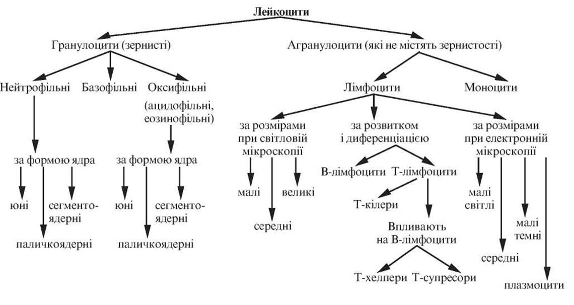
Fig. 46. Classification of leukocytes
Eosinophilic (oxyphilic, acidophilic) granulocytes (granulocytus eosinophilicus). Eosinophils are larger cells than neutrophils. Their diameter ranges from 9 to 10 µm in fresh blood and is about 12–14 µm in smears. The proportion of eosinophilic granulocytes in peripheral blood ranges from 1 to 5% of the total leukocyte count.
The nuclei of mature eosinophils typically have two segments. Juvenile and band-nuclear forms are rarely observed. Among the granules, azurophilic (primary) and eosinophilic (secondary) types are distinguished. The oxyphilia of the granules is due to the presence of a basic protein rich in Arginine, located centrally within the granules as a crystalloid. Additionally, the granules contain the enzyme histaminase. Cytoplasmic organelles are poorly developed.
Eosinophils are motile cells capable of phagocytosis, although their phagocytic activity is lower than that of neutrophils. They have been shown to play a role in foreign protein reactions as well as in allergic and anaphylactic responses, where they participate in histamine inactivation. A specific function of eosinophils is anti-parasitic defense. During parasitic infestations, eosinophil levels can reach up to 90% of the total leukocyte count.
Basophilic granulocytes (granulocytus basophilicus) have a diameter of about 9 µm in fresh blood and 11–12 µm in smears. The proportion of basophils in peripheral blood accounts for 0.5–1% of the total leukocyte count.
Basophil nuclei are poorly segmented, sometimes spherical, and stain less intensely than those of neutrophils or eosinophils. All types of organelles can be detected in the cytoplasm. A characteristic feature is the presence of specific, large metachromatic granules that mask the nucleus. Metachromasia is caused by the presence of the acidic glycosaminoglycan heparin. The granules also contain vasoactive histamine, serotonin, and enzymes. In addition to specific basophilic granules, azurophilic granules (lysosomes) are present in the cytoplasm.
Basophils are sluggish cells with almost no phagocytic capability. They participate in histamine and heparin METABOLISM, the Regulation of Blood clotting and vascular permeability, and immunological Reactions of the body, particularly of an allergic nature. They mediate inflammation and secrete the eosinophil chemotactic factor.
Agranulocytes include lymphocytes and monocytes.
Lymphocytes (lymphocytus). In the blood of adults, lymphocytes account for 20–35%. The size of lymphocytes varies considerably, from 4.5 to 10 µm. Consequently, under a Light Microscope, depending on their size, the following forms of lymphocytes are distinguished: small (4.5–6.0 µm in diameter), medium (7.0–10.0 µm in diameter), and large (10.0 µm or more in diameter).
Lymphocytes possess an intensely stained round or kidney-shaped nucleus surrounded by a relatively narrow rim of basophilic cytoplasm. In some lymphocytes, the cytoplasm contains a small number of azurophilic granules (lysosomes).
Using Electron Microscopy, 4 cell types have been identified within the lymphocyte population of adults:
Small light lymphocytes have a diameter of about 7 µm, with their nuclear-cytoplasmic ratio shifted toward the nucleus. The nucleus is round with potential indentations, and chromatin is condensed along the nuclear periphery. The cytoplasm is light, containing a small number of Ribosomes and Polysomes, poorly developed elements of the granular Endoplasmic reticulum, centrosomes, the Golgi apparatus, and mitochondria. The cytoplasm contains numerous vacuoles and multivesicular bodies, and lysosomes are occasionally found. Typically, organelles are located near the nuclear indentation. This lymphocyte type accounts for about 70–75% of the total human blood lymphocyte count.
Small dark lymphocytes have a diameter of 6.0–7.0 µm, with their nuclear-cytoplasmic ratio even more pronouncedly shifted toward the nucleus. Chromatin is dense, and the nucleolus is large. The cytoplasm exhibits high electron density (dark), surrounds the nucleus in a narrow rim, contains a high number of ribosomes and few mitochondria, and its light matrix stands out against the dark cytoplasmic Background. Other organelles are rare. In human blood, this lymphocyte type constitutes 10–12% of the total count.
Medium lymphocytes have a diameter of about 10.0 µm. The nucleus of these cells is kidney-shaped or round, often displaying finger-like indentations of the nuclear envelope. Nuclear chromatin is looser, with condensed chromatin patches visible only near the nuclear envelope; the nucleolus is well-defined.
The cytoplasm contains elongated tubules of the granular endoplasmic reticulum along with free ribosomes and polysomes. The centrosome and Golgi apparatus are predominantly located near the nuclear membrane invaginations; there are fewer mitochondria, and the number of lysosomes is small. In adult human blood, this cell type comprises 10–12% of the total lymphocyte count.
Plasmacytes (lymphoplasmacytes). A characteristic feature of this cell type is the presence in the cytoplasm of tubules of the granular endoplasmic reticulum arranged concentrically around the nucleus. In adult human blood, this cell type accounts for 1–2% of the total lymphocyte count.
Depending on developmental pathways, differentiation, and The Role of lymphocytes in the body's defense reactions, two MAIN TYPES OF lymphocytes are distinguished: T AND B lymphocytes (see Fig. 57).
T lymphocytes (Thymus-dependent) are derived from Bone Marrow stem cells in the thymus and are responsible for cell-mediated immunity and The regulation of humoral immunity. T lymphocytes are long-lived cells, capable of surviving for several years, or even decades. In peripheral blood, these cells account for 80% of all lymphocytes.
The T-lymphocyte population includes cytotoxic T lymphocytes (killer cells), T helper cells, and T suppressor cells.
B lymphocytes originate from bone marrow stem cells. In birds, this occurs in the bursa of Fabricius (bursa Fabricius); in humans, during the Embryonic period, it takes place in the Liver, while in adults, it occurs in the bone marrow. B lymphocytes are responsible for humoral immunity.
Monocytes (monocytus) are slightly larger than other leukocytes, measuring 9.0–12.0 µm in fresh blood and 18.0–20.0 µm in blood smears. Monocytes belong to the body's macrophage system, also known as the mononuclear phagocyte system, the cells of which originate from bone marrow promonocytes. In the circulating blood, they represent a pool of relatively immature cells en route from the bone marrow to the tissues (their transit time in the blood ranges from 36 to 104 hours).
The cytoplasm of monocytes is less basophilic than that of lymphocytes; when stained with Romanowsky-Giemsa, it appears pale blue, staining slightly darker at the periphery than near the nucleus. It contains a variable number of very fine azurophilic granules (lysosomes), finger-like projections, phagocytic vacuoles, numerous pinocytotic vesicles, short cisternae of granular endoplasmic reticulum, and small mitochondria.
Monocyte nuclei are kidney-shaped, horseshoe-shaped, or occasionally lobulated with numerous projections and indentations. Chromatin appears as fine granules distributed throughout the nucleus. They contain one or more nucleoli. In the peripheral blood of a healthy adult, the monocyte count ranges from 6% to 8%.
Platelets (thrombocytus) are Blood Platelets—anuclear fragments of cytoplasm measuring 2.0–3.0 µm in diameter that have pinched off from giant Cells of the Cytology/practical/86.html">Red bone marrow, known as megakaryocytes.
In fresh blood, platelets appear as small, colorless, round, oval, spindle-shaped, or irregularly shaped bodies. Each platelet consists of a hyalomere and a granulomere.
The hyalomere (hyalomerus) forms the structural foundation of the platelet. Electron microscopy reveals that the hyalomere contains thin filaments and circularly arranged bundles composed of 10–15 microtubules that maintain the plate-like shape of the cells.
The granulomere (granulomerus) consists of granules that cluster in the center of the platelet or are scattered throughout the hyalomere. Although the granulomere stains with Azure Dyes, it contains no chromatin. Electron microscopy shows that the granulomere contains membrane-bound, round, dense granules approximately 0.2 µm in diameter. Two types of granules have been identified: dense, dark granules (a-granules, whose chemical nature remains unclear) and serotonin-containing granules.
Additionally, mitochondria and glycogen granules are found within the granulomere.
Platelets form numerous cytoplasmic extensions of varying size and thickness (pseudopodia or microvilli), through which they interconnect to form a meshwork that serves as one of the key mechanisms in blood coagulation.
When stained with Romanowsky-Giemsa, five types of platelets can be distinguished:
— young forms with a basophilic hyalomere and isolated azurophilic granules;
— mature forms with a weakly oxyphilic hyalomere and distinct azurophilic granulation;
— senescent, dark forms featuring a blue-purple hyalomere and bluish-purple granulation;
— degenerative forms with a grayish-blue hyalomere and bluish-purple granulation;
— giant forms (irritation forms), which are 2–3 times larger than normal platelets and exhibit a pinkish-lilac hyalomere with purple granulation.
The platelet count in 1 L of peripheral blood in an adult is (200-400)∙109. The lifespan of platelets is 5–8 days.
Platelets play a crucial role in blood coagulation. Upon rapid disintegration, they aggregate into clumps—surrounding which fibrin strands are formed—while releasing phospholipids, Lipoproteins, and enzymes.
Last update: 10/08/2026
Editorial and Educational Adaptation: This material has been compiled based on the primary/original source text. The project team performed an editorial review, corrected technical inaccuracies, structured sections, and adapted the content for an educational format.
What was processed:
- elimination of formatting defects (OCR errors, structural breaks, corrupted characters);
- editorial organization of content;
- standardization of terminology in accordance with academic sources;
- verification of factual statements against the original source text.
All mentions of the author, publication year, and origin of the primary text have been preserved in accordance with the source.