Molecular Biology of the Cell - Volume 2 - Alberts B., Bray D., Lewis J., Raff M., Roberts K., Watson J. 1993
Cell adhesion, cell junctions, and the extracellular matrix
Intercellular junctions
Specialized intercellular junctions are particularly numerous and prominent in epithelia, yet they occur in virtually all Tissues at various sites of Cell-cell and cell-matrix contact. For the most part, they are too small to be resolved with a Light Microscope; however, they can be visualized using Electron Microscopy in conventional thin sections or in freeze-fracture replicas. In both cases, the interacting Plasma Membranes (and frequently the underlying Cytoplasm and intercellular space) reveal a highly specialized Structure at these sites. Cell junctions can be divided into three functional groups: (1) occluding junctions, which seal Cells together in an epithelial sheet in such a way as to prevent even small molecules from passing from one side of the sheet to the other; (2) anchoring junctions, which mechanically attach cells (and their cytoskeletons) to neighboring cells or to the Extracellular matrix; and (3) communicating junctions, which mediate the passage of chemical or electrical signals between interacting cells.
The principal types of intercellular junctions within each of these groups are listed in Table 14-1. Tight junctions are the main type of occluding junction; adherens junctions and desmosomes make up the principal category of anchoring junctions; and Gap Junctions, chemical synapses in neural cells, and finally plant plasmodesmata represent the major forms of communicating junctions. Because chemical synapses and plasmodesmata are examined in detail in Chapters 19 and 20, respectively, we will not discuss them in this chapter.
Class="center">Table 14-1. Functional Classification of Cell Junctions
|
I. Occluding (tight) junctions II. Anchoring junctions 1. Actin filament attachment sites (adherens junctions) a) cell-cell (e.g., adhesion belts) b) cell-matrix (e.g., focal contacts) 2. Intermediate filament attachment sites a) cell-cell (desmosomes) b) cell-matrix (hemidesmosomes) III. Communicating junctions 1. Gap junctions 2. Chemical synapses 3. Plasmodesmata (plants only)1 |
1) This is the sole type of junction found between plant cells.
14-4
14-5
14.1.1. Tight junctions establish a permeability barrier in epithelial cell sheets [2]
Despite substantial Structural and Biochemical differences among various types of epithelia, these tissues share at least one common function: they serve as selectively permeable barriers that separate fluids of different chemical composition on either side of the sheet. Tight junctions play a critically important role in maintaining this barrier function, as is well illustrated by the mammalian intestinal epithelium.
The epithelial cells lining the Small Intestine retain the bulk of the gut contents within its lumen. At the same time, however, they must actively pump specific nutrients across The Cell sheet into the extracellular fluid of the underlying Connective Tissue (see Fig. 14-1), from which these substances diffuse into Blood Vessels. This Transepithelial Transport is carried out by two groups of membrane-associated transport Proteins. One group is located on the apical surface (facing the intestinal lumen) and actively transports selected molecules from the lumen into the epithelial cells; the other group resides on the basolateral (basal and lateral) surfaces of the cells, allowing these same molecules to exit the cells and facilitating diffusion into the extracellular fluid on the other side of the epithelium (Fig. 14-2). For this directional transport to succeed, apical transport proteins must be prevented from diffusing into the basolateral membrane, and basolateral proteins must similarly be restricted from the apical surface. Furthermore, the clefts between adjacent epithelial Cells must be sealed tightly enough to prevent transported molecules from diffusing back into the gut lumen through the intercellular spaces "down" the concentration gradients established by transepithelial transport.
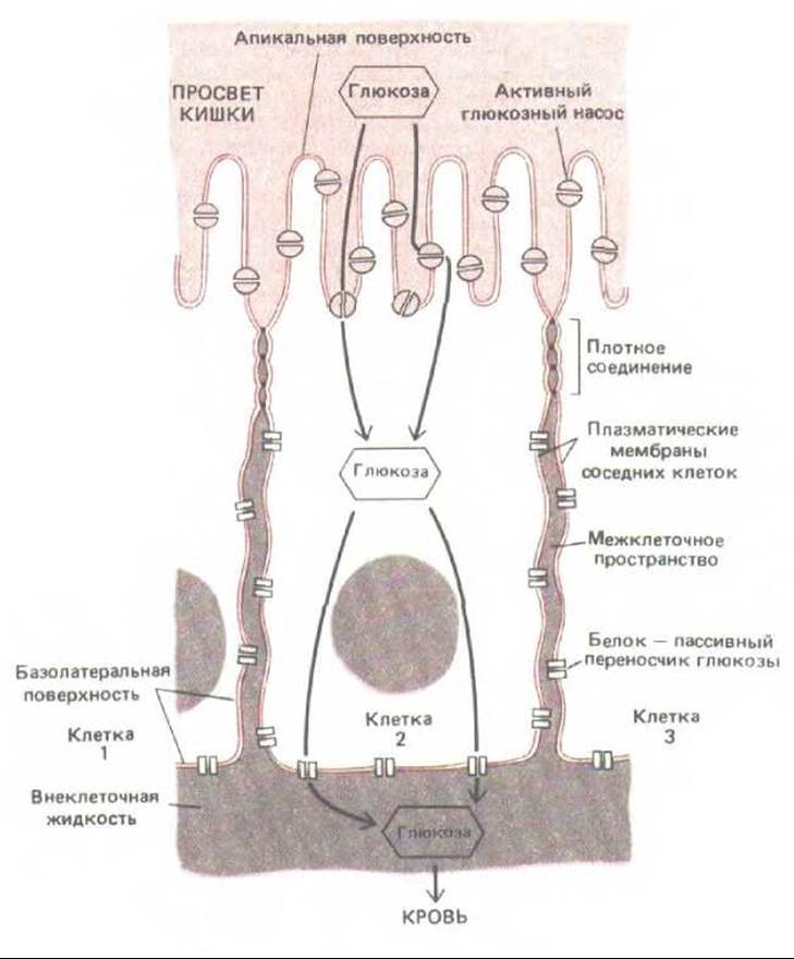
Fig. 14-2. Schematic diagram of an epithelial cell from the small intestine, illustrating how tight junctions demarcate distinct domains of The Plasma Membrane that house different transport proteins. This compartmentalization ensures the net transfer of nutrients from the intestinal lumen across the epithelial layer into the blood. In the example shown here, glucose is actively transported into the cell by apical glucose pumps and then exits via Facilitated Diffusion mediated by passive glucose carrier proteins located in the basolateral membrane domain. Tight junctions appear to restrict proteins to specific Regions of the plasma membrane by acting as diffusion barriers within The Lipid Bilayer; these junctions also block the diffusion of lipid molecules in the outer (but not the inner) leaflet of the lipid bilayer.

Fig. 14-3. A soluble tracer molecule introduced on one side of the epithelial layer is blocked by the tight junctions sealing adjacent cells together. However, this barrier is not absolute, and accumulating evidence indicates that cells can modulate The properties of their tight junctions to regulate the flux of solutes and Water across the epithelium.
Tight junctions between epithelial cells evidently counteract both of these types of diffusion. First, they act as diffusion barriers for Membrane Proteins, restricting them to either the apical or basolateral domains of the plasma membrane (Fig. 14-2). Such undesirable mixing of membrane components occurs when tight junctions are disrupted, for instance, by the removal of extracellular Ca2+ ions, which are required to maintain tight junction integrity. Second, adjacent cells are apposed so tightly that even water-soluble molecules fail to pass between them: when a small, electron-dense tracer molecule is introduced on one side of an epithelial cell sheet, it typically fails to penetrate past the tight junction (Fig. 14-3).

Fig. 14-4. Structure of a tight junction between epithelial Cells of the small intestine. (A) Diagram. (B) Freeze-fracture electron micrograph. (C) Conventional thin-section electron micrograph. Note that the cells are oriented with their apical ends directed downward. In (B), the plane of the micrograph is parallel to the membrane plane, revealing that the tight junction is composed of a branching network of sealing strands that encircles each cell in the sheet. These sealing strands appear as ridges of intramembrane particles on the protoplasmic (P) fracture face (B) or as complementary grooves on the extracellular (E) fracture face. In the conventional thin section (C), the junction appears as a series of focal points of apparent membrane fusion between the outer lipid leaflets of two adjacent membranes; each such contact corresponds to a cross-sectional profile of a sealing strand. [(B and C) from N. B. Gilula, in Cell Communication (R. P. Cox, ed.), pp. 1–29. New York, Wiley, 1974. Reprinted by permission of John Wiley & Sons, Inc.]
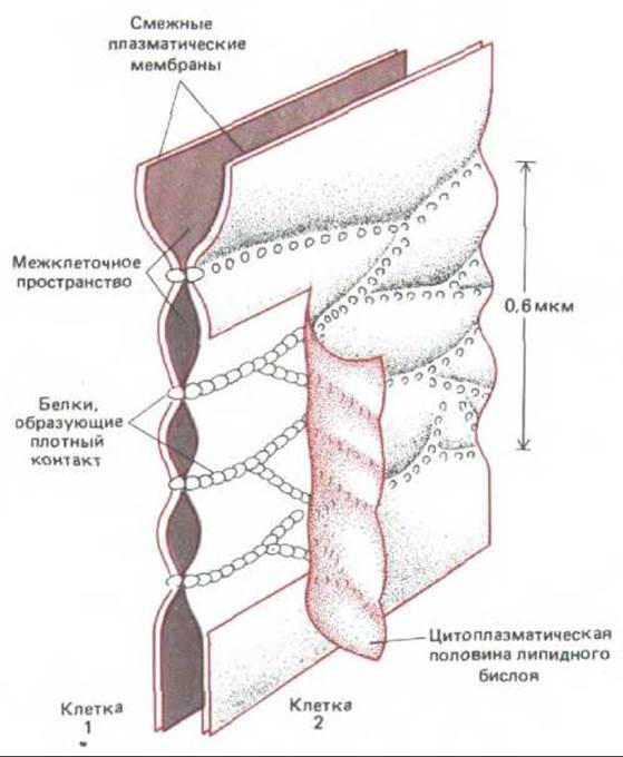
Fig. 14-5. Current model of tight junction ultrastructure. Adjacent plasma membranes are thought to be held together by continuous chains of specialized transmembrane proteins that span the intercellular space and form a tight seal. To reveal these protein strands, the inner lipid monolayer of one membrane is peeled back in this diagram. In freeze-fracture replicas, tight junction proteins remain associated with the inner (cytoplasmic) membrane monolayer rather than the outer monolayer as depicted here, forming the patterned intramembrane particles shown in Fig. 14-4B.
The molecular architecture of tight junctions remains to be fully elucidated, but freeze-fracture electron microscopy reveals that they consist of a network of anastomosing strands that completely encircles the apical end of each cell (Fig. 14-4A and B). In conventional thin-section electron micrographs, they appear as a series of focal contact points between the outer surfaces of two adjacent plasma membranes (Fig. 14-4C). Although all tight junctions are impermeable to macromolecules, their permeability to small molecules varies widely among different epithelia. For instance, tight junctions in the epithelium lining the small intestine are 10,000 times more permeable to inorganic ions than those in the epithelium of the Urinary Bladder. The capacity of a junction to restrict ion movement through the intercellular spaces increases logarithmically with the number of strands in the network, as though each strand functioned as an independent barrier. These strands are thought to be composed of long rows of specific transmembrane proteins contributed by each of the two contacting membranes, which bind directly to one another to occlude the intercellular space (Fig. 14-5).
14.1.2. Anchoring junctions link the cell Cytoskeleton to that of a neighboring cell or to the extracellular matrix
Anchoring junctions are widely distributed across many tissue types. They enable groups of cells—such as epithelia—to function as robust structural units by tying the cytoskeletal elements of individual cells to one another or to the extracellular matrix (Fig. 14-6). They are most abundant in tissues subjected to severe mechanical stress, such as cardiac Muscle, epidermis, and the uterine cervix. They occur in two structurally and functionally distinct forms: (1) adherens junctions, and (2) desmosomes and hemidesmosomes. Adherens junctions serve as anchoring sites for actin filaments, whereas desmosomes and hemidesmosomes anchor Intermediate filaments.
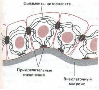
Fig. 14-6. This diagram illustrates how cytoskeletal filaments connect to analogous filaments in neighboring cells and to the extracellular matrix.
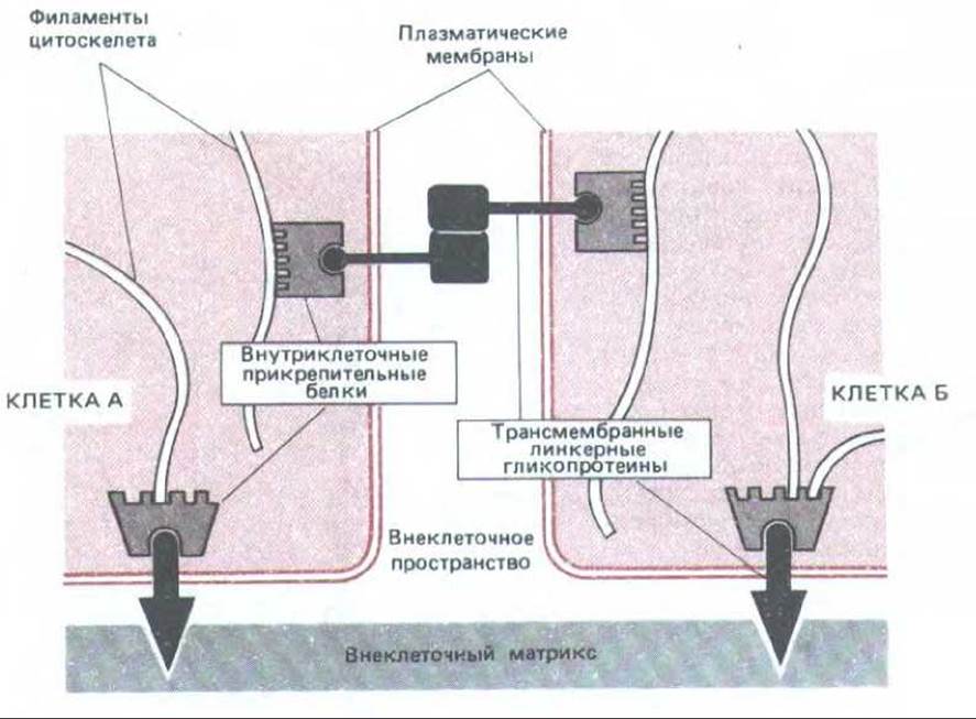
Fig. 14-7. Diagram of the functional role of two groups of proteins forming anchoring junctions: intracellular anchoring proteins and transmembrane linker Glycoproteins. In this example, the extracellular domains of the transmembrane linker glycoproteins holding the cells together interact directly. In other cases, they may be connected by additional proteins located in the extracellular space. Complexes of intracellular anchoring proteins link the linker glycoproteins to the cytoskeleton.
Before discussing the various classes of anchoring junctions, it is useful to briefly outline the General Principles of their structure. As shown in Fig. 14-7, all these junctions consist of Two Types of proteins: (1) intracellular anchoring proteins, which link the junctional complex to specific elements of the cytoskeleton (actin or intermediate filaments), and (2) transmembrane linker glycoproteins, whose intracellular domains bind to one or more intracellular anchoring proteins, while their extracellular domains interact either with the extracellular matrix or with the extracellular domains of transmembrane linker glycoproteins of an adjacent cell.
14.1.3. Adherens junctions link intracellular bundles of actin filaments to similar bundles in other cells or to the extracellular matrix [3]
Intercellular adherens junctions are extremely diverse. In many non-Epithelial Tissues, they take the form of punctate or linear contacts that link actin filaments in the cortical cytoplasm of adjacent cells. In epithelial sheets, they often form a continuous adhesion belt (zonula adherens) around each contacting cell, located near the apical end just below the tight junction. In neighboring cells, the adhesion belts lie directly opposite each other and are held together by a Ca2+-dependent mechanism. The transmembrane linker glycoproteins involved in this junction apparently belong to a family of Ca2+-dependent cell-Cell Adhesion molecules called cadherins (Section 14.3.7). The adherens belt is also referred to as a belt desmosome, though it should be noted that it differs significantly in chemical composition from a true desmosome (see below).

Fig. 14-8. Adherens belts (belt desmosomes) between epithelial cells of the small intestine. This junction encircles each contacting cell; its characteristic feature is the presence of a contractile bundle of actin filaments underlying the cytoplasmic surface of the membrane in the junctional zone.
Within each cell, just beneath the adherens belt, lies a contractile bundle of actin filaments arranged parallel to the plasma membrane; this bundle is attached to the adherens belt via a complex of intracellular proteins containing vinculin (Section 11.2.8). Thus, actin bundles are organized via transmembrane glycoproteins into a dense intercellular network (Fig. 14-8), which is believed to be involved in a fundamental morphogenetic process in animals—the folding of epithelial sheets into tubes and other similar structures (Fig. 14-9).
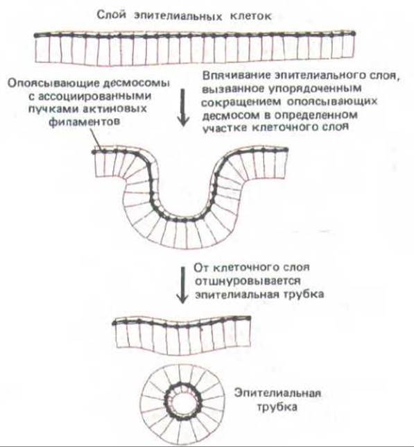
Fig. 14-9. Folding of an epithelial layer into a tube (e.g., during neural tube formation). It is believed that the coordinated contraction of actin filament bundles running along belt desmosomes leads to the constriction of the apical ends of cells in specific regions of the cell layer, causing the layer to roll up into a tube, which then detaches from the epithelium that gave rise to it.
For example, the concerted contraction of such bundles in the neural plate leads to the constriction of the apical end of each epithelial cell, causing the plate to roll up into the neural tube during early vertebrate development (Section 16.1.10).
Cell-matrix adherens junctions link cells and their actin filaments to the extracellular matrix. For instance, when fibroblasts grow on an artificial substrate coated with extracellular matrix molecules, they firmly attach to it at specialized regions of the plasma membrane called focal contacts or adhesion plaques, precisely where actin filament bundles terminate (Section 11.2.8). Many cells in tissues form similar focal contacts with the surrounding extracellular matrix. A large transmembrane linker glycoprotein (which acts on the cell surface as a receptor for the extracellular matrix glycoprotein Fibronectin, see Section 14.2.13) forms one of the linking bridges between the matrix and actin filament bundles in these plaques. The extracellular domain of this fibronectin receptor binds to fibronectin molecules On the surface of the culture dish, whereas its intracellular domain binds to an anchoring protein called talin, which in turn attaches to vinculin; vinculin is then attached to one or two other proteins that bind to actin (see Fig. 11-38).
The fibronectin receptor is just one representative of an extensive family of transmembrane linker glycoproteins called Integrins (Section 14.2.17), which appear to link actin filament bundles to the extracellular matrix. Some integrins have been well studied, demonstrating how transmembrane linker glycoproteins involved in cell-cell adhesion (such as cadherins) can connect bundles of cortical actin filaments of neighboring epithelial cells; however, vinculin is present in adherens belts without talin.
14.1.4. Desmosomes link intermediate filaments of adjacent cells; hemidesmosomes link these filaments to the basal lamina [4]
Desmosomes are punctate intercellular junctions that rivet cells together in various tissues, primarily in epithelia (Fig. 14-10). They also serve as attachment sites for intermediate filaments (Section 11.5), which form a structural cytoplasmic scaffold that resists tension. Thus, the intermediate filaments of neighboring cells are linked by desmosomes into a continuous network permeating the entire tissue. The type of intermediate filaments attached to desmosomes depends on the cell type: in most epithelial cells they are keratin filaments, in cardiac muscle fibers they are desmin filaments, and in certain cells covering The surface of the Brain they are vimentin filaments (see Table 11-5).
Electron microscopy and biochemical studies show that a desmosome consists of (1) a dense cytoplasmic plaque formed by a complex of intracellular proteins responsible for anchoring the cytoskeleton, and (2) transmembrane linker glycoproteins that are linked to the plaque and interact with each other via their extracellular domains, holding adjacent plasma membranes together (Fig. 14-11). The Role of desmosomes in cell-cell adhesion is highlighted in certain forms of the potentially fatal Skin disease Pemphigus, in which patients develop Antibodies against one or more of their own desmosomal linker glycoproteins; this leads to the disruption of desmosomes between epidermal cells and The formation of numerous blisters resulting from tissue fluid leakage into the loosened epithelium. Antibodies disrupt desmosomes only in the skin, implying that desmosomes in other tissues may have a different biochemical nature.
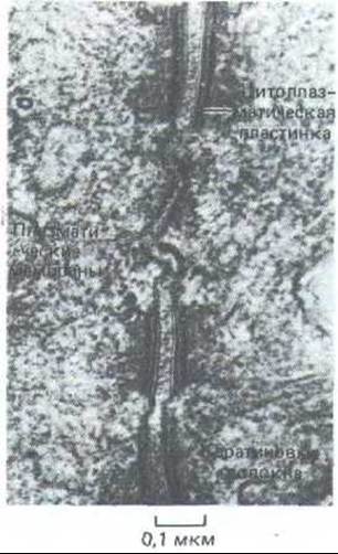
Fig. 14-10. Electron micrograph of three desmosomes between two epithelial cells in the rat intestine. [N. B. Gilula. In: Cell Communication (R.P. Cox, ed.), pp. 1-29. New York, Wiley, 1974. Reprinted by permission of John Wiley & Sons, Inc.]
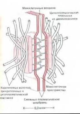
Fig. 14-11. Highly schematic representation of a desmosome. On the inner side of each adjacent plasma membrane lies an electron-dense plaque composed of a mixture of intracellular anchoring proteins called desmoplakins. Each plaque is linked to a dense network of keratin filaments running along the plaque surface. Transmembrane linker glycoproteins called desmogleins bind to the plaques and connect adjacent membranes via their extracellular domains using a Ca2+-dependent mechanism. Although desmosomes and adherens belts differ morphologically and chemically, they share at least one common intracellular protein called plakoglobin.
Hemidesmosomes are morphologically similar to desmosomes but differ in their functional and chemical properties. They do not rivet the plasma membranes of adjacent cells together; instead, they anchor the basal surface of these cells to the underlying basal lamina—a specialized layer of the extracellular matrix at the boundary between epithelium and connective tissue (Section 14.2.15). Furthermore, while keratin filaments associated with desmosomes attach to the latter by their lateral surfaces (Fig. 14-11), many filaments attached to hemidesmosomes terminate directly in the desmosomal plaques (Fig. 14-12).
Both desmosomes and hemidesmosomes act as rivets, distributing compressive and tensile stresses throughout the epithelium and underlying connective tissue.
14-6
14.1.5. Gap junctions allow small molecules to pass directly from cell to cell [5]
This is arguably the most remarkable type of intercellular junction. Gap junctions are among the most widespread—they are extremely abundant in most tissues and present in virtually all animals. In electron micrographs, they appear as regions where the membranes of two adjacent cells are separated by a narrow gap of about 3 nm. Gap junctions are involved in Intercellular Communication, allowing inorganic ions and other small water-soluble molecules to pass directly from the cytoplasm of one cell to the cytoplasm of another, thereby providing electrical and metabolic coupling between cells. Such coupling has profound functional significance, many aspects of which we are only beginning to understand.
Intercellular connections of this type were first demonstrated in 1958 using physiological Methods, but it took more than a decade to show that this physiological coupling correlates with the presence of gap junctions visible in the Electron microscope. The initial evidence for such coupling was obtained from electrophysiological studies of specific pairs of interacting Neurons in the crayfish nerve cord. When a potential difference was applied across two microelectrodes inserted into two interacting cells, an unexpectedly large current flowed across the membrane at their junction point. This indicated that inorganic ions (which carry electrical charges in living tissues) can move freely from one cell to another. Subsequent experiments showed that small fluorescent molecules injected into one cell also readily pass into neighboring cells without leaking into the extracellular space, provided their molecular weight does not exceed 1000–1500. This implied that the effective diameter of the junctional channels must be about 1.5 nm (Fig. 14-13) and that cells exchange small molecules (inorganic ions, sugars, Amino Acids, NUCLEOTIDES, Vitamins, etc.) but not macromolecules (proteins, Nucleic Acids, and Polysaccharides).
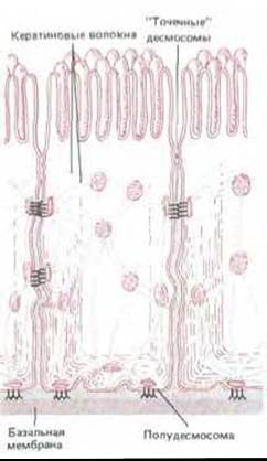
Fig. 14-12. Arrangement of desmosomes and hemidesmosomes in epithelial cells of the small intestine. The networks of keratin filaments in adjacent cells are linked to one another via desmosomes, and to the basal lamina via hemidesmosomes. While keratin filaments attach to the dense plaques of desmosomes by their sides, they anchor by their ends in hemidesmosomes.
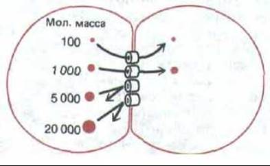
Fig. 14-13. When fluorescent molecules of varying sizes are injected into one of the two cells connected by a gap junction, only those with a molecular weight not exceeding approximately 1000-1500 (depending on cell type) pass into the other cell, whereas larger molecules are excluded. This corresponds to an effective intercellular channel diameter of about 1.5 nm.
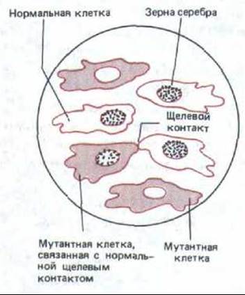
Fig. 14-14. Schematic representation of an autoradiograph demonstrating metabolic coupling between cells linked by gap junctions in an in vitro culture. Mutant cells lack the enzyme thymidine kinase and therefore cannot incorporate radioactive thymidine added to the medium into their DNA. Normal cells are capable of incorporating thymidine into DNA, and thus their nuclei are dotted with black spots (silver grains in the autoradiograph). As shown, in a mixed culture of normal and mutant cells, the radioactive label is also incorporated into the nuclei of those mutant cells that are in contact with normal cells and establish gap junctions with them. This occurs because in the normal cell, radioactive thymidine is phosphorylated by thymidine kinase to form thymidine triphosphate; the radioactive thymidine triphosphate then passes through the gap junctions into the mutant cell and is incorporated into its DNA.
Such an exchange of small intracellular metabolites forms The basis of metabolic cooperation, which can be demonstrated in cultured cells. For example, mutant cell lines lacking the enzyme thymidine kinase can be co-cultured with normal (wild-type) cells possessing this enzyme. Mutant cells on their own are unable to incorporate thymidine into DNA because they cannot carry out the first step of this process—The conversion of thymidine to thymidine triphosphate. If, however, such cells are grown together with wild-type cells in the presence of radioactive thymidine, the label is incorporated into the DNA of the mutant cells that are in direct contact with the wild-type cells. This indicates that some DNA precursor containing radioactive thymidine (evidently thymidine triphosphate) passes directly from the wild-type cells into the contacting mutant cells (Fig. 14-14). Such metabolic cooperation is not observed when similar experiments are performed with cells incapable of forming gap junctions.
There is further evidence indicating that gap junctions are responsible for Electrical and Chemical coupling between contacting cells. Structures typical of such junctions can be found almost everywhere that electrical or chemical coupling is detected. Conversely, coupling is absent in vertebrate cells that lack gap junctions. Furthermore, the passage of current and dye can be blocked by microinjecting antibodies against the major gap junction protein into gap-junction-coupled cells (see below). Finally, when this gap junction protein is incorporated into an artificial lipid bilayer, or when mRNA encoding this protein is injected into frog oocytes, electrophysiological analysis reveals channels with many properties characteristic of gap junction channels.
14.1.6. Gap junction connexons are oligomers of a transmembrane protein that spans the membrane multiple times [6]
Gap junctions are built from transmembrane proteins that form structures called connexons. When the connexons of the plasma membranes of two adjacent cells align, they form a continuous aqueous channel connecting the interiors of the two cells (Fig. 14-15). The connexons are joined in such a way that a gap remains between the adjacent plasma membranes (hence the name "gap junction"), which distinguishes them from tight junctions, where the membranes are closely apposed (cf. Figs. 14-5 and 14-15). In Cytology/cytology/93.html">ELECTRON MICROGRAPHS OF freeze-fracture preparations, each connexon appears as an intramembrane particle, and each gap junction may contain hundreds of clustered connexons (Fig. 14-16).
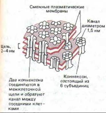
Fig. 14-15. Model of a gap junction based on biochemical studies, electron microscopy, and X-Ray Diffraction Analysis. The plasma membranes of two adjacent cells connected by such a junction are shown. Protein structures called connexons pass through both lipid bilayers; each connexon is thought to consist of six identical protein subunits. The joining of two connexons creates a continuous aqueous channel connecting one cell to the other.
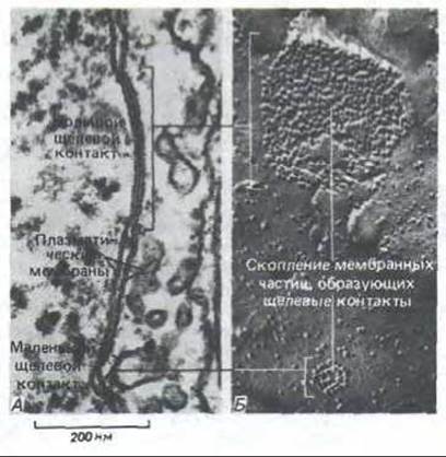
Fig. 14-16. Large and small gap junctions between fibroblasts in culture. Transmission electron micrographs of a thin section (A) and a freeze-fracture replica (B). On the fracture face, each gap junction appears as an aggregate of homogeneous intramembrane particles associated exclusively with the cytoplasmic leaflet of the membrane fracture face. Each intramembrane particle corresponds to a connexon, as shown in Fig. 14-15. [N.B. Gilula. In: Cell Communication (R.P. Cox, ed.), pp. 1-29. New York, Wiley, 1974. Reprinted by permission of John Wiley a. Sons, Inc.]
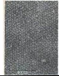
Fig. 14-17. Electron micrograph of a gap junction patch isolated from rat Liver. Negative staining was used to reveal connexons arranged in a hexagonal lattice. The electron-dense central pore of each connexon is approximately 2 nm in diameter. [N. B. Gilula. In: Intercellular Junctions and Synapses (Receptors and Recognition, Series B, Vol. 2; J. Feldman, N.B. Gilula, and J.D. Pitts, eds.), pp. 3-22, London. Chapman a. Hall, 1978.]
Due to their unusual resistance to Proteolytic Enzymes and detergents, gap junctions can be successfully isolated from rodent liver (Fig. 14-17). A gap junction consists predominantly of a single protein with a Molecular Weight of approximately 30,000. DNA Sequencing reveals that its polypeptide chain (comprising about 280 amino acid residues) spans the lipid bilayer of the membrane as four a-helices. Evidently, six such protein molecules assemble to form each connexon, much like the Formation of the Acetylcholine Receptor channel, where the aqueous pore is formed by six a-helices—one from each protein subunit (see Fig. 6-64).
Antibodies against the 30,000-dalton protein react with gap junctions from many tissues and organisms; connexon proteins appear to be similar in all cases (although biochemical and physiological data show that they are not strictly identical). This is consistent with the fact that cells of different types in culture commonly form gap junctions with one another, even when derived from different species.
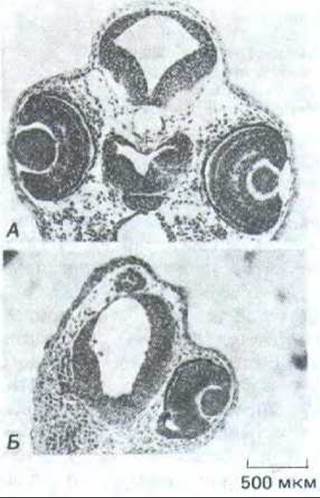
Fig. 14-18. Effect of antibodies directed against the major gap junction protein injected into one of the cells of an early Xenopus embryo. Cross-sections of a normal embryo (A) and an embryo injected at the 8-cell stage (B). Note that the second embryo lacks an eye and has an underdeveloped brain on the injected side. (A. Warner, S. Guthrie, N. B. Gilula, Nature 331: 126-131, 1985. Copyright 1985 Macmillan Journals Limited.)
14-7
14.1.7. Most Cells in Early Embryos Communicate via Gap Junctions [7]
In certain tissues, the role of cell coupling via gap junctions is readily apparent. For example, electrical coupling synchronizes the contractions of Heart muscle cells and smooth muscle cells responsible for gut peristalsis. Similarly, electrical coupling between Nerve Cells allows action potentials to propagate rapidly from cell to cell without the delay characteristic of chemical synapses; this provides a distinct advantage where speed and response reliability are critical, such as in certain escape responses of fish and insects. It is more difficult to understand why gap junctions are necessary in tissues that show no electrical activity. In principle, the exchange of metabolites and ions could coordinate The activity of individual cells in such tissues. For instance, gap junctions might coordinate epithelial cell activities like ciliary beating; moreover, because intracellular messengers such as cyclic AMP can pass Through gap junctions, the response of coupled cells to extracellular signaling molecules could be propagated and coordinated through this pathway.
Cell coupling via gap junctions appears to play a vital role in Embryogenesis. In early vertebrate embryos (beginning at the late eight-blastomere stage in mice), most cells are electrically coupled to one another. However, as specific groups of cells acquire distinct fates and begin to differentiate, they typically uncouple from surrounding tissues. For example, during neural tube closure, neural cells lose their connection with the overlying ectoderm (see Fig. 14-9). Meanwhile, cells within each group remain coupled to one another, thus behaving as a cooperative system that follows a coordinated pathway of development.
One compelling hypothesis is that embryonic cell coupling could mediate long-range signaling within a developing epithelium. For example, small molecules might diffuse through gap junctions from regions where their concentration is maintained at a high level into regions where it remains low, thereby establishing a smooth gradient. Local concentration levels could provide cells with "positional information" to guide their differentiation based on their Location within the embryo. However, whether gap junctions actually perform this function remains unknown.
The potential role of intercellular communication via gap junctions in developmental processes is indicated by experiments in which antibodies against the major gap junction protein were injected into one of the blastomeres of an 8-cell amphibian embryo. The introduced antibodies not only selectively disrupted electrical coupling and prevented dye transfer between the descendants of the treated cell (which was verified after two Cell Division cycles at the 32-cell stage), but also severely impaired embryonic development (Fig. 14-18). It remains unclear how the disruption of cellular coupling at an early stage later leads to developmental defects, but experiments of this kind represent a promising first step in studying the role of gap junctions in embryonic development.
14.1.8. Gap junction permeability can be regulated [8]
Experimental treatments that lower pH or elevate cytosolic free Ca2+ ion concentrations rapidly (within seconds) and reversibly decrease gap junction permeability. In some tissues, permeability may also be regulated by a voltage gradient across the junction or by extracellular chemical signals. These observations indicate that gap junctions are dynamic structures capable of opening or closing in response to cellular changes. Thus, in this respect, they resemble conventional Ion Channels (Section 6.4.14), although transitions between the open and closed states occur much less frequently here than in most ion channels.
The exact role that The regulation of gap junction permeability by potential or pH plays in the normal functioning of cellular assemblies remains unknown. In one case, however, The Significance of control involving Ca2+ ions seems clear. When a cell is injured or dying, its membrane loses its barrier function. Ions such as Ca2+ or Na+ enter the cell, while essential metabolites leak out. If such a cell remained coupled to its healthy neighbors, their internal environment would also be endangered. However, an increase in Ca2+ concentration within the damaged cell triggers the closure of gap junction channels, effectively isolating it and thereby preventing the spread of damage.
An increase in gap junction permeability induced by extracellular chemical signals leads to the propagation of a response to neighboring cells that are not in direct contact with the acting agent. For example, the hormone Glucagon, which stimulates liver cells to break down Glycogen and release glucose into the bloodstream, can also increase gap junction permeability between these cells in the rat. This occurs through an increase in the intracellular concentration of cyclic AMP (cAMP), which activates a cAMP-dependent protein kinase (Section 12.4.1), which in turn likely phosphorylates the major gap junction protein. Glycogen breakdown in liver cells is also driven by elevated cAMP levels; thus, the simultaneous increase in gap junction permeability facilitates the diffusion of cAMP from cell to cell, helping to recruit neighboring cell groups into the Glycogenolysis process. Figure 14-19 summarizes the various types of junctions formed between cells in an epithelium. At the apical end of the cell, the relative position of cell junctions is nearly identical in almost all epithelia: tight junctions occupy the most apical region, followed by the adhesion belt, and then specialized parallel rows of desmosomes; together, these constitute the "junctional complex." Gap junctions and additional desmosomes are arranged less regularly.
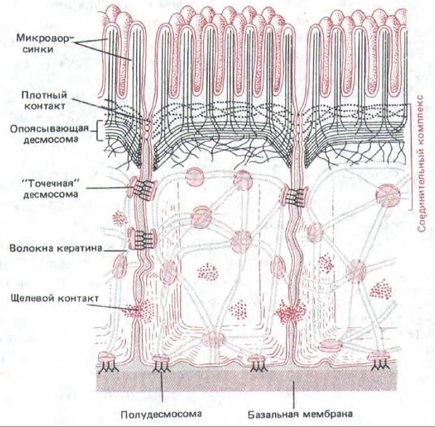
Fig. 14-19. Schematic diagram of the various junctions formed by epithelial cells of the small intestine.
Most cells in tissues are linked to one another and to the extracellular matrix at specialized contact sites called cell junctions. Cell junctions are divided into three functional classes: occluding, anchoring, and communicating junctions. Tight junctions constitute the principal group of occluding junctions and play a major role in maintaining concentration gradients of small hydrophilic molecules across epithelial layers; first, they tightly bind the membranes of adjacent cells, thereby creating a continuous permeability barrier between the Two Sides of the epithelium, and second, they form a barrier within the lipid bilayer that prevents the diffusion of membrane transport proteins between the apical and basolateral domains of the plasma membrane of each epithelial cell.
There are two MAIN TYPES OF anchoring junctions: adherens junctions and desmosomes. Both unite groups of cells into robust structural complexes by linking their cytoskeletal elements. Adherens junctions connect bundles of actin filaments, whereas desmosomes link intermediate filaments. Gap junctions serve for intercellular communication and consist of clusters of channel proteins that allow particles with a molecular mass of less than 1,500 to pass directly from one cell to another. Cells linked by such junctions exchange numerous inorganic ions and other small molecules; that is, they are chemically and electrically coupled. Gap junctions are of great importance for coordinating the functions of electrically active cells and appear to play a similar role in other cell groups as well.
Last update: 12/08/2026
Editorial and Educational Adaptation: This material has been compiled based on the primary/original source text. The project team performed an editorial review, corrected technical inaccuracies, structured sections, and adapted the content for an educational format.
What was processed:
- elimination of formatting defects (OCR errors, structural breaks, corrupted characters);
- editorial organization of content;
- standardization of terminology in accordance with academic sources;
- verification of factual statements against the original source text.
All mentions of the author, publication year, and origin of the primary text have been preserved in accordance with the source.