Review of Medical Physiology - William F. Ganong 2002
Introduction
General Principles and Cellular Basis of Human Physiology
Intercellular Communication
Cells communicate with one another using chemical messengers. Within a specific tissue, some messengers travel from Cell to Cell via Gap Junctions (see above) without ever entering the ECF. In addition, cells are influenced by chemical messengers secreted into the ECF. These messengers bind to protein receptors on The Cell surface or, in some cases, in the nuclear Cytoplasm, triggering a cascade of intracellular changes that produce a physiological effect. Messengers in the ECF mediate three Major Types of intercellular communication: neural communication, in which Nerve Cells release Neurotransmitters at synaptic junctions that influence the postsynaptic cell across a narrow synaptic cleft (see Chapter 4); endocrine communication, in which Hormones and growth factors reach target cells via the bloodstream (see Chapters 18–24); and paracrine communication, in which cellular products diffuse into the ECF to affect neighboring cells that may be some distance away (Fig. 1-34). Cells also secrete chemical messengers that, under certain circumstances, bind to receptors on the same cell that secreted them (autocrine communication). Chemical messengers include amines, Amino Acids, Steroids, Polypeptides, and occasionally other substances. Notably, in different PARTS OF THE body, the exact same chemical messenger may act as a neurotransmitter, a paracrine mediator, a hormone secreted by Neurons into the bloodstream (neurohormone), or a hormone released into the Blood by glandular cells.
Class="center">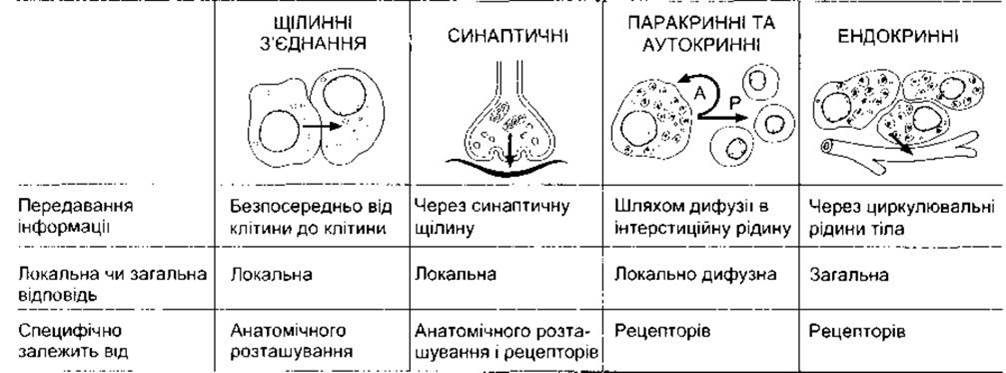
Fig. 1-34. Intercellular communication via chemical mediators
An additional form of intercellular communication is juxtacrine communication. Certain cells extracellularly express multiple repeats of growth factors, such as transforming growth factor-alpha (TGF-a), on transmembrane Proteins that provide cell attachment. Other cells possess TGF-a receptors. Consequently, cell-bound TGF-a can bind to TGF-a receptors on an adjacent cell, tethering the two cells together. This mechanism may be important in establishing local centers of growth within Tissues.
Radioimmunoassay
Antibodies to polypeptides and proteins have now been developed, and specialized technologies also enable the creation of antibodies against other chemical messengers. These antibodies can be used to measure messengers in Body Fluids and tissue extracts using radioimmunoassay (RIA). This technique relies on competition between a labeled Ligand and an added radioactive ligand for binding to the ligand-specific antibody. The greater The amount of the unlabeled ligand present in the sample being analyzed, the more it competes, and the less radioactive ligand binds to the antibody. Radioimmunoassay is widely used in both scientific research and clinical medicine.
Receptors for Hormones, Neurotransmitters, and Other Ligands
Many receptors for chemical messengers have been isolated and characterized. These proteins are not static cellular components; their numbers increase or decrease in response to various stimuli, and their properties change with shifting physiological conditions. When a hormone or neurotransmitter is present in excess, the number of active receptors typically decreases (down-regulation); conversely, when chemical messengers are scarce, the number of active receptors increases (up-regulation). Angiotensin II acting on the adrenal cortex is an exception: it increases rather than decreases the number of receptors in the Adrenal gland. Regarding Membrane Receptors, receptor-mediated endocytosis is responsible for negative regulation; ligands bind to their receptors, and the ligand-receptor complexes move laterally in the membrane to coated pits, where they are drawn into the cell via endocytosis (internalization). This reduces the number of receptors on the cell surface. Some receptors are recycled following internalization, while others are replaced by de novo cellular synthesis. Another type of negative regulation is desensitization, in which receptors undergo chemical modification that renders them less responsive (see Chapter 4).
MECHANISM OF ACTION of Chemical Messengers
The principal mechanisms of intracellular action for chemical messengers are summarized in Table 1-8. Ligands such as acetylcholine bind directly to Ion Channels in The cell membrane, altering their permeability. Thyroid and Steroid Hormones, 1,25-dihydroxycholecalciferol, and retinoids enter cells and act on specific members of a family of structurally related cytoplasmic or nuclear receptors. The activated receptor binds to DNA and enhances the METABOLISM/31.html">Transcription of selected mRNAs. Nearly all other ECF ligands bind to cell-surface receptors, and many stimulate the generation of intracellular messengers—such as cAMP, IP3, and DAG (see below)—which initiate changes in cellular activity. Accordingly, extracellular ligands are referred to as primary messengers, and intracellular mediators as secondary messengers.
Secondary messengers mediate many short-term changes in cellular activity by altering enzymatic activity, stimulating exocytosis, and so on. However, they also remodel the transcription of various genes, doing so in part through transcription factors already present in the activated cell. These factors trigger the transcription of immediate-early genes (see Fig. 1-20). The transcription factors produced by these genes then activate other genes, leading to more long-term effects.
Table 1-8. Principal mechanisms by which chemical messengers in the ECF alter cellular function
Mechanism |
|
Open or close ion channels in the cell membrane |
Acetylcholine at the nicotinic cholinergic receptor, norepinephrine at the cardiac K+ channel |
Act via cytoplasmic or nuclear receptors to enhance transcription of selected mRNAs |
THYROID HORMONES, retinoic acid, steroid hormones |
Activate phospholipase C, leading to intracellular production of DAG, IP3, and other Inositol phosphates |
Angiotensin II, norepinephrine via the a1-adrenergic receptor, vasopressin via the V1 receptor |
Activate or inhibit adenylate cyclase, resulting in increased or decreased cAMP production |
Norepinephrine via the ß-adrenergic receptor (increases cAMP); norepinephrine via the a2-adrenergic receptor (decreases cAMP) |
Increase intracellular cGMP |
ANP, NO (ECF) |
Increase Tyrosine kinase activity of the cytoplasmic domains of transmembrane receptors |
Insulin, EGF, PDGF, M-CSF |
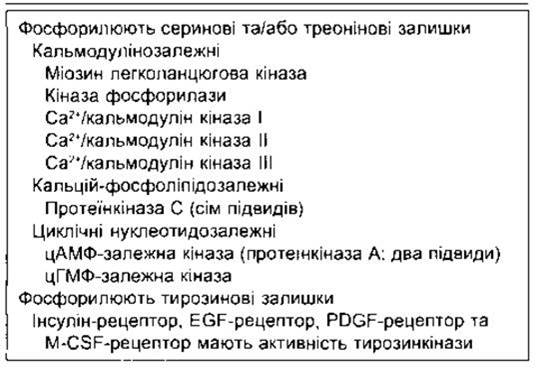
Upon activation, many membrane receptors trigger the release of secondary messengers or induce other intracellular changes via GTP-binding proteins (G proteins, see below). Secondary messengers predominantly activate protein Kinases—Enzymes that catalyze the phosphorylation of tyrosine, Serine, or Threonine residues in proteins. Over 300 protein kinases have been described. Some of those most important in mammals are listed in Table 1-9. The addition of phosphate groups alters protein conformation, thereby modifying their function and, consequently, cellular activity. In some cases, such as the insulin receptor, the intracellular domains of the receptors themselves function as protein kinases and may undergo autophosphorylation. Other receptors, including cytokine receptors, are not protein kinases per se, yet they induce the phosphorylation of numerous intracellular proteins. Phosphatases are likewise crucial, as the removal of phosphate groups deactivates or activates a variety of transport proteins or enzymes.
Table 1-9. Major protein kinases
Stimulation of Transcription
When thyroid and steroid hormones, 1,25-dihydroxycholecalciferol, and retinoids bind to their intracellular receptors, the conformation of the receptor protein changes, exposing the DNA-binding domain (Fig. 1-35). The hormone-receptor complex migrates to the DNA, where it binds to enhancer elements in the untranslated 5' regions of specific genes. Estrogen and triiodothyronine (T3) bind to receptors in The Nucleus; T3 receptors also bind thyroxine (T4), though with lower affinity. The glucocorticoid receptor resides primarily in the cytoplasm, but upon binding its ligand, it translocates directly to the nucleus. The initial cellular Location of other similarly acting receptors remains unclear. In any case, the binding of the hormone-receptor complex to DNA enhances the transcription of mRNAs encoded by the target Gene. These mRNAs are translated on Ribosomes, producing elevated levels of proteins that alter cellular function.
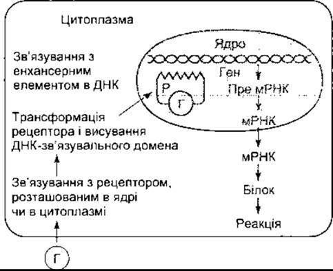
Fig. 1-35. Mechanism of action of steroid and thyroid hormones. H, hormone; R, receptor
In the absence of the steroid, the receptor is complexed with heat Shock protein 90 (Hsp90) and other proteins that mask the DNA-binding domain. The interaction of the steroid with the receptor induces a conformational change, causing the heat shock protein to dissociate and freeing the DNA-binding domain.
Heat shock proteins are a group of intracellular proteins whose expression increases in response to cellular hyperthermia and other stressors. They help cells survive Various Forms of stress, making 'stress proteins' a more accurate designation.
Receptor Structure
The structure of human gluco- and mineralocorticoid receptors is illustrated in Fig. 1-36. Two estrogen receptors (a and ß) and two T3 receptors (a and ß) have been identified; the estrogen receptor-a and T3 receptor-ß are depicted in Fig. 1-36. All of these receptors belong to a large superfamily characterized by a highly conserved, Cysteine-rich DNA-binding domain; a ligand-binding domain located at or near the carboxy-terminal region; and a relatively variable, poorly conserved amino-terminal region. DNA binding is mediated via zinc fingers (see above). Other members of this receptor family include the receptors for progesterone, androgen, and 1,25-dihydroxycholecalciferol. Numerous other gene-regulatory factors operate through this type of receptor across diverse species, ranging from the fruit fly to humans. Over 70 members of this receptor superfamily have now been described. Ligands have been identified for approximately half of these receptors, while the remainder are classified as orphan receptors whose ligands remain unknown. Retinoic acid, a derivative of retinol (vitamin A), plays a widespread role in embryonic development. There are three retinoic acid receptors—a, ß, and y—encoded by two receptor families, RAR and RXR, each possessing a, ß, and y isoforms. T3 receptors form homodimers before binding to DNA; they also form heterodimers with retinoid receptors to modulate transcriptional activity, making this regulatory network quite complex (see Chapter 18).
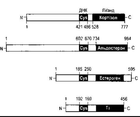
Fig. 1-36. Structure of human glucocorticoid, mineralocorticoid, α-estrogen, and β-T3 receptors. Note that each receptor features a cysteine-rich DNA- and ligand-binding domain at or near the carboxy-terminal group, alongside a highly variable amino terminus of the protein. The numbers indicate amino acid residues.
Rapid Actions of Steroids
Some steroid actions are much faster than those mediated via DNA binding. Examples include the rapid progesterone-induced increase in Ca2+ concentration within sperm heads and rapid steroid-induced alterations in the Functions of various neurons. This has led to the hypothesis that steroids may also bind to Membrane receptors and act through nongenomic mechanisms. There is mounting Evidence for the existence of such receptors. Steroids also bind to GABAA receptors, enhancing their activity (see Chapter 4).
Intracellular Ca2+
The Ca2+ cation regulates an exceptionally wide range of physiological processes, including proliferation, neuronal signaling, memory, contraction, secretion, and Fertilization. Thus, Ca2+ plays a critical role. The concentration of free Ca2+ is maintained at approximately 100 nmol/L; in the extracellular fluid, it is about 12,000 times higher than in the cytoplasm, amounting to 1,200,000 nmol/L, so the concentration and electrical gradients are strongly inwardly directed. A significant portion of intracellular Ca2+ is sequestered by the Endoplasmic reticulum and other Organelles (Fig. 1-37), providing a pool from which Ca2+ can be released to increase free Ca2+ concentration in the cytoplasm. As a result of this elevation, cytoplasmic Ca2+ binds to and activates calcium-binding proteins, which in turn activate a cascade of protein kinases.
Ca2+ enters the cell through a variety of channels, including voltage-gated and ligand-gated channels. Voltage-gated Ca2+ channels, of which there are at least four types, are activated by depolarization, whereas ligand-gated Ca2+ channels are activated by numerous different neurotransmitters and hormones. In addition, there are stretch-activated Ca2+ channels.
Voltage-gated Ca2+ channels are frequently classified into T-type (from transient) or L-type (from long-lasting) based on the duration of their activation during depolarization. Subtypes of such channels have also been described, though their emergence is attributed to mRNA Alternative Splicing and subunit combinations.
Ca2+ is pumped out of the cell in exchange for two H+ by means of a Ca2+-H+-ATPase, and is also transported across the cell membrane by an antiporter driven by a Na+ gradient, which exchanges three Na+ ions for every Ca2+.
Many secondary messengers elevate cytoplasmic Ca2+ concentrations by releasing Ca2+ from intracellular pools—primarily The endoplasmic reticulum—by enhancing Ca2+ influx into the cell, or through a combination of both mechanisms. IP3 is the primary secondary messenger responsible for Ca2+ release from the endoplasmic reticulum. In addition, cyclic adenosine diphosphate ribose (cADPR)—a NAD+ metabolite acting on ryanodine receptors (see Chapter 3)—may also be involved. In many tissues, a rapid leakage of Ca2+ from internal stores into the cytoplasm triggers the opening of Ca2+ channels and Ca2+ influx (store-operated Ca2+ entry). This enables more sustained responses and helps replenish intracellular stores.
Notably, cellular responses to a slight increase in cytoplasmic Ca2+ may differ significantly from responses to a substantial increase. This is crucial because, in many cases, release from a pool in one region of the cell produces a high local Ca2+ concentration that subsequently spreads to other areas, resulting in a more diffuse yet lower concentration. Furthermore, cytoplasmic Ca2+ concentrations often fluctuate within a normal physiological range, and hormones or other extracellular messengers typically alter the frequency rather than the amplitude of these oscillations.
Calcium-binding proteins
Numerous Ca2+-binding proteins have been described, including troponin, calmodulin, and calbindin. Troponin is involved in skeletal Muscle contraction (see Chapter 3). Calmodulin consists of 148 amino acid residues (Fig. 1-38) and contains four Ca2+-binding domains. It is unique in that the 115th residue is trimethylated and extraordinarily conserved, being present in both PLANT AND ANIMAL proteins. Upon binding Ca2+, calmodulin activates five different calmodulin-dependent kinases (see Table 1-9). One of these is Myosin light-chain kinase, which phosphorylates myosin, thereby triggering smooth muscle contraction. Another is phosphorylase kinase, which activates phosphorylase (see Chapter 17). Additionally, Ca2+/calmodulin-dependent kinases I and II are involved in synaptic function, while Ca2+/calmodulin-dependent kinase III plays a role in Protein Synthesis. Another calmodulin-activated protein is calcineurin, a phosphatase that deactivates Ca2+ channels through dephosphorylation. It also participates in T-cell activation and is inhibited by certain immunosuppressants (see Chapter 27).
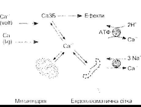
Fig. 1-37. Ca2+ metabolism in mammalian cells. Cytoplasmic Ca is in equilibrium with Ca2+ bound to the Mitochondria and endoplasmic reticulum. Calcium-binding proteins (CBPs) bind Ca2+ and, once activated, trigger various physiological effects. Ca2+ enters the cell via voltage-gated (volt) and ligand-gated (lig) Ca2+ channels. Ca2+ is extruded from the cell by the Ca2+-H+-ATPase and the Na+-Ca2+ exchanger.
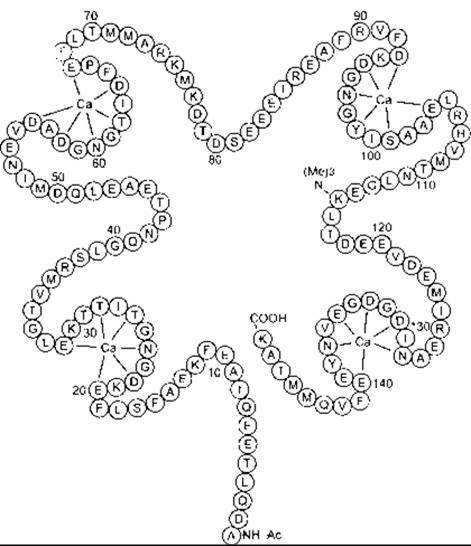
Fig. 1-38. Structure of calmodulin from bovine Brain. Amino acid residues are designated by single letters (Table 17-2). Note the four calcium domains (colored residues) flanked on each side by a-helix regions (reprinted with permission from Cheung WY: Calmodulin. An Overview. Fed Proc 1982, 41:2253)
G Proteins
Typically, signal Transduction into an intracellular biological response is mediated by nucleotide-regulatory proteins that bind GTP (G proteins). GTP is a guanosine analogue of ATP (see Chapter 17). When a signal reaches a G protein, the protein exchanges GDP for GTP. The GTP-protein complex triggers the response. The intrinsic GTPase activity of the protein converts GTP to GDP, restoring the resting state. GTPase activity is enhanced by GTPase-activating proteins (GAPs) (regulators of G-protein signaling).
Small G proteins are involved in a multitude of cellular functions. Members of the Rab family regulate The rate of vesicle transport between the endoplasmic reticulum, Golgi apparatus, Lysosomes, endosomes, and Plasma Membrane (see above). Another family of small GTP-binding proteins, Rho/Rac, mediates interactions between the Cytoskeleton and The Plasma Membrane, while yet another, Ras, regulates growth by relaying signals from the cell membrane to the nucleus. Members of these three families are related to the product of the ras proto-oncogene.
Another family of G proteins—large heterotrimeric G proteins—links cell surface receptors to catalytic units that catalyze the intracellular formation of second messengers, or couples receptors directly to ion channels. These G proteins consist of three subunits: a, ß, and y (Fig. 1-39). The a subunit is bound to GDP. When a ligand binds to a G-protein-coupled receptor, this GDP is exchanged for GTP, and the a subunit dissociates from the joined ß and y subunits. The dissociated a subunit exerts numerous biological effects. The ß and y subunits do not dissociate from each other, and ßy activates a variety of diverse effectors. The intrinsic GTPase activity of the a subunit converts GTP to GDP, leading to the reassociation of the a and ßy subunits and the termination of effector activation.
Heterotrimeric G proteins Relay signals from over 1,000 receptors, with ion channels and enzymes serving as their intracellular effectors. Examples are listed in Table 1-10. At least 16 different a subunits, 6 different ß subunits, and 12 different y subunits can assemble in numerous combinations. Based on their effect on effectors and Sequence Homology, they are divided into five families: Gs, Gi, Gt, Gq, and G13.
Many G proteins undergo modification through the attachment of specific Lipids, meaning they become lipidated (see Fig. 1-6). Trimeric G Proteins can be myristoylated, palmitoylated, or prenylated, whereas small G proteins are exclusively prenylated.

Fig. 1-39. Heterodimeric G proteins. Top: overview of the overall reaction. Bottom: when a ligand (square) binds to a serpentine receptor in the cell membrane, GTP replaces GDP on the a subunit. The GTP-a dissociates from the ßy subunit, and both GTP-a and ßy activate various effectors to elicit physiological effects. Subsequently, the intrinsic GTPase activity of GTP-a converts GTP to GDP, and the a, ß, and y subunits reassociate.
Table 1-10. Major Ligands for G-Protein-Coupled Receptors
Class |
Ligand |
Neurotransmitters |
Adrenaline Noradrenaline Dopamine 5-Hydroxytryptamine Histamine Acetylcholine Adenosine Opioids |
Tachykinins |
Substance P Neurokinin A Neuropeptide K |
Other Peptides |
Angiotensin II Arginine vasopressin Oxytocin VIP, GHRH, TRH, PTH |
Glycoprotein hormones |
TSH, FSH, LH, hCG |
Arachidonic acid derivatives |
Thromboxane A2 |
Others |
Odorants Tastants Endothelins Platelet-activating factor Cannabinoids Light |
Serpentine receptors
All heterotrimeric G-protein-coupled receptors are proteins that span the cell membrane seven times (serpentine receptors). These receptors can be palmitoylated. An extraordinarily large number of them have been cloned, and their functions are numerous and diverse. The structures of two of them are shown in Fig. 1-40. Predominantly small ligands bind to amino acid residues within the membrane, whereas large polypeptide and protein ligands bind to extracellular domains that are larger and better developed in receptors for polypeptides and proteins. It is typically amino acid residues in the third cytoplasmic loop, closest to the carboxy-terminal group, that interact with G proteins.
Inositol trisphosphate and diacylglycerol as second messengers
The Link Between a membrane-bound ligand junction acting via Ca2+ and the rapid increase in cytoplasmic Ca2+ concentration is often provided by inositol trisphosphate (inositol 1,4,5-trisphosphate, IP3). When one of these ligands binds to its receptor, receptor activation triggers the activation of phospholipase C on the inner surface of the membrane via a Gq protein. Eight isoforms of phospholipase C (PLC) have been identified to date, with PLCβ1 and PLCβ2 forms being activated by G proteins. They catalyze the Hydrolysis of phosphatidylinositol 4,5-bisphosphate (PIP2), yielding IP3 and diacylglycerol (DAG) (Fig. 1-41). Tyrosine kinase-linked receptors (see below) can also produce IP3 and DAG by activating PLCβ1. IP3 diffuses into the endoplasmic reticulum, where it triggers the release of Ca2+ into the cytoplasm (Fig. 1-42). The IP3 receptor is similar to the ryanodine receptor, which is a Ca2+ channel in the sarcoplasmic reticulum of Skeletal Muscle (see Chapter 3), except that the IP3 receptor is twice as large. DAG is also a second messenger; it remains in the cell membrane, where it activates one of the seven subspecies of protein kinase C (see Table 1-9). Examples of ligands acting through these second messengers are listed in Table 1-8.
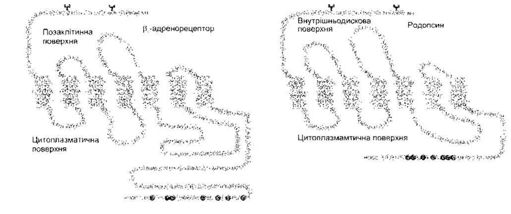
Fig. 1-40. STRUCTURE OF THE β2-adrenoceptor and rhodopsin. Individual amino acid residues are designated by single-letter codes, and those shaded dark represent phosphorylation sites. Y-shaped symbols on N residues indicate glycosylation sites. Note the extracellular amino terminus, the extracellular carboxy terminus, and the light-shaded seven membrane-spanning regions of each protein (reproduced with permission from Nature 1986;321:869. Macmillan Magazines, 1986).

Fig. 1-41. Phosphatidylinositol metabolism in cell membranes. Phosphatidylinositol is phosphorylated to yield phosphatidylinositol 4-phosphate (PIP), and subsequently phosphatidylinositol 4,5-bisphosphate (PIP2). Phospholipase Cβ1 and β2 catalyze The breakdown of PIP2 into inositol 1,4,5-trisphosphate (IP3) and diacylglycerol. Other inositol phosphates and phosphatidylinositol derivatives may also be formed. IP3 is dephosphorylated to inositol, and diacylglycerol is metabolized to cytidine diphosphate (CDP) diacylglycerol. Subsequently, CDP-diacylglycerol and inositol combine to form phosphatidylinositol, completing the cycle (modified from Berridge MJ: Inositol triphosphate and diacylglycerol as second messengers. Biochem J 1984;220:345).
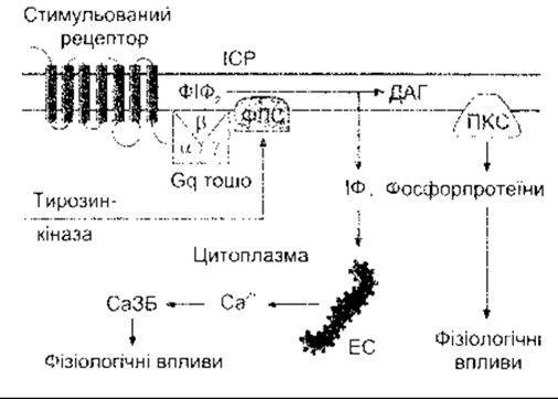
Fig. 1-42. Schematic diagram of the release of inositol trisphosphate (IP3) and diacylglycerol (DAG) as second messengers. Binding of a ligand to a G-protein-coupled receptor activates phospholipase C (PLC) β1 or β2. Activation of receptors by intracellular tyrosine kinase domains can activate PLCγ1. Consequently, the hydrolysis of PIP2 produces IP3, which releases Ca2+ from the endoplasmic reticulum (ER), and DAG, which activates protein kinase C (PKC). CaB, Ca2+-binding proteins; ISF, interstitial fluid.
The precursor of PIP2 is phosphatidylinositol (see Fig. 1-41). This phospholipid is present in relatively small amounts in the inner leaflet of the cell membrane. It is first converted into phosphatidylinositol 4-phosphate (PIP), and subsequently into the PIP2 derivative, which is hydrolyzed to yield IP3 and DAG. Other inositol phosphates are also formed in cells, although their exact functions remain unclear. IP3 is metabolized through sequential dephosphorylation steps to inositol. DAG is converted into phosphatidic acid, and then into cytidine diphosphate (CDP) diacylglycerol, which combines with inositol to form phosphatidylinositol, completing the cycle.
Cyclic AMP
Cyclic AMP (cAMP) also serves as a second messenger (Fig. 1-43). Some of the numerous ligands that act through this compound are listed in Table 1-8. Cyclic AMP is cyclic adenosine-3',5'-monophosphate. It is synthesized from ATP through the action of the enzyme Adenylyl Cyclase and converted into inactive 5'-AMP by the action of the enzyme phosphodiesterase. Cyclic AMP activates cyclic nucleotide-dependent protein kinase (protein kinase A), which, much like protein kinase C, catalyzes the phosphorylation of proteins, altering their conformation and activity. A classic example is the activation of phosphorylase kinase in the Liver by adrenaline via cAMP and protein kinase A (see Fig. 17-13). Additionally, the active catalytic subunit of PKA translocates to the nucleus and phosphorylates cAMP-response element-binding protein (CREB). This transcription factor subsequently binds to DNA and alters the transcription of A number of genes.
Cyclic AMP is metabolized by phosphodiesterase. This phosphodiesterase is inhibited by methylxanthines, such as caffeine and theophylline, thereby potentiating the hormone and transmitter effects mediated via cAMP.
Activation of adenylyl cyclase
The mechanism by which ligands cause changes in intracellular cAMP concentration involves five components: the catalytic unit, adenylyl cyclase, which catalyzes The conversion of ATP to cAMP; stimulatory and inhibitory receptors; and stimulatory and inhibitory G proteins that couple the receptor to the catalytic unit (Fig. 1-44). Like receptors, adenylyl cyclase is a transmembrane protein that spans the membrane 12 times. Eight isoforms of this enzyme have been described, and their combination with many different forms of G proteins allows cAMP pathways to be tailored to the specific needs of a tissue. When an appropriate ligand binds to a stimulatory receptor, the Gsα subunit activates an adenylyl cyclase. Conversely, when an appropriate ligand binds to an inhibitory receptor, the Giα subunit inhibits adenylyl cyclase. Receptors are specific and respond only to a single ligand or a group of related ligands. Nevertheless, heterotrimeric G proteins mediate the stimulatory and inhibitory effects produced by many different ligands. Furthermore, there is crosstalk between the phospholipase C system and the adenylyl cyclase system, and several adenylyl cyclase isoforms are stimulated by calmodulin. Finally, the effects of protein kinase A and protein kinase C are widespread. Given such complexity, how are specific responses to specific stimuli achieved? The answer lies in the anchoring of G proteins, adenylyl cyclase, and protein kinase to the cytoskeleton in such a way that local microdomains are formed. Some of these anchorages are mediated by lipid products (see Fig. 1-6).
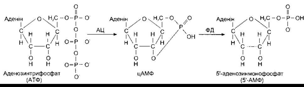
Fig. 1-43. Formation and metabolism of cAMP. AC, adenylyl cyclase; PDE, phosphodiesterase.
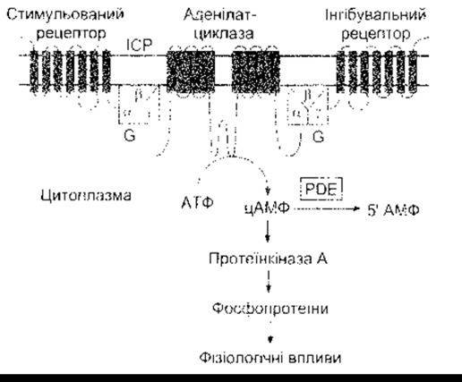
Fig. 1-44. The cAMP system. Activation of adenylyl cyclase catalyzes the conversion of ATP to cAMP. Cyclic AMP activates protein kinase A, which phosphorylates proteins, producing physiological effects. Stimulatory ligands bind to stimulatory receptors and activate adenylyl cyclase via Gs. Inhibitory ligands inhibit adenylyl cyclase via inhibitory receptors. ISF, interstitial fluid.
A fraction of cyclic AMP leaves the cell upon stimulation by certain hormones, but these amounts are small compared to the intracellular concentration, and only negligible amounts of extracellular cAMP enter cells.
Two Bacterial toxins exert profound effects on adenylyl cyclase through G-protein-mediated mechanisms. The A subunit of cholera toxin catalyzes the ADP-ribosylation of an arginine residue within the α subunit of Gs. This inhibits its GTPase activity, leading to prolonged stimulation of adenylyl cyclase (see Chapter 25). Pertussis toxin catalyzes the ADP-ribosylation of a cysteine residue near the carboxy-terminal end of the Gi α subunit. This inhibits Gi function.
Aside from causing disease states, both toxins are widely used as research tools in fundamental studies of G-protein function. The drug forskolin stimulates adenylyl cyclase activity through a direct action on the enzyme.
Guanylyl cyclase
Another cyclic physiologically important nucleotide is cyclic guanosine monophosphate (cyclic GMP, cGMP). Cyclic GMP plays a crucial role in Vision. Light acts on rhodopsin, which is contained in the rod photoreceptor Cells of the retina. Rhodopsin is coupled to phosphodiesterase via Gt, and activation of phosphodiesterase accelerates the conversion of cGMP to 5"-GMP (see Chapter 8). A similar process occurs in cones. In addition, there are cGMP-Gated ion channels, and cGMP activates cGMP-dependent kinase (see Table 1-9), triggering a variety of physiological effects.
Guanylyl cyclases are a family of enzymes that catalyze The formation of cGMP. They exist in two forms (Fig. 1-45). One form possesses an extracellular amino-terminal receptor domain, a single transmembrane domain, and a cytoplasmic carboxy-terminal domain that contains a tyrosine kinase-like region and the catalytic domain of guanylyl cyclase. Three such guanylyl cyclases have been characterized. Two serve as receptors for ANP (ANP-A and ANP-B; see Chapter 24), and the third binds Escherichia coli enterotoxin and the gastrointestinal polypeptide guanylin (see Chapter 26). The other form of guanylyl cyclase is soluble, contains heme, and is entirely intracellular. There are several isoforms of this intracellular enzyme. They are activated by nitric oxide (NO) and NO-donating compounds. Nitric oxide acts as endothelium-derived relaxing factor (EDRF) and an intercellular messenger. It is generated from arginine and plays a vital role in regulating blood vessel diameter (see Chapter 31). NO is also involved in numerous other functions, including penile erection (see Chapter 23) and synaptic transmission in the brain (see Chapter 4). Recent evidence suggests that In addition to cGMP-mediated effects, NO may also act directly by activating Ca2+-dependent K+ channels.
Phosphatases
Cells contain numerous phosphatases that remove phosphate groups from proteins. These enzymes are frequently closely related to, or associated with, tyrosine kinases and serine/threonine kinases. Two representatives of this group are shown in Fig. 1-45.
Growth Factors
Growth factors are becoming increasingly important in many aspects of physiology. They are polypeptides and proteins divided into three groups. The first group includes agents that promote the proliferation or development of various cell types: nerve growth factor (see Chapter 2), insulin-like growth factor I (IGF-I; see Chapter 22), activins and inhibins (see Chapter 23), and epidermal growth factor (EGF). Over 20 members of this group have been described. The second group consists of cytokines. These factors are produced by macrophages and lymphocytes and are essential for regulating The Immune System (see Chapter 27). More than 20 cytokines have also been identified. The third group comprises colony-stimulating factors, which regulate the proliferation and maturation of red and white Blood Cells. This group is likewise discussed in Chapter 27.
Receptors for EGF, platelet-derived growth factor (PDGF), and many other growth- and proliferation-promoting factors feature a single membrane-spanning domain with an intrinsic intracellular tyrosine kinase domain (see Fig. 1-45). When a ligand binds to the receptor, the tyrosine kinase domain undergoes autophosphorylation. Upon ligand binding, several of these receptors dimerize, and their intracellular tyrosine kinase domains cross-phosphorylate one another. One signaling pathway activated by phosphorylation proceeds via the ras proto-oncogene product and a cascade of MAP kinases directly to the formation of nuclear transcription factors that alter Gene Expression. This important direct pathway from the cell surface to the nucleus is schematically illustrated in Fig. 1-46. Note that Ras is one of the small G proteins requiring GTP binding for its activation. Receptors for cytokines and colony-stimulating factors differ from other growth factor receptors in that most lack tyrosine kinase domains in their cytoplasmic regions, while others possess a very small or nonexistent carboxy-terminal cytoplasmic segment. Nevertheless, they stimulate cytoplasmic tyrosine kinase activity. In some cases, this leads to association with the membrane-anchored protein gp130 (see Chapter 27). Cytokine receptors, in particular, activate cytoplasmic Janus kinases (JAKs) (see Fig. 1-47), which in turn phosphorylate signal transducers and activators of transcription (STAT) proteins. Phosphorylated STATs form homo- and heterodimers and translocate to the nucleus, where they act as transcription factors. Currently, four JAKs and seven STATs are known in mammals. The JAK-STAT pathway represents another major direct signaling route from the cell surface to the nucleus. However, it should be noted that the Ras and JAK-STAT pathways are highly complex and exhibit cross-talk with each other as well as with the phospholipase C and cAMP pathways.
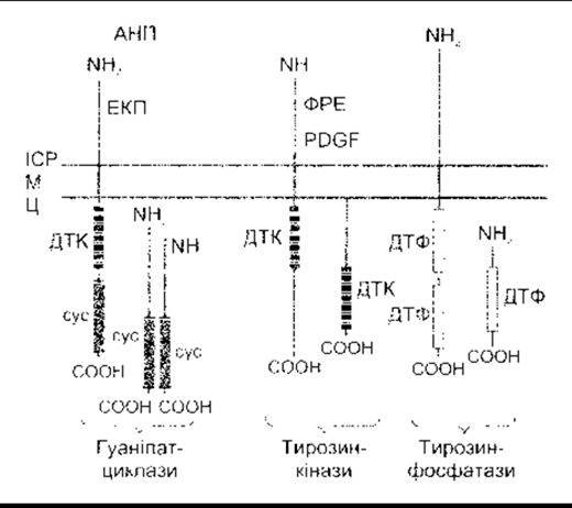
Fig. 1-45. Schematic representation of guanylyl cyclases, tyrosine kinases, and tyrosine phosphatases. ANP, atrial natriuretic peptide; C, cytoplasm; Cyc, cyclase domain; EGF, epidermal growth factor; ISF, interstitial fluid; M, cell membrane; PDGF, platelet-derived growth factor; TKD, tyrosine kinase domain; TPD, tyrosine phosphatase domain; ECE, Escherichia coli enterotoxin (Modified and reproduced with permission from Koesling D, Böhme E, Schultz G: Guanylyl cyclases, a growing family of signal transducing enzymes. FASEB J 1991;5:2785).
Receptors of another family bind transforming growth factor-beta (TGF-β) and related polypeptides. These receptors exhibit serine/threonine kinase activity, and their effects are mediated by SMADs—intracellular proteins that, upon phosphorylation, translocate to the nucleus, bind to DNA, and cooperate with other proteins to initiate the transcription of various genes.
Finally, as noted above, Integrins also initiate the phosphorylation of proteins that enter the nucleus and alter gene transcription.
Receptor and G-Protein Diseases
An increasing number of disorders are caused by Mutations in genes encoding receptors. For instance, receptor "loss-of-function" mutations have been identified in the 1,25-dihydroxycholecalciferol receptor (see Chapter 21) and the insulin receptor (see Chapter 19). Certain diseases result from The production of autoantibodies against receptors. Specifically, antibodies against TSH receptors cause Graves' disease (see Chapter 18), whereas antibodies against nicotinic acetylcholine receptors cause myasthenia gravis (see Chapter 44).
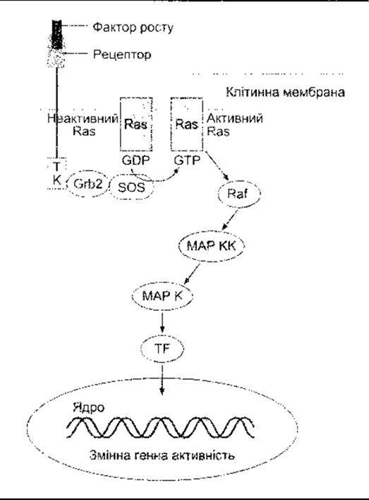
Fig. 1-46. One of the direct pathways by which growth factors alter gene activity. TK, tyrosine kinase domain; Grb2, growth factor receptor-bound protein 2; Sos, son of sevenless (Ras guanine nucleotide exchange factor); Ras, ras gene product; MAPK, mitogen-activated protein kinase; MAPKK, MAPK kinase; TF, transcription factor. Cross-talk exists between this pathway and the cAMP pathway, as well as an interconnection with the IP3-DAG pathway.
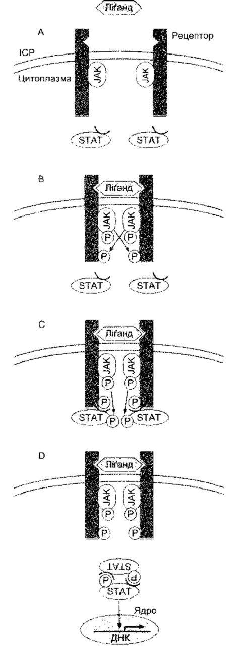
Fig. 1-47. Signal transduction via the JAK-STAT pathway. A: Ligand binding leads to receptor dimerization. B: Activation and tyrosine phosphorylation of JAKs. C: JAKs phosphorylate STATs. D: STATs dimerize and bind to a specific DNA response element (Modified from Takeda K, Kishimoto T, Akira S: STAT6: Its role in interleukin 4-mediated biological functions. J Mol Med 1997;75:17).
Mutant receptors may either gain or lose function. This is reflected in Table 1-11, which lists disorders caused by alterations in G protein-coupled receptors and G protein subunits. An example of a receptor loss-of-function mutation is nephrogenic diabetes insipidus, which arises from the inability of the V2 vasopressin receptor to mediate urinary concentration (see Chapters 14 and 38). Conversely, a gain-of-function mutation in the Ca2+-sensing receptor (see Chapter 21) leads to excessive suppression of Parathyroid hormone and causes familial hypocalciuric hypercalcemia.
Loss- or gain-of-function mutations causing disease can also affect G proteins. In one form of pseudohypoparathyroidism, mutant Gsα loses its ability to respond to parathyroid hormone, resulting in symptoms of hypoparathyroidism despite normal circulating parathyroid hormone levels. A fascinating disorder characterized by both gain- and loss-of-function features is familial male-limited precocious Puberty (testotoxicosis). In this condition, an activating mutation in Gsα causes excessive testosterone secretion and prepubertal sexual maturation. However, the mutant protein is Temperature-sensitive and active only at the relatively low temperature of the Testes (33°C, see Chapter 23). At 37°C—the normal temperature of the rest of the body—gain of function is replaced by loss of function, leading to hypoparathyroidism and blunted responsiveness to TSH. Another mutation in Gsα is associated with patches of Skin pigmentation with irregular borders and hypercorticism in McCune-Albright syndrome. This mutation occurs during embryonic development, producing a mosaic of normal and abnormal cells. Yet another mutation in Gsα impairs its intrinsic GTPase activity, rendering it chronically active in somatotroph cells of the anterior pituitary and leading to persistently elevated cAMP levels. This, in turn, causes the cells to become neoplastic, forming pituitary adenomas that result in acromegaly (in 40% of all cases of acromegaly) (see Chapter 22).
Last update: 10/08/2026
Editorial and Educational Adaptation: This material has been compiled based on the primary/original source text. The project team performed an editorial review, corrected technical inaccuracies, structured sections, and adapted the content for an educational format.
What was processed:
- elimination of formatting defects (OCR errors, structural breaks, corrupted characters);
- editorial organization of content;
- standardization of terminology in accordance with academic sources;
- verification of factual statements against the original source text.
All mentions of the author, publication year, and origin of the primary text have been preserved in accordance with the source.