BIOCHEMISTRY - L. Stryer - 1984
VOLUME 3
PART V. MOLECULAR PHYSIOLOGY
CHAPTER 35. HORMONE ACTION
35.2. Cyclic AMP Is Synthesized by Adenylate Cyclase and Cleaved by Phosphodiesterase
cAMP is formed from ATP through the action of the membrane-bound enzyme adenylate cyclase:
Class="center">
This reaction is slightly endergonic, with a ∆G°' of approximately 1.6 kcal/mol. The energy for cAMP synthesis is provided by the subsequent Hydrolysis of pyrophosphate. A specific phosphodiesterase degrades cAMP by hydrolysis to yield AMP:

This is a highly exergonic reaction with a ∆G°' of about —12 kcal/mol. In the absence of phosphodiesterase, cAMP is an extremely stable compound.
35.3. cAMP Serves as a Second Messenger in the Action of Many Hormones
Sutherland's work led to METABOLISM/2.html">THE CONCEPT OF cAMP acting as a second messenger in the MECHANISM OF ACTION of certain Hormones. The first messenger is the hormone itself. The Essence of this concept is as follows:
1. Cell Plasma Membranes contain Hormone Receptors.
2. The Interaction of a hormone with its specific receptor on The Plasma Membrane leads to The stimulation of adenylate cyclase, which is also associated with the plasma membrane.
3. As a result of adenylate cyclase activation, the intracellular level of cAMP increases.
4. The action of cAMP is exerted within The Cell and consists of altering The rate of one or more processes.
An important feature of this second-messenger hypothesis is that it does not require the hormone to enter the cell. The action of the hormone itself is restricted to The cell membrane. The biological effect of the hormone is mediated intracellularly by cAMP; the hormone does not act directly. The validity of this concept has been tested using A number of experimental criteria, namely:
1. Cellular adenylate cyclase must be stimulated by those hormones that target the given cell. Hormones that fail to elicit a specific biological response in the cell should not increase The activity of this enzyme.
2. The concentration of cAMP in target Cells must change in proportion to the biological response of these cells to hormonal stimulation; that is, it must exhibit both temporal and quantitative dependence on hormone concentration.
3. Phosphodiesterase inhibitors, such as theophylline or caffeine, must act synergistically with those hormones whose effects are mediated by cAMP as a second messenger.
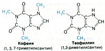
4. The addition of cAMP or a related compound to target cells should mimic the biological action of the hormone. (In practice, cAMP itself is not used in such experiments because it penetrates cells poorly; however, less polar derivatives of cAMP, notably dibutyryl-cAMP, readily enter cells and exert their effects.)
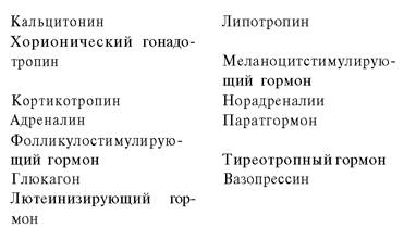
Experiments have demonstrated that cyclic AMP serves as a second messenger not only for epinephrine and Glucagon, but also for numerous Other Hormones (Table 35.1). cAMP influences an exceptionally wide range of cellular processes. For instance, its action accelerates The breakdown of stored fuel reserves, enhances Hydrochloric acid secretion by the gastric mucosa, triggers the dispersion of melanin pigment granules, and diminishes platelet aggregation.
Table 35.1. Hormones whose action is mediated by cAMP
35.4. Coupling of Hormone Receptors to Adenylyl Cyclase is Mediated by a Guanine Nucleotide-Binding Protein
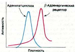
How does the binding of a hormone such as epinephrine or glucagon to its specific receptor lead to the activation of adenylyl cyclase? The hormone-binding sites are located on the outer surface of the plasma membrane, whereas the catalytic sites of adenylyl cyclase face the Cytosol. In essence, the hormone-binding and catalytic sites belong to distinct Proteins that can be separated by centrifuging the plasma membrane in a detergent solution (Fig. 35.4). Adenylyl cyclase (185 kDa) and the epinephrine receptor (75 kDa) are both large integral Membrane Proteins. The epinephrine receptor is also referred to as the β-adrenergic receptor because it binds a variety of pharmacologically active compounds.
Fig. 35.4. Separation of Adenylyl Cyclase and the β-adrenergic receptor by centrifugation of the solubilized (in a detergent solution) plasma membrane on a sucrose density gradient
The binding of a hormone to its specific receptor activates adenylyl cyclase not directly, but indirectly through a third protein known as a G protein1 (named for guanyl, as this protein binds guanine NUCLEOTIDES). This regulatory protein (42 kDa) exists in two forms. The G protein–GTP complex activates adenylyl cyclase, whereas the G protein–GDP complex does not. The G protein transitions from its inactive GDP-bound form to the active GTP-bound form via the exchange of bound GDP for GTP. This GTP–GDP exchange is catalyzed by the hormone–receptor complex rather than by the free receptor. Thus, the signal Transduction pathway proceeds from the hormone receptor to the G protein and subsequently to adenylyl cyclase (Fig. 35.5).
1 In Russian literature, the designation N protein is more commonly used. — Translator's Note.
Fig. 35.5. Signal transduction pathway during adenylyl cyclase activation triggered by hormone binding to its specific receptor. The G protein plays a crucial role in both the activation and inactivation of adenylyl cyclase
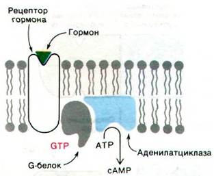
How is adenylyl cyclase turned off? The G protein possesses an additional property that enables it to Relay signals from hormone receptors to adenylyl cyclase: the GTP bound to the G protein is slowly hydrolyzed to ADP. In other words, the G Protein Functions as a GTPase. Consequently, this regulatory protein contains a built-in mechanism for inactivating adenylyl cyclase. The fraction of the G protein complexed with GTP—and, correspondingly, the degree of adenylyl cyclase activation—depends on The ratio of the rate of GDP-for-GTP exchange to the rate of GTP hydrolysis. The rate of GTP–GDP exchange increases significantly when the G protein binds to the hormone–receptor complex. Therefore, at low hormone concentrations, almost all of the G protein remains in the GDP-bound form, and consequently, nearly all of the adenylyl cyclase is inactive. In some cells, adenylyl cyclase activation also depends on Ca2+ concentration. Activation in these cells requires not only the GTP-bound form of the G protein, but also a complex of Ca2+ with calmodulin (a 17 kDa protein). Thus, the regulatory cycles governing adenylyl cyclase activity are beginning to be understood.
Last update: 06/08/2026
Editorial and Educational Adaptation: This material has been compiled based on the primary/original source text. The project team performed an editorial review, corrected technical inaccuracies, structured sections, and adapted the content for an educational format.
What was processed:
- elimination of formatting defects (OCR errors, structural breaks, corrupted characters);
- editorial organization of content;
- standardization of terminology in accordance with academic sources;
- verification of factual statements against the original source text.
All mentions of the author, publication year, and origin of the primary text have been preserved in accordance with the source.