BIOCHEMISTRY - L. Stryer - 1984
VOLUME 3
PART V. MOLECULAR PHYSIOLOGY
CHAPTER 35. HORMONE ACTION
35.5. Cyclic AMP Activates Protein Kinases
How does cAMP exert its effects on such a wide array of distinct cellular processes? Is there a common thread running through these diverse responses? Indeed there is, and it was uncovered once again by studying the Regulation of Glycogen METABOLISM—the very pathway where cAMP first came to light. Edwin Krebs and Donal Walsh established that cAMP activates protein kinase in Skeletal Muscle. Protein kinase phosphorylates both glycogen synthase (converting it to an inactive state) and phosphorylase kinase (converting it to an active state). In this manner, cAMP stimulates glycogen breakdown while inhibiting its synthesis in muscle (Section 16.15). A remarkably similar mechanism operates in the Liver. In essence, all known effects of cAMP are mediated by the activation of protein Kinases. Every Cell type examined to date contains protein kinases that are activated by concentrations of cAMP on the order of 10-8 M. These kinases modulate The activity of various target Proteins through phosphorylation.
The mechanism by which cyclic AMP activates muscle protein kinase is particularly intriguing. This enzyme is composed of two distinct types of subunits: a regulatory (R) subunit (49 kDa), which binds cAMP, and a catalytic (C) subunit (38 kDa). In the absence of cAMP, the regulatory and catalytic subunits associate to form an R2C2 complex that is devoid of enzymatic activity. The binding of cAMP to each of the Regulatory Subunits triggers the dissociation of the R2C2 complex into one R2 subunit and two C subunits. The free catalytic subunits possess full enzymatic activity. Consequently, the binding of cAMP to the regulatory subunit relieves the inhibition of the catalytic subunit, with cAMP functioning as an allosteric effector. In its subunit architecture—featuring separate regulatory and catalytic domains—protein kinase bears a striking resemblance to aspartate transcarbamoylase (Section 22.14).
Class="center">Fig. 35.6. cAMP activates protein kinase by inducing the dissociation of the enzyme's regulatory and catalytic subunit complex

35.6. Cyclic AMP: An Evolutionarily Ancient Starvation Signal
As discussed earlier (Section 28.6), cAMP exerts regulatory control over bacterial Cells, where it stimulates the Transcription of specific genes. It is evident that cAMP has a long evolutionary history as a signaling molecule. In Bacteria, cAMP acts as a starvation signal: its appearance indicates a shortage of glucose and triggers the synthesis of Enzymes required to utilize alternative Energy Sources. In certain mammalian cells, such as those of The Liver and muscle, cAMP retains its ancestral function as a starvation signal, though its action is now directed toward stimulating protein kinase rather than enhancing the transcription of specific genes. Another major difference is that in higher organisms, cAMP has evolved into a second messenger, operating within intracellular rather than Intercellular Communication pathways.
Why did cAMP evolve to become a second messenger? Three factors appear to have been crucial.
1. cAMP is generated from the ubiquitously available ATP through a straightforward reaction driven by The energy released from subsequent Pyrophosphate Hydrolysis.
2. Although derived from a molecule that occupies a central hub in Intermediary Metabolism, cAMP itself stands apart from the Major Metabolic Pathways. It serves purely as an integrator of cellular metabolism, acting neither as a biosynthetic precursor nor as an intermediate in energy production. As a result, the intracellular concentration of cAMP can be regulated with high precision. Furthermore, cAMP is chemically stable unless degraded by a specific phosphodiesterase.
3. cAMP possesses an optimal Complement of functional groups that ensure tight and specific binding to receptor proteins (such as the regulatory subunit of muscle protein kinase) and elicit the appropriate allosteric responses.
Crucially, the utilization of cAMP as a second messenger brings about a dramatic Amplification of the hormonal signal. For instance, many Hormones circulate in the Blood at concentrations around 10-10 M. Within stimulated target cells, the concentration of cAMP is significantly higher because each activated molecule of adenylate cyclase synthesizes numerous molecules of cAMP. The subsequent phosphorylation of many protein molecules by a single cAMP-activated protein kinase represents yet another tier of signal amplification. The enzymatic cascade governing Glycogen Metabolism serves as a prime example of how minuscule initial stimuli can provoke profound shifts in cellular metabolism.
Fig. 35.7. Three-dimensional model of cAMP

35.7. Cholera Toxin Stimulates Adenylate Cyclase by Inhibiting the GTPase Activity of G Proteins
The direct involvement of cAMP in pathological states was definitively established through The Study of cholera. The CAUSATIVE AGENT OF this potentially lethal disease is *Vibrio cholerae*, a Gram-negative bacterium propelled by a single polar flagellum. The primary clinical manifestation of cholera is severe watery diarrhea. Within a matter of hours, the body can lose several liters of fluid; if this fluid loss is left uncorrected, Shock and death rapidly ensue. This devastating diarrhea is not caused directly by the bacterium itself, but rather by a bacterial exotoxin. Cholera toxin (also known as choletergen) hyperactivates adenylate cyclase in the mucosa of the Small Intestine, which in turn drives intracellular cAMP levels exceptionally high. This surge stimulates active Ion transport across the intestinal epithelium, leading to a massive efflux of Na+ and Water into the intestinal lumen.
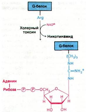
Cholera toxin is an 87-kDa protein composed of an A1 and an A2 peptide linked by a disulfide bridge, along with five B Peptides. The toxin gains entry into The Cell by binding to the ganglioside GM1 (Section 20.7) on the cell surface, a carbohydrate-rich sphingolipid recognized by the toxin's B chains. Once inside the cell, the A1 subunit (23 kDa) covalently modifies the G protein that regulates adenylate cyclase activity. Specifically, the toxin's A1 subunit catalyzes The transfer of ADP-ribose from NAD to an Arginine side chain on the G protein (Fig. 35.8). This ADP-ribosylation shuts down the intrinsic GTPase activity of the G protein. In other words, the modified G protein is stripped of its built-in shut-off mechanism for turning off adenylate cyclase (see Fig. 35.5). Consequently, the G protein becomes locked in its GTP-bound state, leaving adenylate cyclase chronically active even in the absence of hormone stimulation. A comparable effect can be achieved *in vitro* by adding guanylyl imidodiphosphate—a nonhydrolyzable analog of GTP—to unmodified G protein.
Fig. 35.8. Cholera toxin catalyzes the ADP-ribosylation of the G protein that regulates adenylate cyclase activity
Like GTP, this analog activates the G protein but cannot be converted to GDP. The binding of guanylyl imidodiphosphate to unmodified G protein activates adenylate cyclase just as effectively as the binding of GTP to a G protein that has lost its GTPase activity. The Mechanism of cholera toxin further underscores the physiological importance of the GTPase activity inherent to G proteins. It is worth recalling that the debilitating effects of diphtheria toxin are likewise rooted in the ADP-ribosylation of a protein possessing GTPase activity (Section 29.28).

Last update: 06/08/2026
Editorial and Educational Adaptation: This material has been compiled based on the primary/original source text. The project team performed an editorial review, corrected technical inaccuracies, structured sections, and adapted the content for an educational format.
What was processed:
- elimination of formatting defects (OCR errors, structural breaks, corrupted characters);
- editorial organization of content;
- standardization of terminology in accordance with academic sources;
- verification of factual statements against the original source text.
All mentions of the author, publication year, and origin of the primary text have been preserved in accordance with the source.