Human Anatomy and Physiology (with age-related characteristics of the child's body) - Sapin M.R., Sivoglazov V.I. 2002
Structure of the Human Body
Tissues
Connective Tissues
Connective Tissue is composed of Cells and Extracellular matrix, which always contains a significant number of connective tissue fibers. Having diverse structures and locations, connective tissue performs mechanical (supportive), trophic (providing Nutrition to Cells and Tissues, e.g., Blood), and protective (mechanical protection and phagocytosis) Functions.
Based on the structural and Functional Characteristics of the extracellular matrix and cells, connective tissue proper, Skeletal Tissues, and blood are distinguished.
Connective tissue proper
Connective tissue proper accompanies Blood Vessels down to the capillaries, fills the spaces between and within Organs, and underlies Epithelial Tissue. Connective tissue proper is subdivided into Fibrous connective tissue and Connective tissue with special properties (reticular, adipose, and pigment tissues).
Fibrous connective tissue, in turn, is subdivided into loose and dense, and the latter into irregular and regular. The Classification of fibrous connective tissue is based on The ratio of cells to extracellular fibrous structures, as well as the arrangement of the connective tissue fibers.
Loose fibrous connective tissue is present in all organs near Blood and Lymphatic vessels, as well as nerves, and forms the stroma of many organs (Fig. 6). The primary cellular elements of loose fibrous connective tissue are fibroblasts. Extracellular structures are represented by the ground substance and the collagenous and elastic fibers embedded within it. The ground substance is a homogeneous colloidal mass consisting of acidic and neutral Polysaccharides complexed with Proteins. These polysaccharides are known as Glycosaminoglycans and Proteoglycans, including hyaluronic acid. The liquid portion of the ground substance is tissue fluid.
Collagen and elastic fibers provide connective tissue with its mechanical and high-strength properties. The basis of collagen fibers is the protein collagen. Each collagen fiber consists of individual collagen fibrils about 7 nm thick. Collagen fibers are characterized by high mechanical tensile strength. They aggregate into bundles of varying thickness.
Class="center">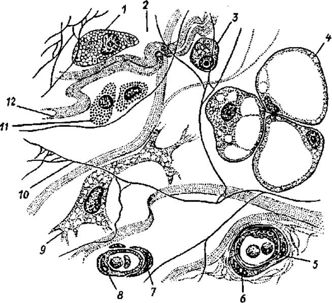
Fig. 6. Structure of loose fibrous connective tissue:
1 — macrophage, 2 — amorphous intercellular (ground) substance, 3 — plasmacyte (plasma Cell), 4 — lipocyte (fat cell), 5 — blood vessel, 6 — myocyte, 7 — pericyte, 8 — endotheliocyte, 9 — fibroblast, 10 — elastic fiber, 11 — tissue basophil, 12 — collagen fiber
Elastic fibers determine the elasticity and extensibility of connective tissue. They consist of the amorphous protein Elastin and filamentous, branching fibrils.
The cells of connective tissue include young, functionally active fibroblasts and mature fibrocytes. Fibroblasts participate in The formation of the extracellular matrix and collagen fibers. Fibroblasts are spindle-shaped, have basophilic Cytoplasm, and are capable of mitotic division. Fibrocytes differ from fibroblasts in their poorly developed membrane Organelles and low metabolic rate.
Connective tissue contains specialized cells, including Blood Cells (leukocytes) and cells of The Immune System (lymphocytes, plasma cells). Mobile cellular elements, such as macrophages and mast cells, are found in loose connective tissue.
Macrophages are actively phagocytic cells, 10—20 μm in size, containing numerous organelles for intracellular Digestion and the synthesis of various antibacterial substances, and possessing multiple microvilli on The Cell membrane surface.
Mast cells (tissue basophils) synthesize and accumulate BIOLOGICALLY ACTIVE SUBSTANCES (heparin, serotonin, dopamine, etc.) in their cytoplasm. They act as regulators of local Homeostasis in connective tissue. Loose fibrous connective tissue also contains fat cells (adipocytes) and pigment cells (pigmentocytes).
Dense Fibrous Connective tissue consists predominantly of fibers, a small number of cells, and amorphous ground substance. It is classified into dense irregular and dense regular fibrous connective tissue. The former (irregular) is formed by numerous fibers of various orientations and has complex systems of intersecting bundles (e.g., the reticular layer of the Skin). In dense regular fibrous connective tissue, the fibers are arranged in a single direction, aligned with the direction of tensile force (Muscle tendons, ligaments).
Connective tissue with special properties is represented by reticular, adipose, mucous, and pigment tissues.
Reticular connective tissue consists of reticular cells and reticular fibers. The fibers and branching reticular cells form a loose network. Reticular tissue forms the stroma of Hematopoietic and Immune organs, creating a microenvironment for the blood and lymphoid cells developing within them.
Adipose tissue consists predominantly of fat cells. It performs thermoregulatory, trophic, and shaping functions. Fat is synthesized by the cells themselves, so the specific function of Adipose tissue is lipid storage and METABOLISM. Adipose tissue is located mainly under the skin, in the omentum, and in other fat depots. During starvation, adipose tissue is utilized to meet the body's energy demands.
Mucous connective tissue, consisting of large branching cells (mucocytes) and an extracellular matrix rich in hyaluronic acid, is present in the umbilical cord, protecting the umbilical blood vessels from compression.
Pigment connective tissue contains A large number of pigment cells—melanocytes (found in the iris of the eye, age spots, etc.)—whose cytoplasm contains the pigment melanin.
Skeletal tissues
Skeletal tissues include Cartilage and Bone Tissues, which primarily perform supportive and mechanical functions in the body, as well as participate in Mineral Metabolism.
Cartilage tissue consists of cells (chondrocytes, chondroblasts) and extracellular matrix. The cartilage matrix, which is in a gel state, is formed mainly by glycosaminoglycans and proteoglycans. Cartilage contains a large amount of Fibrillar Proteins (mainly collagen). The extracellular matrix is highly hydrophilic.
Chondrocytes are round or oval in shape, located in specialized cavities (lacunae), and produce all Components of the extracellular matrix. Chondroblasts are young cartilage cells. They actively synthesize the cartilage matrix and are capable of division. Peripheral (appositional) growth of cartilage occurs due to chondroblasts.
The layer of connective tissue covering The surface of the cartilage is called the perichondrium. The perichondrium consists of an outer fibrous layer, composed of dense fibrous connective tissue containing blood Vessels and nerves, and an inner chondrogenic layer, which contains chondroblasts and their precursors, prechondroblasts. The perichondrium provides appositional growth of the cartilage, while its vessels facilitate diffuse nutrition of the cartilage tissue and the removal of Metabolic waste products.
Based on the structural CHARACTERISTICS OF THE extracellular matrix, cartilage is classified into hyaline, elastic, and fibrocartilage.
Hyaline cartilage is characterized by its translucency and bluish-white color. This cartilage is found at the junctions of the Ribs with the Sternum, on the articular surfaces of bones, at the epiphyseal plates (junctions of the epiphysis and diaphysis) of long bones, in the laryngeal Skeleton, and in the walls of the Trachea and Bronchi.
Elastic cartilage contains a large number of elastic fibers in its extracellular matrix alongside collagen fibers. The auricle of the ear, certain small cartilages of the Larynx, and the epiglottis are composed of elastic cartilage.
Fibrocartilage contains a large amount of collagen fibers in its extracellular matrix. The anuli fibrosi of intervertebral discs, articular discs, and menisci are composed of fibrocartilage.
Bone tissue is composed of bone cells and an extracellular matrix containing various salts and connective tissue fibers. The arrangement of bone cells, the orientation of fibers, and the distribution of salts provide bone tissue with hardness and strength. The organic components of bone are referred to as ossein (from Latin os — bone). The Inorganic Components of bone include salts of calcium, phosphorus, magnesium, and others. The combination of organic and inorganic substances makes bone both strong and resilient. In childhood, bones contain more organic substances than in adulthood, which is why bone fractures are rare in children. In elderly and aged individuals, The amount of organic substances in bones decreases, making the bones more fragile and brittle.
The cells of bone tissue are osteocytes, osteoblasts, and osteoclasts.
Osteocytes are mature, non-dividing, branching bone cells ranging from 22 to 55 µm in length, with a large ovoid Nucleus. They are spindle-shaped and reside within bone cavities (lacunae). Bone canaliculi containing the processes of osteocytes branch out from these cavities.
Osteoblasts are young bone tissue cells with a rounded nucleus. Osteoblasts develop from the osteogenic (deep) layer of the periosteum.
Osteoclasts are large multinucleated cells up to 90 µm in diameter. They are involved in bone resorption and cartilage calcification.
Two Types of bone tissue are distinguished: lamellar and woven (coarse-fibered) bone. Lamellar (fine-fibered) bone tissue consists of bone lamellae composed of mineralized extracellular matrix, with bone cells and collagen fibers embedded within. The fibers in adjacent lamellae have different orientations. Lamellar bone tissue forms the compact (dense) and spongy (cancellous) bone of the skeleton. Compact bone forms the diaphyses (shafts) of long bones and the Superficial layer of their epiphyses (ends), as well as the outer layer of flat and other bones. Spongy bone forms trabeculae (spicules) within the epiphyses and other bones, situated between the plates of compact bone. The trabeculae of spongy bone are oriented in various directions that correspond to the lines of compression and tension of the bone tissue (Fig. 7).
Compact bone is formed by concentric lamellae, numbering from 4 to 20, which surround blood vessels running through the bone. The thickness of a single concentric lamella ranges from 4 to 15 µm. The tubular cavity through which vessels up to 100–110 µm in diameter pass is called the osteonic (Haversian) canal. The entire structure surrounding this canal is called an osteon, or Haversian system (the Structural and functional unit of bone). Differently arranged bone lamellae between adjacent osteons are called interstitial lamellae. The inner layer of compact bone is formed by inner circumferential lamellae. These lamellae are products of the bone-forming function of the endosteum—a thin connective tissue membrane lining the internal surface of the bone (the walls of the medullary cavity and the spaces of spongy bone). The outer layer of compact bone is formed by outer circumferential lamellae, produced by the inner osteogenic layer of the periosteum. The outer layer of the periosteum is coarse-fibered and fibrous. This layer is rich in nerve fibers and blood vessels, which not only nourish the periosteum but also penetrate the bone through nutrient foramina on the bone surface. The periosteum is firmly anchored to the bone surface by thin connective tissue fibers (Sharpey's fibers) that penetrate from the periosteum into the bone.

Fig. 7. Structure of a long bone.
1 — periosteum, 2 — compact bone, 3 — outer circumferential lamellae layer, 4 — osteons, 5 — inner circumferential lamellae layer, 6 — medullary cavity, 7 — trabeculae of spongy bone

Fig. 8. Blood cells:
1 — basophilic granulocyte, 2 — acidophilic granulocyte, 3 — segmented neutrophilic granulocyte, 4 — erythrocyte, 5 — monocyte, 6 — platelets, 7 — lymphocyte
Last update: 10/08/2026
Editorial and Educational Adaptation: This material has been compiled based on the primary/original source text. The project team performed an editorial review, corrected technical inaccuracies, structured sections, and adapted the content for an educational format.
What was processed:
- elimination of formatting defects (OCR errors, structural breaks, corrupted characters);
- editorial organization of content;
- standardization of terminology in accordance with academic sources;
- verification of factual statements against the original source text.
All mentions of the author, publication year, and origin of the primary text have been preserved in accordance with the source.