Human Histology - O.D. Lutsyk 2003
Systemic Histology
Hematopoietic and Immune System Organs
The hematopoietic and immune defense system (Fig. 4.11) includes the Cytology/practical/86.html">Red Bone Marrow, Thymus, accumulations of lymphoid elements in the walls of the alimentary canal and respiratory tract, Lymph Nodes, hemolymph nodes, and the Spleen. Of these, the first two are considered central, while all others are peripheral Hematopoietic Organs. The peripheral part of the hematopoietic and immune defense system also includes mucosa-associated lymphoid tissue (MALT) and Skin-associated lymphoid tissue (SALT). The former includes accumulations of lymphocytes and other immunocytes (plasma Cells, macrophages) in the mucous membranes of the digestive tract (GALT - Gut-Associated Lymphoid Tissue), Bronchi (BALT - Bronchus-Associated Lymphoid Tissue), urogenital tract, and excretory ducts of the salivary and Mammary Glands. Lymphocytes are distributed diffusely in loose Fibrous Connective Tissue or form lymphatic nodules (solitary or aggregated). In the Digestive System, the largest number of nodules is observed in the Tonsils, ileum, and Appendix. Skin-associated lymphoid tissue includes intraepidermal lymphocytes and dendritic cells that form an immune protective barrier.
Class="center">
Fig. 4.11. General Organization OF THE hematopoietic and immune defense system
The function of the central Organs of the immune defense system is associated with The formation of all types of formed elements of Blood and providing conditions for antigen-independent proliferation of lymphocytes. In the peripheral Organs of Immunogenesis, Blood Cells that have completed their life cycle are eliminated (destroyed), and effector cells (T- and B-lymphocytes) undergo antigen-dependent specialization to provide Immunity—the defense of the body against genetically foreign material.
All hematopoietic organs are structurally based on reticular tissue, which forms the framework and microenvironment for maturing formed elements of the blood. In addition to blood Cell proliferation, hematopoietic organs store BLOOD AND LYMPH and clear them of foreign particles. The critical importance of the normal functioning of this system for the body is evidenced by the fact that two of the most dangerous and virtually incurable pathological conditions—Acquired Immunodeficiency Syndrome (AIDS) and malignant neoplasms—are directly associated with damage to the organs of The Immune System. The lack of effective treatments for these diseases highlights The complexity of immune defense processes and the close interconnection of all hematopoietic organs.

Fig. 4.12. Red bone marrow: A - localization zones of red bone marrow in an adult; B - Structure OF THE cancellous Bone tissue of a vertebra, forming the coarse stroma of the red bone marrow
Red bone marrow (medulla ossium rubra) is the central hematopoietic organ (Fig. 4.12), which contains hematopoietic stem cells and where the proliferation and differentiation of myeloid and lymphoid lineages occur: erythrocytes, platelets, granulocytes, monocytes, B-lymphocytes, and T-lymphocyte progenitors are formed. In the adult body, red bone marrow is located in the epiphyses of long bones and in the cancellous substance of flat bones. The total mass of red bone marrow is 4-5% of body weight, which, for a body weight of 70 kg, is 3-3.5 kg. Bone marrow has a semi-liquid consistency and is dark red in appearance.
The trabeculae of cancellous bones provide support (coarse stroma) for the reticular tissue, which, in turn, serves as a framework (fine stroma) for hematopoietic cells—stem, progenitor, and subsequent classes of Cells of the erythroid, megakaryocytic, granulocytic, monocytic, and lymphocytic lineages. Hematopoietic cells typically form hematopoietic islands (Fig. 4.13), which contain cells of a particular histogenetic Lineage. Proliferation and maturation processes are most intense near the endosteum. Red bone marrow is highly vascularized; the presence of sinusoidal blood capillaries (sinusoids) allows mature blood cells to enter the Circulation. It should be noted that under normal conditions, the sinusoidal capillaries of the bone marrow are impermeable to immature blood cells. This selective permeability is likely related to the specific Chemical Composition and cytotopography of carbohydrate determinants On the surface of mature versus poorly differentiated cells.
The formation of red bone marrow begins In the second month of embryonic development in the clavicle of the embryo. During the 5th to 7th months of Embryogenesis, the red bone marrow Functions as the primary hematopoietic organ, with erythropoiesis being the predominant process during this period. In childhood, red bone marrow fills the diaphyses and epiphyses of long bones, as well as flat bones. At 12-18 years of age, the red bone marrow in the diaphyses of long bones is replaced by yellow bone marrow, which contains numerous adipocytes. Normally, yellow bone marrow does not perform hematopoietic functions; however, in cases of significant blood loss, centers of myeloid hematopoiesis may appear within it. In old age, both red and yellow bone marrow acquire a gelatinous consistency and transform into gelatinous bone marrow.
The thymus is a central organ of immunogenesis where the proliferation and maturation (antigen-independent differentiation) of T-lymphocytes take place. The thymus produces thymosin, thymulin, thymopoietin, and other regulatory Peptides that ensure the proliferation and maturation of T-lymphocytes in central and peripheral organs of immunogenesis, as well as several other BIOLOGICALLY ACTIVE SUBSTANCES: an Insulin-like factor (lowers blood sugar levels), a Calcitonin-like factor (lowers blood calcium levels), and a growth factor (promotes body growth).
The thymus is located behind the Sternum. Its mass in an adult is 10-30 g, and in newborns, it is about 12-14 g. The shape of the thymus is polygonal, characterized by significant individual and age-related Variability. At 18 years of age, the dimensions of the thymus are 19x7x2 cm. Externally, the thymus is covered by a connective tissue capsule, from which septa grow into the organ, dividing it into lobules. The connective tissue of the thymic capsule is separated from its parenchyma by a porous basement membrane, which forms characteristic channels extending deep into the organ at the sites of blood vessel entry.
The thymic lobule (Figs. 4.14, 4.15) is the Structural and functional unit of the organ. The basis of the lobule is a framework of so-called epithelioreticulocytes—special star-shaped epithelial cells whose processes contact each other to form a meshwork. The spaces between the epithelioreticulocytes are filled mainly with T-lymphocytes and, to a lesser extent, macrophages. A small portion of the cellular elements of the thymus consists of fibroblasts, myofibroblasts, and mast cells. The central region of the thymic lobule, which stains lighter than the peripheral region on histological preparations, is called the medulla; the dark periphery of the lobule is called the cortex.
In the cortex of the thymic lobule, small and medium lymphocytes are compactly arranged, surrounded by macrophages and epithelioreticulocytes, as well as T-lymphoblasts; the latter are localized mainly in the subcapsular zone. Epithelioreticulocytes, macrophages, and dendritic cells of the subcapsular zone of the thymus are often called thymic nurse cells because they create the microenvironment and necessary conditions for the maturation of T-lymphocytes (thymocytes). T-lymphocyte progenitors migrate from the red bone marrow to the thymic cortex. Here, they undergo proliferation, maturation under METABOLISM/18.html">The Influence of thymosin produced by epithelioreticulocytes, and selective phagocytosis of some newly formed cells by macrophages. Only 3-5% of the generated cells exit the thymus. The remaining cells die by apoptosis. Lymphocyte Selection occurs with the participation of epithelioreticulocytes. Cells that do not react to self-Proteins of the Major Histocompatibility Complex (MHC) survive, while cells with receptors for the body's self-Antigens perish. Selected (non-phagocytosed) T-lymphocytes migrate into the medulla, from where they can enter the peripheral circulation.
The medulla of the thymic lobule is formed by differentiated T-lymphocytes that express CD4 (helper/inducer) or CD8 (killer/suppressor) receptors, as well as T-cell receptors (TCR), on their membrane.
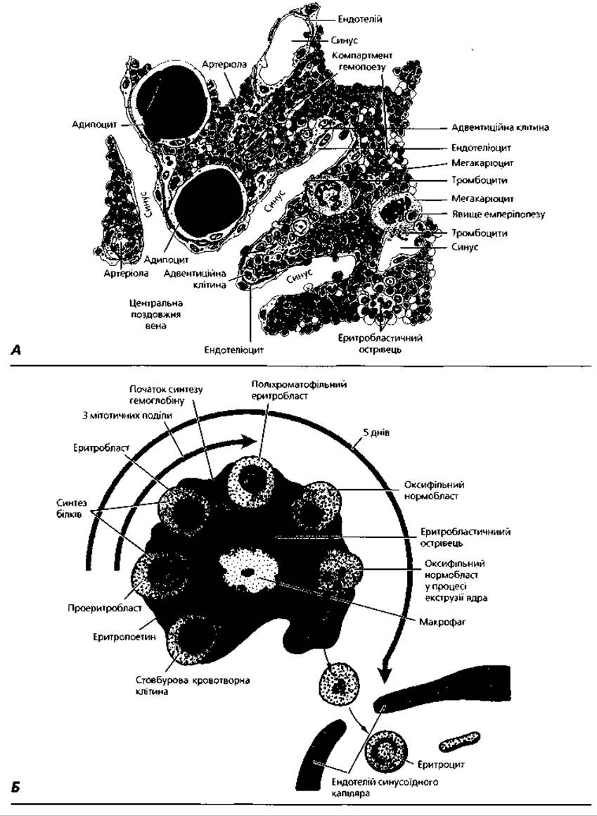
Fig. 4.13. A - semi-schematic representation of the microstructure of a red bone marrow area located near the central longitudinal vein of the bone; B - diagram of an erythroblastic island of the red bone marrow
Lymphocytes of the medulla are surrounded by epithelioreticulocytes and macrophages and are arranged less compactly compared to those in the cortex (up to 90% of thymic lymphocytes are contained in the cortex and only 10% in the medulla). Medullary lymphocytes enter the bloodstream via venules and efferent Lymphatic vessels. A characteristic morphological feature of the thymus is the presence of special concentric layers of epithelial cells in the medulla, known as Hassall's corpuscles. They are formed during the degeneration and mutual layering of star-shaped medullary epithelioreticulocytes. Hassall's corpuscles stain acidophilically; keratin granules, thick bundles of fibrils, and large vacuoles are found in the Cytoplasm of the cells forming them. Acidophilic cellular debris is located in the center of the thymic corpuscles. There is a certain correlation between the appearance of Hassall's corpuscles and the acquisition of immunocompetence by T-lymphocytes.
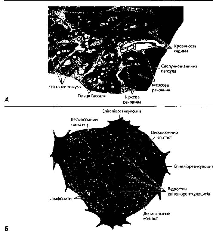
Fig. 4.14. Thymus: A - semi-schematic representation of the thymus structure of a four-month-old infant, x 28; B - relationship between epithelioreticulocytes and lymphocytes within a thymic lobule
The cortex and medulla of thymic lobules have specific microcirculatory bed structures. In particular, lymphocytes of the cortex are separated from the lumen of blood capillaries by the so-called blood-thymus barrier. It is formed by a continuous layer of epithelioreticulocytes resting on the basement membrane, which accompany all Vessels of the microcirculatory bed and limit the pericapillary space, as well as by the wall of the blood capillaries. The blood-thymus barrier prevents antigens from the vascular bed from reaching the maturing lymphocytes in the cortex, which possess cytoreceptors for the body's self-antigens, thereby preventing The Development of autoimmune reactions (damage to the body's own Cells and Tissues). The blood-thymus barrier is absent in the medulla, which creates conditions for T-lymphocyte recirculation.
The human thymus is formed during the fifth week of embryogenesis as an epithelial thickening of the third and fourth pairs of pharyngeal pouches. At the end of the second month, the first lymphocytes colonize the epithelial stroma of the thymus. In the third month, lobules appear, in which the cortex and medulla can be distinguished, and Hassall's corpuscles become visible. The organ reaches its maximum mass in early childhood. Throughout a person's life, changes occur in the thymus, known as age-related involution. This process consists of the gradual replacement of the parenchymal elements of the thymus with adipose and loose connective tissue, along with a decrease in the organ's mass. Hassall's corpuscles persist longer. Four phases are distinguished in the age-related involution of the thymus: rapid (up to 10 years of age), slow (from 10 to 25 years), accelerated (from 25 to 40 years), and delayed (after 40 years). The rate of age-related thymic involution is largely determined by the hormonal status of the body. In older individuals, the thymus is completely replaced by adipose tissue and transforms into a fat body.

Fig. 4.15. Light Microscopy of the thymus: A - general organization of the lobules, x 32; B - fragment of a child's thymic lobule, x 118
The absence of age-related thymic involution is a manifestation of a severe pathology known as status thymicolymphaticus. This condition is usually accompanied by insufficiency of the glucocorticoid function of the adrenal cortex and proliferation of lymphoid tissue in organs. In the presence of status thymicolymphaticus, the body's resistance to infections and intoxications drops sharply, and the risk of malignant neoplasms increases.
Under the influence of adverse factors (stress) on the body—such as injuries, starvation, intoxication, or infections—the so-called accidental involution of the thymus is observed. It is accompanied by the mass death of lymphocytes under the action of corticosteroids, as well as an increase in the number and size of Hassall's corpuscles. Initially, lymphocytes disappear from the cortex, which, at this stage of involution, blurs the distinction between the cortex and medulla of the thymic lobules. Accidental thymus involution is a morphological manifestation of the body's protective reactions.
Lymphoid nodules, or follicles (noduli lymphatici), in the wall of the Human digestive tract and respiratory tract are considered a dissociated analogue of the bursa of Fabricius in birds, i.e., a central organ of B-lymphocytopoiesis. In them, B-lymphocytes arriving from the red bone marrow acquire immunocompetence (receive receptors for various antigens). Lymphoid nodules (Fig. 4.16) are spherical clusters of B- and T-lymphocytes within the loose connective tissue of the lamina propria of the mucosa and in the submucosa of the corresponding PARTS OF THE digestive and respiratory tracts; T-lymphocytes within them play an auxiliary role in the maturation of B-lymphocytes. After acquiring immunocompetence, B-lymphocytes can enter the peripheral bloodstream. Some of these cells, upon returning, transform into plasma cells, which, in close cooperation with the epithelial lining cells of the digestive and respiratory tracts, produce class A IMMUNOGLOBULINS (Antibodies).
Lymph nodes (noduli lymphatici) are bean-shaped thickenings along the course of lymphatic vessels, where antigen-dependent proliferation of B- and T-lymphocytes occurs, their acquisition of immunocompetence takes place, and lymph is cleared of foreign particles. The total mass of lymph nodes is 1% of body weight, which is about 700 g. Lymph nodes form more than 50 groups. According to their topography, they are divided into somatic (body), visceral (Internal Organs), and mixed nodes, which collect lymph from both viscera and other organs. The size of lymph nodes ranges from 5 to 10 mm.
The lymph node (Figs. 4.17, 4.18) is covered by a connective tissue capsule, from which connective tissue septa—trabeculae—extend into the organ. Smooth Muscle cells have been found in the capsule of some lymph nodes, participating in the Formation of the node's supportive-contractile apparatus. The parenchyma of the node is formed by B- and T-lymphocytes, for which reticular tissue forms the framework (delicate stroma). The cortex and medulla of the lymph node are distinguished.
The cortex is formed by lymphatic nodules (follicles) located under the capsule—spherical clusters of B-lymphocytes with a diameter of 0.5–1 mm. In addition to B-lymphocytes, the lymphoid follicles of the lymph node include both typical macrophages and a special variety called dendritic cells. Externally, the nodule is covered by reticuloendothelial cells—cells that combine the Morphology of reticular cells with the function of endothelium, as they line the sinuses of lymph nodes. Among the reticuloendothelial cells, there is a significant number of fixed macrophages, the so-called littoral cells. Each nodule contains a light (reactive, or germinal) center, where lymphocyte proliferation occurs and B-lymphoblasts are predominantly localized, and a dark peripheral zone, in which small and medium lymphocytes are compactly arranged. An increase in the number and size of the reactive centers of lymph node nodules indicates antigenic stimulation of the body.
The medulla of the lymph node is formed by medullary cords—ribbon-like clusters of B-lymphocytes, plasma cells, and macrophages, extending from the hilum of the node toward the lymphatic nodules. Externally, the medullary cords, just like the nodules of the cortex, are covered by reticuloendothelial cells. Between the medullary cords and nodules, and correspondingly between the medulla and cortex of the lymph node, there is a diffuse accumulation of T-lymphocytes called the paracortical zone. In this zone, there are postcapillary venules with cuboidal endothelial cells, where homing* occurs. Macrophages within the paracortical zone are represented by a variety of so-called interdigitating cells, which contact each other through finger-like processes and produce substances that stimulate the proliferation of T-lymphocytes. Thus, the cortex and medulla are bursa-dependent, while the paracortical layer is the thymus-dependent zone of the lymph node.
Between the layers of reticuloendothelial cells covering the lymphatic nodules and medullary cords on one side, and the connective tissue stroma (capsule and trabeculae) on the other, there are slit-like spaces called sinuses. The sinus System of the lymph node includes the marginal sinus (located between the capsule and the nodules), cortical sinuses (between the nodules and trabeculae), medullary sinuses (between the medullary cords and trabeculae), and the hilar sinus (in the region of the concave part—the hilum of the lymph node). Through the sinus system, lymph circulates from the marginal sinus, where afferent lymphatic vessels empty, through intermediate sinuses toward the hilar sinus, from which lymph drains via The system of efferent lymphatic vessels. During this process, the lymph is cleared due to the phagocytosis of foreign particles by littoral macrophages; it is enriched with immunocompetent T- and B-lymphocytes, memory cells, and immunoglobulins (antibodies).
* Lymphatic nodules of the alimentary canal and other tubular organs, as well as lymph nodes, contain high endothelial venules that express so-called vascular addressin on their surface, which is recognized by the CD44 molecule of lymphocytes circulating in the blood. As a result, lymphocytes are arrested in these areas. This is referred to by the word "homing," which translates as "returning home."

Fig. 4.16. A — diagram of the MAIN TYPES OF Structural organization of unencapsulated lymphoid tissue and encapsulated Lymphoid organs; B — semi-schematic representation of The structure of the palatine tonsil

Fig. 4.17. Lymph node: A — General structural plan; B — diagram of Blood Circulation (right side of the diagram) and lymph circulation (left side of the diagram); blood is supplied to and drains from the lymph node through its hilum; lymph enters from afferent lymphatic vessels on the convex side of the lymph node and drains through the hilum

Fig. 4.18. Light microscopy of a lymph node: A — general structural plan, x 30; B — lymphoid follicle with a light reactive center, x 200; C — medullary cord surrounded by reticular stroma, x 200
The mechanisms of lymph node functioning involve a close relationship among all its structural components. Littoral cells and typical macrophages of lymphatic nodules phagocytose foreign particles that pass with lymph through the sinus system of the lymph node. In this process, with the participation of lysosomal Enzymes of macrophages, the antigens of phagocytosed particles are converted from a particulate form to a molecular form capable of inducing an Immune Response: lymphocyte proliferation, transformation of B-lymphocytes into plasma cells (antibody producers), and T-lymphocytes into effectors (T-killer cells) and memory cells. Antigen-activated B-lymphocytes migrate from lymphatic nodules to medullary cords, where they transform into plasma cells—producers of antibodies. Memory cells enter the vascular bed, forming effector cells upon repeated encounter with the antigen. Dendritic cells of cortical nodules are a type of macrophage capable of fixing antibody-antigen complexes on their surface (acting as so-called antigen-presenting cells). Upon contact with dendritic cells, B-lymphocytes are stimulated to produce antibodies. Interdigitating cells of the paracortical zone secrete biologically active substances that stimulate the proliferation and maturation of T-lymphocytes, transforming them into effector cells (T-killer cells).
Development of lymph nodes. The first Lymph Nodes in the human embryo are detected at the end of the second month of embryonic development as zones of local clusters of mesenchymal cells around lymphatic vessels. The capsule and trabeculae form from the outer layer of mesenchyme, while the reticular stroma of the nodes forms from the inner layer. The egress of lymphoblasts and lymphocytes from the bone marrow leads to the formation of medullary cords and lymphatic nodules at the end of the fourth month of embryogenesis. Somewhat later, the thymus-dependent paracortical zone is populated, and the lymph nodes are enriched with macrophages. At the end of the fifth month, lymph nodes acquire morphological features characteristic of the adult Organism. They complete their development During the first three years of a child's life. Reactive centers in lymphatic nodules appear upon immunization of the body during life and the establishment of its protective functions. In old age, the number of reactive centers in lymph node nodules decreases, the phagocytic activity of macrophages declines, some nodes atrophy, and they are replaced by adipose tissue.
Hemolymph nodes (noduli lymphatici haemales) are a special variety of lymph nodes in whose sinuses blood, rather than lymph, circulates, and which perform the function of both lymphoid and myeloid hematopoiesis. In humans, hemolymph nodes are localized in the perinephric fat, around the Abdominal Aorta, and less frequently in the posterior Mediastinum. Structurally, they resemble typical lymph nodes, but they are smaller and have less developed medullary cords and cortical nodules. With age, involution of hemolymph nodes is observed: the cortex and medulla are replaced by adipose tissue or loose fibrous connective tissue.
The spleen (splen, lien) is an unpaired organ located in the Abdominal cavity. The spleen has an oblong shape and is localized in the left hypochondrium. Its mass is 100–150 g, and its dimensions are 10x7x5 cm. Proliferation and antigen-dependent differentiation of lymphocytes, as well as the elimination of erythrocytes and platelets that have completed their life cycle, take place in the spleen. The spleen also functions as a blood and iron reservoir, produces biologically active substances (splenin, erythropoiesis-inhibiting factor), and is a universal hematopoietic organ during the Embryonic period. The spleen is covered by a connective tissue capsule, from which septa—trabeculae—grow into the organ. In addition to connective tissue rich in Collagen and elastic fibers, the capsule and trabeculae contain bundles of smooth muscle cells and constitute the supportive-contractile apparatus of the spleen. Red and white pulp are distinguished in the splenic parenchyma (Figs. 4.19–4.22).
White pulp constitutes about 20% of the organ's mass and is formed by lymphocytes, plasma cells, macrophages, dendritic and interdigitating cells, for which reticular tissue serves as the stroma. Spherical clusters of these cell types are called splenic lymphatic nodules (follicles). The diameter of the nodules is 0.3–0.5 mm, and they are surrounded by reticuloendothelial cells.
The lymphatic nodule (the so-called Malpighian corpuscle) of the spleen has four zones: periarterial, mantle, marginal, and a light (reactive, or germinal) center. The reactive centers of splenic and lymph node nodules are structures identical in anatomy and function. They contain B-lymphoblasts, typical macrophages, dendritic, and reticular cells. The appearance of reactive centers in nodules is a response to antigenic stimulation. The periarterial zone is an accumulation of T-lymphocytes around the nodular artery, or the so-called central artery of the spleen. The periarterial zone is enriched with interdigitating antigen-presenting cells—macrophages capable of fixing antibody-antigen complexes on their surface and inducing the proliferation and maturation of T-lymphocytes. The periarterial zone of splenic nodules is an analogue of the thymus-dependent paracortical zone of lymph nodes. The dark mantle zone is formed by compactly arranged small B-lymphocytes and a small number of T-lymphocytes, plasma cells, and macrophages. The marginal zone—the transition site from white to red pulp—is formed by B- and T-lymphocytes and macrophages, and is surrounded by sinusoidal hemocapillaries. After maturation, lymphocytes migrate from the germinal center and periarterial zone to the mantle and marginal zones, subsequently entering the bloodstream.

Fig. 4.19. Spleen: A — diagram of the relationship between the main Structural components of the organ; B — semi-schematic representation of a histological specimen of the spleen, x 60

Fig. 4.20. Spleen: A — diagram of the relationship between the structural Components of the organ and blood circulation within it; B — ULTRASTRUCTURE OF THE red pulp. Both theories—open and closed circulation—are illustrated

Fig. 4.21. Light microscopy of the spleen: A - silver impregnation to reveal the supportive-contractile apparatus and histoarchitecture of the organ, x 30; B, C - white and red pulp, x 100

Fig. 4.22. Scanning Electron microscopy of splenic venous sinuses (sinusoids): A - longitudinally sectioned sinusoid with adjacent Billroth's cords, x 1 600; B - fragment of a sinusoid, internal view: slit-like spaces between rod-shaped endothelial cells 2-3 μm wide act as a barrier preventing old erythrocytes with a rigid Plasmalemma structure from passing from the red pulp into the sinusoid, while functionally viable cells with a flexible membrane can penetrate. M - macrophage; N - neutrophil; L - lymphocyte, x 5300
Periarterial lymphatic sheaths (PALS) are elongated accumulations of lymphocytes that surround the white pulp Arteries like sleeves and merge on one side into the splenic lymphoid nodules (Fig. 4.21). In the central part of the sheath, closer to the vessel lumen, B-lymphocytes and plasma cells are concentrated, while T-lymphocytes are located at the periphery.
The red pulp, which accounts for about 80% of the splenic mass, is an accumulation of formed blood elements located either surrounded by reticular cells or within the system of splenic venous sinuses. The Regions of the red pulp located between the sinuses are called Billroth's cords. They are the sites where B-lymphocytes differentiate into plasma cells, and monocytes differentiate into macrophages. Splenic macrophages are capable of recognizing and destroying senescent or damaged erythrocytes and platelets. During this process, Hemoglobin from the destroyed erythrocytes is recycled, serving as a source of iron for bilirubin and transferrin synthesis. Molecules of the latter are captured from the circulation by red bone marrow macrophages and used in The process of erythropoiesis.
The Vascular System of the spleen has several unique features that ensure the performance of the organ's functions (Figs. 4.20, 4.22). The splenic artery enters the splenic hilum and branches into a system of vessels located within the splenic trabeculae, known as trabecular arteries. Trabecular arteries branch into the Arteries of the white pulp, around which lymphocytes aggregate to form periarterial lymphatic sheaths and lymphoid nodules. The segments of the white pulp arteries passing through the lymphoid nodules are called central arteries, as they serve as centers for lymphocyte migration during the development of lymphoid nodules in ontogeny. The central arteries continue as red pulp arteries, which branch into penicillar arterioles that terminate in ellipsoid capillaries. Ellipsoid capillaries are surrounded by distinctive "sheaths" composed of clusters of reticular cells, macrophages, and lymphocytes. Ellipsoid capillaries connect to the splenic venous sinuses (Fig. 4.20). However, some capillaries may open directly into the red pulp, forming the open circulation system of the spleen. Venous sinuses (sinusoids) can serve as blood reservoirs. From the venous sinuses, blood flows into the pulp Veins, then into the trabecular veins, and finally into the splenic vein.
Splenic development begins at THE START OF the second month of embryogenesis as vascularized mesenchymal cell condensations in the dorsal mesentery. Reticular tissue develops from the mesenchyme and is subsequently colonized by hematopoietic stem cells. During the third month of embryogenesis, the periarterial thymus-dependent zone differentiates in the spleen; germinal centers and marginal Zones of the nodules form during the fifth month, and the red pulp becomes distinguishable by the sixth month. Concurrently (from the third to the fifth month of embryogenesis), myeloid hematopoiesis increases in the spleen, and it functions as a universal hematopoietic organ. From the sixth month until birth, myeloid hematopoiesis declines and is replaced by lymphocytopoiesis. In adulthood, the spleen exhibits significant regenerative capacity: experimental studies have demonstrated its ability to regenerate even after the loss of 80-90% of its parenchyma. Splenic mass decreases slightly between the ages of 20 and 30, remaining stable between 30 and 60 years of age. In old age, there is Atrophy of the red and white pulp, proliferation of the connective tissue stroma, a decrease in the proportion of macrophages and lymphocytes among parenchymal elements, an increase in granulocytes and mast cells, and the appearance of megakaryocytes. The recycling of iron from erythrocytes destroyed in the spleen also deteriorates.
Intercellular interactions in providing the body's immune defense. An adequate response to foreign substances entering the body (antigenic stimulation) requires the interaction and cooperation of various TYPES OF IMMUNE system cells. These include cells of macrophage lineage: blood monocytes, connective tissue histiocytes-macrophages, bone marrow, peritoneal, and alveolar macrophages, Langerhans cells of the skin, Kashchenko-Hofbauer cells of the Placenta, Kupffer cells of the Liver, dendritic and interdigitating cells of the LYMPH NODES AND spleen, osteoclasts of bone tissue, and microglial cells of The Nervous system. There is also a group of so-called microphages, which include blood neutrophilic granulocytes, as well as cells that can exhibit phagocytic properties under certain conditions, such as endothelial cells. The third group comprises various populations of T AND B lymphocytes (killer T cells, helper T cells, suppressor T cells, plasma cells, and memory T and B cells). The total mass of cells directly involved in the body's immune defense constitutes about 1% of body weight.
Helper T cells are the first to respond to the invasion of foreign particles into the body: antigenic determinants bind to specific receptors on their surface. The resulting antigen-receptor complex detaches from The surface of the helper T cell plasmalemma and is bound by the surface receptors of a macrophage. In the next stage, the antigens processed by macrophages are presented to B lymphocytes, which, under the influence of antigenic stimulation and the activating effect of helper T cells, differentiate into plasma cells. The latter synthesize protein molecules of immunoglobulins (antibodies) that selectively bind to antigens and cause their inactivation. Following contact with the antigen, helper T cells produce specific chemical substances that stimulate the proliferation of killer T cells. The latter have The ability to destroy The Cell walls of Bacteria and cells carrying antigenic determinants on their surface.
At each of these stages, partial inactivation of the foreign material may occur, as well as its modification and transfer to other cell populations to mount an immune response. Alternatively, an antigen-bearing particle can be recognized and engulfed by a macrophage without T-lymphocyte involvement, degraded by its lysosomal enzymes, and the resulting antigenic determinants presented to T and B lymphocytes, stimulating their differentiation into effector cells (killer T cells and plasma cells) and memory cells. Cells of the immune system—immunocytes—produce physiologically active substances called interleukins, the BIOLOGICAL EFFECTS OF which are summarized in Table 24.
Table 24. Interleukins
|
Abbreviation used in English literature |
Producer cells |
Main functions |
|
IL-1 |
Macrophages, keratinocytes |
Proinflammatory endogenous pyrogen; activates fibroblasts, granulocytes, osteoclasts; sensitizes T lymphocytes to signals |
|
IL-2 |
T lymphocytes |
Stimulates proliferation of T and B lymphocytes and immunocompetent cells |
|
IL-3 |
T lymphocytes |
Stimulates proliferation of multipotent hematopoietic cells |
|
IL-4 |
T lymphocytes, mast cells |
Regulates B-lymphocyte isotype switching to IgG and IgE |
|
IL-5 |
T lymphocytes, mast cells, possibly B lymphocytes |
Stimulates proliferation and differentiation of eosinophils; activates IgA production |
|
IL-6 |
T lymphocytes, macrophages, fibroblasts |
Proinflammatory factor; stimulates differentiation of B lymphocytes and thymocytes |
|
IL-7 |
Reticular stroma of red bone marrow |
Stimulates differentiation and maturation of B lymphocytes |
|
IL-8 |
Keratinocytes, fibroblasts, monocytes |
Stimulates activation and chemotaxis of neutrophils |
|
IL-9 |
T lymphocytes |
Stimulates proliferation of T lymphocytes, thymocytes, and mast cells |
|
IL-10 |
T lymphocytes, mast cells, possibly B lymphocytes |
Inhibits cytokine synthesis in many cells; stimulates proliferation of mast cells |
Terms to remember
1. Red bone marrow. 2. Yellow bone marrow. 3. Gelatinous bone marrow. 4. Thymus. 5. Thymosin. 6. Epithelioreticulocyte. 7. Thymic lobule. 8. Cortex of the thymic lobule. 9. Medulla of the thymic lobule. 10. Thymic corpuscle (Hassall's corpuscle). 11. Blood-thymus barrier. 12. Age-related thymic involution. 13. Adipose body. 14. Thymicolymphatic status. 15. Accidental thymic involution. 16. Lymphoid nodules of the digestive tract and respiratory tract. 17. Lymph node. 18. Cortex of the lymph node. 19. Lymphoid nodule of the lymph node. 20. Germinal (reactive) center of the lymphoid nodule. 21. Reticuloendothelial cell. 22. Littoral cell. 23. Dendritic cell. 24. Paracortical (thymus-dependent) zone of the lymph node. 25. Interdigitating cell. 26. Medulla of the lymph node. 27. Medullary cord. 28. Lymph node sinus. 29. Hemolymph node. 30. Spleen. 31. Supportive-contractile apparatus of the spleen. 32. White pulp of the spleen. 33. Lymphoid nodule of the spleen (Malpighian corpuscle). 34. Central artery of the spleen. 35. Periarterial zone. 36. Germinal (reactive) center. 37. Mantle zone. 38. Marginal zone. 39. Periarterial lymphatic Sheath of the spleen. 40. Artery of the white pulp. 41. Red pulp of the spleen. 42. Splenic cord (Billroth's cord). 43. Penicillar arteriole of the spleen. 44. Ellipsoid capillary. 45. Ellipsoid (pericapillary sheath) of the spleen. 46. Splenic venous sinus (sinusoid). 47. Pulp vein. 48. Trabecular vein of the spleen.
Last update: 09/08/2026
Editorial and Educational Adaptation: This material has been compiled based on the primary/original source text. The project team performed an editorial review, corrected technical inaccuracies, structured sections, and adapted the content for an educational format.
What was processed:
- elimination of formatting defects (OCR errors, structural breaks, corrupted characters);
- editorial organization of content;
- standardization of terminology in accordance with academic sources;
- verification of factual statements against the original source text.
All mentions of the author, publication year, and origin of the primary text have been preserved in accordance with the source.