Human Histology - O.D. Lutsyk 2003
Systemic Histology
Cardiovascular System
The Vascular System is a complex network of branching tubes of various diameters that transport Blood to all Organs, regulate organ blood supply, facilitate METABOLISM between the blood and surrounding Tissues, and drain Lymph from tissues into the Venous system. Approximately 20% of the body's total fluid volume circulates within human Blood Vessels. Closely linked to the vascular system is The Heart, which acts as a pump to propel blood. Thus, The Cardiovascular system comprises the heart, blood vessels, and lymphatic vessels (Fig. 4.1).
Blood vessels are classified into Arteries, arterioles, blood capillaries (hemocapillaries), venules, Veins, and arteriolovenular anastomoses. Blood flows away from the heart through arteries and is oxygenated (except for the pulmonary artery). Blood flows toward the heart through veins and is deoxygenated (except for the Pulmonary veins). Capillaries are situated between arteries and veins. Additionally, there are so-called rete mirabile (miraculous capillary networks): an arterial rete mirabile in the Kidney, where capillaries are located between two arteries, and a venous rete mirabile in The Liver and Pituitary Gland, where capillaries lie between two veins. Arteriolovenular anastomoses allow blood to bypass the capillary bed, shunting it directly from the arterial to the venous system.
The microvascular bed (microcirculatory bed) is a system of small vessels that includes arterioles, capillaries, venules, and arteriolovenular anastomoses (Fig. 4.2). This functional complex of blood vessels, surrounded by lymphatic capillaries and vessels, along with the adjacent Connective Tissue, performs crucial Functions such as regulating organ blood supply, transcapillary exchange, drainage, and blood pooling. Each organ, depending on its function, exhibits specific structural and spatial features of its microcirculatory bed. Microcirculatory vessels are highly plastic and responsive to changes in blood flow. They can pool formed elements of blood, undergo spasm to allow only plasma to pass, or alter their permeability to interstitial fluid.
Blood capillaries (vasa haemocapillaria) (Fig. 4.3) perform The primary function of the Circulatory system, which is the exchange of substances between blood and tissues; they act as a blood-tissue barrier and facilitate microcirculation. Hemodynamic conditions in capillaries are characterized by low pressure (25–30 mmHg at the arterial end and 8–12 mmHg at the venous end) and slow blood flow velocity (0.5 mm/s). These are the thinnest vessels. The Latin word "capillaris" means "Hair-like," although the vast majority of capillaries are thinner than a human hair. The capillary lumen is sometimes smaller than the diameter of red Blood Cells (3–5 µm), though There are also large capillaries with diameters exceeding 20–30 µm, known as sinusoid capillaries and lacunae. The average length of a capillary is 0.7–0.8 mm, with a cross-sectional area of 30 µm2. Capillaries are the most numerous vessels in the body.
Class="center">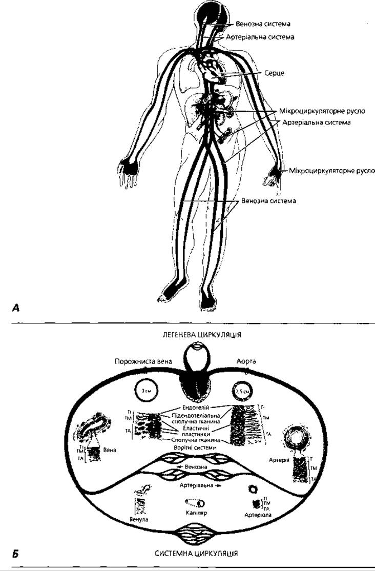
Fig. 4.1. A — general principle of cardiovascular system Organization; B — diagram illustrating different segments of the hemocirculation system: TI (Tunica intima), inner coat, intima; TM (Tunica media), middle coat, media; TA (Tunica adventitia), outer coat, adventitia

Fig. 4.2. Microcirculatory bed: A — general principle of organization: arrows indicate the direction of BLOOD AND LYMPH flow; B — Different types of arteriolovenular junctions: 1 — typical capillary bed; 2 — arteriolovenular anastomosis; 3 — arterial rete mirabile (renal glomerulus); 4 — venous rete mirabile (liver, pituitary gland); C — diagram of the Cytology/cytology/26.html">Structure of Different capillary types; D — Water movement through the connective tissue adjacent to the microcirculatory bed

Fig. 4.3. Blood capillaries: A — light Cell/15.html">Microscopy of connective tissue capillaries, x 800; B — cross-section of the omentum: the capillary lumen is comparable in size to red blood cells and significantly smaller than the size of adipocytes, x 1000; C — transmission Electron microscopy of a cerebellar capillary, cross-section, x 4000
In most cases, capillaries form a network, but they can also form loops (for example, in dermal papillae and synovial villi of joints) as well as glomeruli (in the kidney). Different organs have varying degrees of capillary network development. For instance, there are 40 capillaries per 1 mm2 in the Skin, and about 1000 in Muscles. The Gray matter of the Central Nervous system, Endocrine glands, skeletal muscles, heart, and adipose tissue exhibit highly developed capillary networks.
The Capillary Wall is extremely thin: it is formed by endothelium, a basement membrane, and pericytes. The endothelium is the inner layer of cells lining the capillaries, as well as all other blood vessels and the heart. It is a layer of flat, polygon-shaped, elongated cells with irregular, wavy margins that are clearly visible when impregnated with silver. The Cell width is 8–19 µm, and the length ranges from 10–22 to 75–175 µm or more (up to 500 µm in the aorta). The thickness of the cell varies in its different regions, which is why the following zones are distinguished in endothelial cells:
1) the nuclear zone, 4–8 µm thick; the Nucleus of the endothelial cell is elongated and oval, and the cell may contain 2–3 or more nuclei;
2) the organelle zone, 2–3 µm thick, which contains Organelles and inclusions; together with the nuclear zone, the organelle zone serves as the trophic center of the cell;
3) the peripheral zone — the thinnest part of the endothelial cell (up to 200 nm), which is crucial for metabolism between blood and tissues; the peripheral zone may contain openings called fenestrae, 50–60 nm in size, which in some cases are closed by a Diaphragm.
The luminal surface (facing the blood flow) of endothelial cells is covered with a glycoprotein layer. Pinocytotic vesicles and caveolae are located along the inner and outer surfaces of the cells, indicating active transendothelial transport of various substances. Endothelial cells may possess individual microvilli and form valve-like structures that increase the endothelial surface area and change their size depending on The activity of transendothelial transport. Endothelial cells are joined together by tight junctions (zonulae occludentes) and Gap Junctions (nexuses).
Endothelial cells produce a range of physiologically active substances. These include nitric oxide, which induces relaxation of smooth myocytes, thereby causing vasodilation, and endothelins, which have the opposite, vasoconstrictive effect. Prostacyclins and thrombomodulins, secreted by the vascular endothelium under physiological conditions, inhibit platelet aggregation. In the event of vascular wall damage, The production of prostacyclins and thrombomodulins is suppressed, while the release of thromboplastin, platelet-activating factor, and von Willebrand factor is activated, promoting platelet aggregation and blood clotting. With the involvement of other physiologically active substances — selectins — endothelial cells facilitate the adhesion of blood neutrophils and acidophils to their surface and their subsequent migration to the site of inflammation. Selectins accumulate in the Cytoplasm of endothelial cells as specific electron-dense rod-like inclusions — the so-called Weibel-Palade bodies. Under normal conditions, the vascular endothelium is impermeable to blood components. However, under The Influence of various factors, particularly histamine, endothelial cells lose contact with each other and contract. This leads to the leakage of water and Plasma Proteins into the interstitial space, resulting in edema.
The basement membrane of blood capillaries, 35–50 nm thick, has a fine fibrillar structure and contains Collagen, glycosaminoglycans, and Lipids. It plays an important role in the Transport of substances across the capillary wall. Its state determines capillary permeability. The basement membrane ensures the anchorage of endothelial cells and provides external support for their Cytoskeleton. The basement membrane can be continuous or contain openings (pores).
Pericytes are connective tissue cells with processes that envelop capillaries from the outside. Pericytes can be located within duplications of the basement membrane. In areas where the basement membrane contains pores, pericytes form tight junctions with the endothelium, thus creating an integrated system. Another name for pericytes is adventitial cells, or Marchand cells. They are considered poorly differentiated cellular elements that provide physiological regeneration and The formation of new capillaries.
Depending on The structure of the endothelium, the basement membrane, and the diameter of the lumen, capillaries are classified into:
1) somatic type, up to 10 µm in diameter, which have non-fenestrated endothelium and a continuous basement membrane; they are localized in the skin, Muscle tissue, heart, and Brain;
2) visceral type capillaries, which contain fenestrated endothelium and a continuous basement membrane; they are localized in the renal glomeruli, villi of the Small Intestine, and endocrine glands;
3) sinusoid type capillaries, which contain fenestrae in the endothelium and Pores in the basement membrane; they are located in Hematopoietic organs and the liver.
Arteriolovenular anastomoses (AVAs). This part of the microcirculatory bed provides a direct passage of arterial blood into the veins, bypassing the capillaries. AVAs exist in almost all organs. Their diameter ranges from 30 to 500 µm, and their length reaches up to 4 mm. Two groups of anastomoses are distinguished:
1) true AVAs, or shunts, through which pure arterial blood is shunted into the venous bed; they are subdivided into simple true anastomoses and true anastomoses equipped with contractile structures;
2) atypical AVAs, or partial shunts, where mixed blood flows.
True simple anastomoses contain a transition zone from the arteriole to the venule, which corresponds to the area where the tunica media of the arteriole ends (Fig. 4.5, C). Blood flow is regulated by the muscle Cells of the tunica media of the arteriole itself, without any specialized contractile apparatus. True anastomoses of the second subgroup possess specialized contractile devices in the subendothelial layer in the form of cushions or ridges formed by longitudinally arranged muscle cells. Contraction of these muscular cushions, which protrude into the lumen of the anastomosis, shuts off the blood flow.
Epithelioid-type AVAs, which can be simple or complex, also belong to this subgroup. Simple ones have an inner longitudinal and an outer circular layer of smooth muscle cells in their tunica media; as they approach the venous end, these are replaced by short, oval, pale cells resembling epithelial cells. In the venous segment, the wall of such an arteriolovenular anastomosis is dramatically thinned and contains a small number of circularly arranged muscle cells in its tunica media. The tunica adventitia is composed of loose connective tissue. In complex, or glomerular, epithelioid-type anastomoses, unlike simple ones, the afferent arteriole divides into 2-4 branches that transition into the venous segment. These branches are enclosed in a single common connective tissue sheath.
Atypical arteriolovenular anastomoses, or partial shunts, are connections between arterioles and venules via a short capillary-type vessel, which is why the blood entering the venous bed is not purely arterial.
Direct connections between the arterial and venous systems, bypassing the capillaries, play a crucial role in regulating blood pressure, organ blood supply, arterialization of venous blood, mobilization of stored blood, and regulating the flow of interstitial fluid into the venous bed.
Arteries (arteriae) (Figs. 4.4, 4.5) function to transport blood to organs and regulate their blood supply. Hemodynamic conditions in arteries are characterized by high blood flow velocity and high blood pressure (0.5-1 m/s and 120 mmHg in the aorta, respectively). Based on their diameter and structural features, arteries are classified into three types: 1) muscular type (medium and small caliber); 2) mixed, muscular-elastic type (medium caliber); 3) elastic type (large caliber).
Mixed-type arteries. The structure of a mixed-type artery can serve to illustrate the General structural plan of the vascular wall. The wall of a mixed-type artery, like that of other arteries and veins, consists of three tunics: the inner intima (tunica interna, intima), the middle media (tunica media), and the outer adventitia (tunica externa, adventitia).
The tunica intima is composed of the endothelium, the subendothelial layer, and the internal elastic membrane. The endothelium was described above in the section on capillaries. The subendothelial layer is a layer of loose irregular connective tissue containing thin elastic and collagen fibers running mostly longitudinally, as well as poorly differentiated, irregularly shaped stellate connective tissue cells. The ground substance is rich in sulfated glycosaminoglycans. The internal elastic membrane is situated between the subendothelial layer and the tunica media. In histological specimens, this fenestrated elastic lamina appears as a wavy, shiny band; postmortem contraction of the muscular layer gives it this wavy appearance.
The tunica media consists of two main elements: smooth muscle cells arranged in an oblique spiral, and elastic fibers, which also run mostly helically, as well as radially and arcuately. The ratio of smooth myocytes to elastic fibers in the tunica media of a mixed-type artery is approximately 1:1. This tunic also contains a small number of reticular fibers, fibroblasts, and a ground substance rich in acid glycosaminoglycans. At the boundary between the tunica media and tunica adventitia lies the external elastic membrane, which is similar in structure to the internal elastic membrane but somewhat thinner. All elastic elements are interconnected, forming a single elastic framework of the artery that provides the vessel with elasticity during stretching and resilience during compression, prevents collapse, and thus ensures continuous blood flow.

Fig. 4.4. Diagrammatic structure of arteries and veins: A - three-dimensional reconstruction of a medium-caliber artery wall in vivo. B - cross-section of a medium-caliber artery in fixed histological material: left - hematoxylin and eosin staining, right - Weigert's Elastin staining. Postmortem contraction of smooth myocytes leads to wall thickening and folding of the tunica intima, which is clearly visible when compared to fragment A; C - comparative Morphology of the arterial (left) and venous (right) walls

Fig. 4.5. Light microscopy of arteries and veins: A - comparative morphology of a small artery (top) and a vein (bottom), x 300; B - neurovascular bundle demonstrating Vessels and nerves of various calibers, x 400; C - arteriolovenular anastomosis: the arrow indicates the direction of blood flow, x 250; D - segment of a large-caliber vein wall with characteristic longitudinal bundles of smooth myocytes within its tunica adventitia, x 100
Smooth myocytes of the blood vessel wall contain a specific channel protein system for K+ and Ca2+ ions—the so-called calcium trigger—which ensures relaxation of smooth myocytes and reduction of blood pressure.
The tunica adventitia consists of loose irregular Fibrous connective tissue, with fibers oriented mostly longitudinally. The inner layer of this tunic also contains smooth myocytes. The tunica adventitia contains vasa vasorum and nervi vasorum.
Muscular arteries. As the caliber of arteries decreases, the structure of their wall changes. The primary changes occur in the tunica media, where the relative content of elastic fibers decreases while the content of smooth myocytes increases. This is due to changing hemodynamic conditions: muscular arteries are located far from the heart, where blood pressure decreases, requiring additional effort to maintain it, which is achieved through the contraction of the muscular elements of these vessels. In addition to these Changes in the tunica media, as the arterial caliber decreases, the thickness of all tunics decreases, the subendothelial layer and internal elastic membrane become thinner, and the external elastic membrane disappears.
The smallest muscular arterial vessels (arterioles) belong to the microcirculatory bed and transition into capillaries; their diameter does not exceed 50-100 μm. All three tunics are preserved in these vessels, but they are poorly developed. The tunica media is formed by one or two layers of smooth muscle cells. In precapillary arterioles, muscle cells are solitary, and the distance between them increases in the distal regions, but unlike in capillaries, they are always present. Through contractions of the wall's smooth myocytes, as well as precapillary sphincters, arterioles regulate blood inflow to organs.
Elastic arteries. The aorta is an example of an elastic artery. Its tunica media is dominated by elastic elements that form 40-50 elastic fenestrated membranes interconnected by elastic fibers. Muscle cells are relatively scarce here and are oriented obliquely relative to the elastic fibers. This specific structure is dictated by the high pressure and high velocity of blood in elastic arteries, providing them with high elasticity to dampen blood pulsations.
Other Structural Features of the aortic wall include: large endothelial cells (500x150 μm); the presence of numerous poorly differentiated stellate cells in the subendothelial layer; the presence of longitudinally oriented smooth myocytes in the tunica intima; and the absence of an internal elastic membrane, which is replaced by a dense plexus of elastic fibers where inner circular and outer longitudinal layers can be distinguished.
Veins (venae) (Figs. 4.4, 4.5) provide blood return to the heart, blood pooling, and drainage. The general structural plan of the venous wall is similar to that of arteries. However, their structure exhibits significant differences due to different hemodynamic conditions, namely low blood pressure and low blood flow velocity.
These factors account for the following differences in the structure of veins compared to arteries:
1) the wall of a vein is thinner than that of the corresponding artery;
2) collagen fibers predominate among the Structural elements of the vein, while elastic fibers are poorly developed;
3) absence of an external elastic membrane and poor development (or complete absence) of an internal elastic membrane;
4) the lumen of a vein in a specimen is more often irregularly shaped, whereas in an artery it is rounded;
5) the adventitia has the greatest relative thickness in veins, whereas the media is the most developed layer in arteries;
6) the presence of Valves in some veins.
The Classification of veins takes into account the presence and degree of development of muscular elements in their wall. According to this classification, veins are divided into non-muscular (fibrous) and muscular types.
Non-muscular veins are composed of endothelium with cell boundaries that are more sinuous than in other blood vessels, and a basement membrane on which the endothelium rests. The tunica media is absent here. The outer tunic of these veins is fused with the connective tissue layers of the organs in which they are located. These veins include the VEINS OF THE dura and pia mater, retina, bones, Spleen, and Placenta.
Muscular veins are subdivided into veins with weakly developed muscular elements and veins with strongly developed muscular elements. The former are located in the upper body and upper limbs, while the latter are found in the lower body and lower limbs. The structural differences between these veins are due to different hemodynamic conditions: in the former, blood flows under the influence of gravity, whereas in the latter, it moves in the opposite direction. This explains the varying content of muscular elements in their walls. Veins with strongly developed muscular elements are characterized by the presence of smooth muscle cells in all three tunics; in the tunica intima and tunica adventitia, the myocytes are arranged longitudinally, while in the tunica media, they are arranged circularly. A characteristic feature of these veins is also the presence of valves.
Valves are pocket-like folds of the venous intima that open toward the heart. They prevent the backflow of blood and ensure normal cardiac function by reducing blood fluctuations. The core of the valve consists of fibrous connective tissue, which is elastic on the luminal side and collagenous on the wall side. Endothelial cells covering the valves on the side of the bloodstream are elongated longitudinally, whereas those on the opposite side are oriented transversely to the length of the valve.
According to their caliber, veins are classified into large, medium, and small. In different organs, veins may exhibit structural features unique to that specific organ.
Development of blood vessels. The first blood vessels develop from the mesenchyme of the yolk sac wall at the end of the second or beginning of the third week of Embryogenesis. This process occurs through the formation of so-called blood islands. Mesenchymal cells at the periphery of the island lose connection with the central cells and differentiate into endothelial cells of the primary blood vessel, while the central cells round up and become blood cells. By the end of the third week of embryogenesis, the embryonic vessels connect with the vessels of extraembryonic organs.
Lymphatic vessels (vasae lymphaticae) are part of The Lymphatic system, which also includes Lymph Nodes. Lymphatic vessels are closely associated with blood vessels, especially in the microcirculatory bed (Fig. 4.2, A, D). It is here that interstitial fluid is formed and enters the lymphatic bed. Lymphatic vessels are classified into lymphatic capillaries, intra- and extraorgan lymphatic vessels that drain lymph from organs, and the main lymphatic trunks of the body, which include the Thoracic duct and the Right lymphatic duct. The latter empty into the deep jugular veins.
Lymphatic capillaries represent the initial segment of the lymphatic system. Interstitial fluid enters them from tissues along with metabolic products, and in pathological cases, foreign particles, microorganisms, and malignant tumor cells. Lymphatic capillaries form a system of blind-ended, flattened endothelial tubes that anastomose with each other, permeating organs or accompanying blood capillaries.
Compared to blood capillaries, the wall structure of lymphatic capillaries has the following features (Fig. 4.2, C): large endothelial cells (three to four times larger than in blood capillaries); a discontinuous basement membrane, with pericytes being absent; the presence of anchoring filaments that anchor the lymphatic endothelial cells to the collagen fibers of the surrounding connective tissue; and a lymphatic capillary diameter that is several times larger than that of the corresponding blood capillaries.
Collecting lymphatic vessels are similar in structure to veins, which is explained by the low pressure and slow flow rate of the fluid, as well as the direction of its movement—from organs to the heart—in both types of vessels. Distinctive structural features of lymphatic vessels include the presence of valves and a well-developed tunica adventitia. Depending on their diameter, lymphatic vessels are divided into small, medium, and large, and depending on their wall structure, into muscular and non-muscular. The latter include small lymphatic vessels with a diameter of 30-40 мкм, whose walls contain no muscle cells and consist only of endothelium and a connective tissue adventitia. Medium and large lymphatic vessels have three well-developed tunics: intima, media, and adventitia.
The structural Features of the main lymphatic trunks can be illustrated by the thoracic duct. Its wall has a variable structure at different levels. It is most developed at the level of the diaphragm, where it has three clearly demarcated tunics and resembles the INFERIOR VENA CAVA. The tunica adventitia of the thoracic duct is 3-4 times thicker than the tunica intima and tunica media. The thickness of the muscular layers of the thoracic duct decreases in the direction of lymph flow, and its wall at the site of entry into the jugular vein is 2-3 times thinner than at the level of the diaphragm. Up to nine semilunar valves, formed by the intima, are found along the thoracic duct, with isolated, transversely oriented smooth muscle cells located within their cusps.
The heart (cor) is a modified segment of the vascular tube that has developed into a hollow muscular organ divided into four chambers with valves. Its function is to propel blood. The mass of the human heart is 200-350 g, and it has a conical shape with a rounded apex and base. The dimensions of the heart are approximately 13x10x7 cm, and it is located above the diaphragm in the middle Mediastinum. The heart wall is formed by three layers: the inner endocardium, the middle myocardium, and the outer epicardium. The heart lies within a fibrous sac, the Pericardium. Between the pericardium and epicardium, There is a small amount of fluid that acts as a lubricant to facilitate cardiac movements.
The endocardium lines the Chambers of the heart, covers the papillary muscles and chordae tendineae, and forms the heart valves. The thickness of the endocardium is greater in the left chambers of the heart, especially on the interventricular septum, as well as near the outflow tracts of the aorta and pulmonary artery. The endocardium consists of four layers. The endothelial layer rests on a thick basement membrane, and the connective tissue subendothelial layer is rich in poorly differentiated cells. The third, musculoelastic layer, is formed by smooth myocytes interwoven with elastic fibers. The outer connective tissue layer is located at the boundary with the myocardium. It consists of connective tissue containing thick elastic, collagen, and reticular fibers, and contains blood vessels. The endocardium is nourished primarily by blood from the heart chambers. Heart valves are structured as thin plates of fibrous Connective tissue with few cells, covered by endothelium.
The myocardium, or cardiac muscle, consists of Cardiac muscle tissue and layers of loose connective tissue containing vessels and nerves. Structurally, cardiac muscle tissue is striated (Figs. 4.6, 4.7). The striation has the same nature as in Skeletal Muscle, meaning it is caused by the optical heterogeneity of myofibrils, which consist of Two Types of myofilaments. Cardiac muscle is built of fibers that anastomose with each other to form a network. The muscle fibers of cardiac muscle are formed by individual uninucleated or binucleated muscle cells arranged in a chain, which appear rectangular in cross-section. These cells are called typical, or contractile, cardiomyocytes.
Contractile cardiomyocytes (Fig. 4.6, B) have a length of 50 to 120 мкм and a width of 15-20 мкм. The Nucleus is located in the center of the cell, unlike the peripheral localization of nuclei in skeletal muscle fibers. Compared to skeletal muscle fibers, cardiac myocytes contain a large amount of sarcoplasm and relatively few myofibrils. Sarcoplasm of cardiomyocytes contains a significant number of Mitochondria. The sarcoplasmic reticulum is not as highly developed as in skeletal muscle and does not form large terminal cisternae. In cardiac muscle cells, T-tubules invaginate at the level of the Z-discs, so their number corresponds to the number of sarcomeres. T-tubules are twice as wide as in skeletal muscle and, additionally, differ in being lined by a basement membrane that lies external to the sarcolemma. A typical triad pattern is also absent here because the cisternae of the sarcoplasmic reticulum contacting the T-tubules are small and do not form complete rings around the myofibrils. The function of T-tubules in cardiac muscle is the same as in skeletal muscle, namely, conducting action potentials into the cell and ensuring the simultaneous contraction of all myofibrils.
Contractile cardiomyocytes connect with each other to form so-called intercalated discs (Figs. 4.6, 4.7). In histological specimens, they appear as dark bands running across the fiber. Under an Electron microscope, the intercalated disc has a step-like profile with a variable structure. In the transverse Regions of the intercalated disc, there are Three types of Intercellular junctions. The first consists of desmosome-like junctions that provide a strong connection between cells; thin myofilaments also attach in these areas. The second consists of small gap junctions (nexuses) scattered in the transverse regions, which provide metabolic coupling between adjacent cells. In the longitudinal regions of the intercalated disc, There are many large gap junctions, which play a major role in conducting impulses to typical cardiac myocytes. The third type consists of fasciae adherentes.
The second variety of myocardial cells, conducting cardiomyocytes, form the Conduction system of the Heart (Fig. 4.8). The latter consists of the sinoatrial node, the atrioventricular node, and the atrioventricular bundle of His with its branches (Purkinje fibers), which transmit impulses to the contractile muscle cells. Among the conducting cardiac myocytes, three types of cells can be distinguished based on their morphological and functional characteristics.
Cells of the first type are called pacemaker cells (P-cells). They have an unstable Resting Potential and are capable of depolarizing at a frequency of 70 times per minute, meaning these cells generate impulses for contraction. The source of these impulses is the so-called calcium oscillator. Its principle of operation lies in periodic changes in the concentration of Ca2+ ions in the Cytosol-smooth sarcoplasmic reticulum cisternae system, due to the presence of the Ca2+-ATPase channel protein. Pacemaker cells are localized in the central part of the sinoatrial node. Morphologically, they are characterized by small size, a polygonal shape with a maximum diameter of 8-10 мкм, and a small number of myofibrils that lack organized orientation. The sarcoplasmic reticulum is poorly developed, the T-system is absent, and there are many pinocytotic vesicles and caveolae.

Fig. 4.6. Structural organization of cardiac muscle: A - Longitudinal section of human myocardium, semi-schematic, x 500; B - three-dimensional reconstruction of an isolated contractile cardiomyocyte; C - diagram of the intercalated disc region of two adjacent cardiomyocytes

Fig. 4.7. Cardiac muscle: A - light microscopy, longitudinal section of fibers, x 700; B - transmission electron microscopy of two adjacent cardiomyocytes, longitudinal section, x 15,000

Fig. 4.8. SCHEMATIC STRUCTURE OF the cardiac conduction system
The second type of cells are transitional cells, whose functional role is to transmit excitation from P-cells to the bundle cells and contractile elements of the myocardium. These cells are localized at the periphery of the sinoatrial node and make up most of it. Morphologically, they are thin, elongated cells, smaller in diameter than typical cardiac myocytes. They contain slightly more myofibrils than P-cells, but fewer than contractile cardiomyocytes, and their arrangement is less organized.
The third type of cells are the cells of the conduction system bundle and its branches (so-called Purkinje fibers). They transmit excitation from transitional cells to the contractile ventricular cardiomyocytes. Structurally, Purkinje fibers are distinguished by their large size—over 15 µm in diameter. They contain few myofibrils, which are located at the periphery of the fiber and oriented in various directions. Under a Light Microscope, they appear as light bands against the Background of darker contractile muscle (Fig. 4.9, A). All cells of the cardiac conduction system contain a large amount of Glycogen. Among Enzymes, those of anaerobic Glycolysis predominate.

Fig. 4.9. Light microscopy of conducting cardiomyocytes (Purkinje fibers), x 400
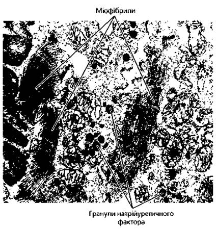
Fig. 4.10. Electron micrograph of a cardiac myoendocrine cell with natriuretic factor granules near the nuclear pole, x 7000
Atrial cardiomyocytes have a well-developed rough Endoplasmic reticulum and Golgi complex, which are involved in the synthesis of atrial natriuretic factor. The latter has a diuretic effect (promotes the Excretion of Water and salts from the body), is capable of increasing hematocrit, and lowering blood pressure. Atrial natriuretic factor accumulates in the cytoplasm of atrial cardiomyocytes in the form of specific electron-dense granules with a diameter of 300-400 nm (Fig. 4.10).
Epicardium and pericardium. The outer layer of the heart, or epicardium, is the visceral layer of the pericardium. The epicardium consists of a thin plate of connective tissue fused with the myocardium and covered by mesothelium. The connective tissue stroma of the epicardium contains a Superficial layer of collagen fibers, a layer of elastic fibers, a Deep Layer of collagen fibers, and a deep collagenous-elastic layer. In the pericardium, the connective tissue stroma is more developed than in the epicardium. The surface of the pericardium facing the pericardial cavity is also covered by mesothelium. Accumulations of adipose cells occur along the course of blood vessels.
Development of the heart. The heart develops from several embryonic primordia. The endocardium and blood vessels develop from the mesenchyme. The myocardium and epicardium develop from the visceral mesoderm (the so-called myoepicardial plate). The nerve ganglia and nerve fibers of the heart originate from the neuroectoderm. The heart primordium is formed in an embryo of 1.5 mm in length at the beginning of the third week of development. The Differentiation of the histological elements of the heart, which begins in the Embryonic period, is completed only at 16-20 years of age.
Key terms
1. Artery. 2. Arteriole. 3. Blood capillary. 4. Venule. 5. Vein. 6. Arteriolovenular anastomosis. 7. Rete mirabile arteriosum. 8. Rete mirabile venosum. 9. Microcirculatory bed. 10. Endothelium. 11. Fenestrated endothelium. 12. Pericyte. 13. Somatic capillary. 14. Visceral capillary. 15. Sinusoidal capillary. 16. True arteriolovenular anastomosis (shunt). 17. Atypical arteriolovenular anastomosis (half-shunt). 18. Muscular artery. 19. Mixed (musculoelastic) artery. 20. Elastic artery. 21. Subendothelial layer. 22. Internal elastic membrane. 23. External elastic membrane. 24. Venous valve. 25. Non-muscular vein. 26. Muscular vein. 27. Muscular vein with highly developed muscular elements. 28. Muscular vein with poorly developed muscular elements. 29. Lymphatic vessel. 30. Lymphatic capillary. 31. Intra- and extraorgan lymphatic vessels. 32. Thoracic duct. 33. Right lymphatic duct. 34. Anchoring fibrils. 35. Efferent lymphatic vessel. 36. Main lymphatic trunks. 37. Heart. 39. Endocardium. 39. Myocardium. 40. Epicardium. 41. Pericardium. 42. Musculoelastic layer of the endocardium. 43. Outer connective tissue layer of the endocardium. 44. Heart valve. 45. Cardiac muscle. 46. Cardiomyocyte. 47. Typical (contractile) cardiomyocyte. 48. Atypical (conducting) cardiomyocyte. 49. Atrial myoendocrine cells. 50. Cardiac conduction system. 51. Sinoatrial node. 52. Atrioventricular node. 53. Atrioventricular bundle. 54. Pacemaker cell (P-cell). 55. Transitional conducting cardiomyocyte. 56. Purkinje fibers.
Last update: 09/08/2026
Editorial and Educational Adaptation: This material has been compiled based on the primary/original source text. The project team performed an editorial review, corrected technical inaccuracies, structured sections, and adapted the content for an educational format.
What was processed:
- elimination of formatting defects (OCR errors, structural breaks, corrupted characters);
- editorial organization of content;
- standardization of terminology in accordance with academic sources;
- verification of factual statements against the original source text.
All mentions of the author, publication year, and origin of the primary text have been preserved in accordance with the source.