Human Anatomy - Kotsan I. Y. 2009
Cardiovascular system
Circulatory system
Conduction system of the heart
An essential role in the rhythmic activity of The Heart and the coordination of its individual chambers is played by the cardiac conduction system (systema conducens cardiacum), which ensures the heart's automaticity and rhythm.
The cardiac cycle, lasting 0.8 s, consists of three phases: atrial contraction, ventricular contraction, and a general pause. Atrial contraction (systole) lasts 0.1 s, ventricular contraction lasts 0.3 s, and the general pause lasts 0.4 s. During the general pause, both the atria and ventricles are relaxed, allowing Blood from the venae cavae and Pulmonary Veins to flow into the right and left atria, respectively. This is followed by atrial contraction. The contraction process begins near the opening of the SUPERIOR VENA CAVA into the right atrium and spreads across both atria, thereby forcing blood from the atria through the atrioventricular orifices into the ventricles. Next, a wave of contraction initiates in the heart walls, spreading to both ventricles and ejecting blood into the openings of the pulmonary trunk and aorta, while the atrioventricular Valves close. A pause then ensues, and the cycle repeats.
This sequential contraction and relaxation of the various PARTS OF THE heart is closely linked to its Structure and the presence of the conduction system through which the impulse propagates. The conduction system is formed by a network of interconnected atypical Muscle Cells known as conducting cardiomyocytes. Conducting cardiomyocytes differ from other muscle cells: they are densely innervated by fibers of the Autonomic Nervous system, contain fewer myofibrils, Cell/35.html">Mitochondria, and Ribosomes, but possess a greater volume of sarcoplasm. Furthermore, they are capable of spontaneously generating and conducting excitation. Conducting cardiomyocytes form specialized bundles and nodes that occupy specific locations immediately beneath the endocardium.
The centers of the conduction system are two nodes: the sinoatrial node and the atrioventricular node (Fig. 197).
Class="center">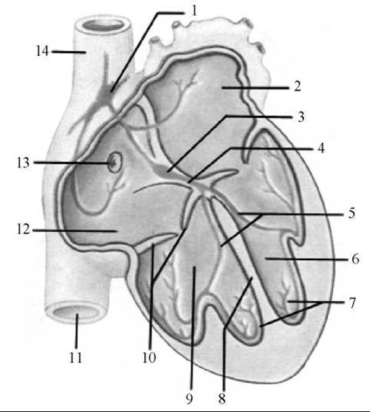
Fig. 197. The cardiac conduction system (diagram)
1 — sinoatrial node (node of Keith and Flack); 2 — left atrium; 3 — atrioventricular node (node of Aschoff and Tawara); 4 — atrioventricular bundle (bundle of His); 5 — right and left Branches of the atrioventricular bundle; 6 — left ventricle; 7 — Purkinje conducting muscle fibers; 8 — interventricular septum; 9 — right ventricle; 10 — right atrioventricular valve; 11 — INFERIOR VENA CAVA; 12 — right atrium; 13 — opening of the coronary sinus; 14 — superior vena cava
The sinoatrial node (nodus sinuatrialis) (node of Keith and Flack) is elongated, approximately 4 cm in length, and located in the wall of the right atrium between the opening of the superior vena cava and the right auricle. This node sends branches to the atrial myocardium and to the atrioventricular node. Three interatrial pathways emerge from the sinoatrial node: the anterior internodal tract of Bachmann, the middle internodal tract of Wenckebach, and the posterior internodal tract of Thorel, which connect it to the atrioventricular node. However, debate continues regarding whether impulse transmission from the sinoatrial node to the atrioventricular node is mediated by these specific pathways or by typical atrial cardiomyocytes. The sinoatrial node serves as the primary pacemaker of the heart, regulating its rhythm and heart rate. A normal resting heart rate is considered to be 70 beats per minute (130 beats/min in newborns).
The atrioventricular node (nodus atrioventricularis) (node of Aschoff and Tawara) is situated in the lower part of the interatrial septum, anterior to the opening of the coronary sinus. If the sinoatrial node is damaged, this node assumes The Role of a secondary pacemaker with an accelerated rate of impulse generation. A short atrioventricular bundle (fasciculus atrioventricularis), or bundle of His, extends from the atrioventricular node into the substance of the interventricular septum, connecting the atrial myocardium to the ventricular myocardium. In the upper portion of the interventricular septum, this bundle divides into right and left branches (crus dextrum et crus sinistrum). The left branch is larger than the right and is further divided into anterior and posterior fascicles. The branches and fascicles run subendocardially along both sides of the septum and transition into Purkinje fibers, which ramify throughout all Regions of the ventricular myocardium. The atrioventricular bundle transmits impulses from the atria to the ventricles, thereby establishing Coordinated regulation of the systolic rhythm of both the atria and ventricles.
Last update: 08/08/2026
Editorial and Educational Adaptation: This material has been compiled based on the primary/original source text. The project team performed an editorial review, corrected technical inaccuracies, structured sections, and adapted the content for an educational format.
What was processed:
- elimination of formatting defects (OCR errors, structural breaks, corrupted characters);
- editorial organization of content;
- standardization of terminology in accordance with academic sources;
- verification of factual statements against the original source text.
All mentions of the author, publication year, and origin of the primary text have been preserved in accordance with the source.