Human Anatomy - Kotsan I. Ya. 2009
Cardiovascular system
Circulatory system
Structure of the heart chambers
The right atrium (atrium dextrum) is cube-shaped and has a fairly large additional cavity known as the right auricle (auricula dextra). It is separated from the left atrium by the interatrial septum (septum interatriale). The septum clearly features an oval-shaped depression, the fossa ovalis, which is a remnant of the foramen ovale that connects the right and left atria during embryonic development. The fossa ovalis is bounded superiorly and anteriorly by the limbus fossae ovalis.
The right atrium contains the opening of the SUPERIOR VENA CAVA (ostium venae cavae superioris) and the opening of the INFERIOR VENA CAVA (ostium venae cavae inferioris). Between these openings lies a small tuberculum intervenosum, which is considered a remnant of the valve that directs Blood flow from the superior vena cava into the right atrioventricular orifice during the Embryonic period. Along the inferior margin of the opening of the inferior vena cava runs a small, crescent-shaped fold known as the valve of the inferior vena cava (valvula venae cavae inferioris). During embryonic development, this valve directs blood flowing from the inferior vena cava toward the foramen ovale and, through it, into the left atrium. The expanded posterior region of the right atrial cavity that receives both venae cavae is called the sinus venarum cavarum.
Inferiorly, the right atrium communicates with the right ventricle via the right atrioventricular orifice (ostium atrioventriculare dextrum). Between the right atrioventricular orifice and the opening of the inferior vena cava lies the ostium sinus coronarii. The coronary sinus is a wide venous vessel that receives blood from most of the large cardiac Veins. At the opening of the coronary sinus, a thin, sickle-shaped fold is visible, known as the valve of the coronary sinus (valvula sinus coronarii). Adjacent to the opening of the coronary sinus are the minute openings of the smallest cardiac veins (foramina venarum minimarum), which empty independently into the right atrium in varying numbers.
The internal wall of the right atrium is smooth, except for the upper part of the anterior wall and the wall of the right auricle, which feature longitudinal muscular ridges called pectinate Muscles (musculi pectinati). Superiorly, the pectinate muscles terminate at the crista terminalis, which separates the sinus venarum cavarum from the right atrial cavity. On the exterior surface of The Heart, the crista terminalis corresponds to the sulcus terminalis, located between the opening of the superior vena cava and the right auricle.
The right ventricle (ventriculus dexter) is located to the right and anterior to the left ventricle, resembling a three-sided pyramid with its apex directed downward. The inferior wall of the ventricle, which rests against the tendinous center of the Diaphragm, is flattened, while the anterior wall is convex anteriorly. The medial (left) wall is formed by the interventricular septum (septum interventriculare), which comprises two parts: a larger inferior muscular part (pars muscularis) and a smaller superior membranous part (pars membranacea). The membranous part of the septum is thin, consisting solely of a fibrous layer covered on both sides by the endocardium. Developmental heart anomalies in the form of ventricular septal defects are frequently found in the region of the membranous part. In the upper, widest section of the ventricle, There are two openings: posteriorly, the right atrioventricular orifice (ostium atrioventriculare dextrum), through which venous blood passes from the right atrium into the ventricle, and anteriorly, the pulmonary trunk orifice (ostium trunci pulmonalis), through which blood enters the pulmonary trunk.
The right atrioventricular orifice is closed by the right atrioventricular (tricuspid) valve (valva atrioventricularis dextra (tricuspidalis)), which is anchored to a dense connective-tissue fibrous ring whose tissue continues into the cusps of the valve. The cusps resemble triangular tendinous plates. Their bases are attached along the circumference of the atrioventricular orifice, while their free edges project into the ventricular cavity. The anterior cusp (cuspis anterior) is attached to the anterior semicircle of the orifice, the posterior cusp (cuspis posterior) to the posterolateral semicircle, and the septal cusp (cuspis septalis) to the medial semicircle. The atrial surface of the cusps is smooth, whereas the ventricular surface is uneven and features projections from which chordae tendineae originate. These tendinous cords attach by their opposite ends to cone-shaped muscular projections of the ventricular wall known as papillary muscles: the anterior, posterior, and septal papillary muscles (musculi papillares anterior, posterior et septalis). Together with the chordae tendineae, the papillary muscles hold the valve cusps in place and prevent the backflow of blood into the atrium during ventricular contraction.
The anterosuperior part of the right ventricle, which narrows funnel-like toward the pulmonary trunk, is called the conus arteriosus. The lower margin of the conus arteriosus, the supraventricular crest (crista supraventricularis), marks the boundary between the conus and the rest of the right ventricle. The wall of the right ventricle is smooth in the region of the conus arteriosus, whereas the remaining internal surface features muscular ridges known as trabeculae carneae. The function of the trabeculae carneae is to improve cardiac hemodynamics and reduce thrombus formation by creating parietal blood flow vortices. The septomarginal trabecula carries the right branch of the bundle of His of the heart's conduction system.
In the region of the pulmonary trunk orifice lies the pulmonary valve (valva trunci pulmonalis), which consists of three semilunar cusps: anterior, right, and left (valvulae semilunares anterior / dextra / sinistra). At the midpoint of the free edge of each semilunar cusp is a poppy-seed-sized nodule. These noduli valvularum semilunarium promote a tighter closure of the semilunar cusps. Between the wall of the pulmonary trunk and each semilunar cusp lies a small, pocket-like sinus of the pulmonary trunk (sinus trunci pulmonalis). When the ventricular musculature contracts, the semilunar cusps are pressed by the blood flow against the walls of the pulmonary trunk, allowing blood to pass freely from the ventricle; upon relaxation, when ventricular pressure drops, the reverse blood flow fills the sinuses and opens the cusps, whose edges then meet to prevent blood from flowing back into the heart.
The left atrium (atrium sinistrum) has an irregular cubic shape and is separated from the right atrium by the interatrial septum. The fossa ovalis on this septum is more pronounced from the right atrial side. The anterior wall of the atrium features a forward-projecting, cone-shaped extension called the left auricle (auricula sinistra), which contains pectinate muscles (musculi pectinati). Posteriorly and superiorly on the internal wall of the atrium, four pulmonary vein openings (ostia venarum pulmonalium) are visible—two on the right and two on the left. The left atrium communicates with the left ventricle through the left atrioventricular orifice (ostium atrioventriculare sinistrum).
The left ventricle (ventriculus sinister) is cone-shaped with its base directed upward. The walls of the left ventricle are 2–3 times thicker than those of the right ventricle, a difference attributable to the greater workload performed by the left ventricle in pumping blood through the systemic Circulation. In the upper part of the ventricle, there are two openings: posteriorly and to the left lies the left atrioventricular orifice, and to the right of it is the aortic orifice (ostium aortae). The left atrioventricular orifice is closed by the left atrioventricular (bicuspid, mitral) valve (valva atrioventricularis sinistra (bicuspidalis, mitralis)), which consists of two cusps: the anterior cusp (cuspis anterior) and the posterior cusp (cuspis posterior). The internal surface of the ventricle features numerous large trabeculae carneae and two papillary muscles—the anterior papillary Muscle (musculus papillaris anterior) and the posterior papillary muscle (musculus papillaris posterior)—with their thick chordae tendineae attached to the cusps of the atrioventricular valve. The aortic valve (valva aortae) has a Structure similar to that of the pulmonary valve. It is formed by three semilunar cusps: the right semilunar cusp (valvula semilunaris dextra), the left semilunar cusp (valvula semilunaris sinistra), and the posterior semilunar cusp (valvula semilunaris posterior) (Fig. 194). Between each cusp and the aortic wall lies an aortic sinus (sinus aortae). The aortic cusps are thicker, and the noduli valvularum semilunarium located at the midpoint of their free edges are larger than those in the pulmonary trunk.
Class="center">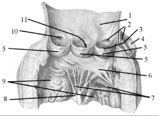
Fig. 194. Aortic valve
1 — aorta opened and spread out; 2 — left coronary artery; 3 — nodule of the semilunar cusp; 4 — left auricle; 5 — semilunar cusps; 6 — left atrioventricular (mitral) valve; 7 — chordae tendineae; 8 — papillary muscles; 9 — left ventricle; 10 — opening (ostium) of the right coronary artery; 11 — aortic sinus (cavity bounded by the semilunar cusp and the aortic wall)
The heart wall consists of three layers: the inner layer—endocardium, the middle layer—myocardium, and the outer layer—epicardium.
The endocardium (endocardium) lines the heart chambers from the inside, covering the papillary muscles and chordae tendineae. It is composed of endothelium resting on a thick basement membrane, a subendothelial Connective Tissue layer, and a myoelastic layer. The endothelium and subendothelial layer are continuous with the tunica intima of the vessels emerging from the heart. The heart Valves and folds are formed by folds (duplications) of the endocardium.
The myocardium (myocardium) is the thickest and functionally most powerful part of the heart wall (Figs. 195, 196). It is formed of Cytology/practical/59.html">Striated cardiac muscle tissue, which differs from Skeletal Muscle tissue in that it does not consist of separate multinucleated fibers, but rather of striated muscle Cells—cardiomyocytes—interconnected by numerous intercalated discs. Through these discs, they are linked into muscle complexes or fibers that form a narrow-meshed network. This narrow-meshed muscular network ensures the complete, rhythmic contraction of the atria and ventricles.
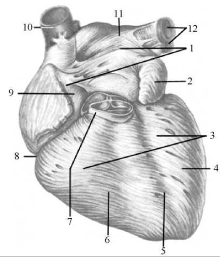
Fig. 195. Myocardium of the atria and ventricles (anterior view). Epicardium removed
1 — atrial myocardium; 2 — left auricle; 3 — ventricular myocardium; 4 — left ventricle; 5 — anterior interventricular sulcus; 6 — right ventricle; 7 — pulmonary trunk; 8 — coronary sulcus; 9 — right atrium; 10 — superior vena cava; 11 — left atrium; 12 — left Pulmonary veins
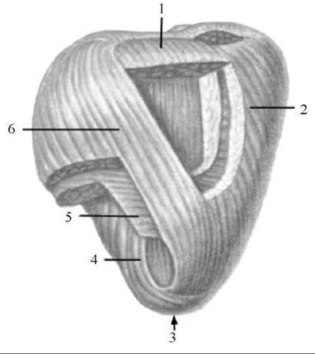
Fig. 196. Layers of the ventricular myocardium of the heart (posterior view)
1 — myocardial fibers (superficial layer) originating from the left fibrous ring; 2 — superficial (longitudinal) layer of the myocardium in the right ventricular wall; 3 — vortex of the heart; 4 — deep (longitudinal) layer of the myocardium in the left ventricular wall; 5 — middle (circular) layer of the myocardium in the left ventricular wall; 6 — superficial (longitudinal) layer of the myocardium in the left ventricular wall
The muscle fibers of the atria and ventricles originate from fibrous rings that completely separate the atrial myocardium from the ventricular myocardium. Consequently, atrial contraction occurs independently of ventricular contraction. The right and left fibrous rings (anuli fibrosi dexter/sinister), located around the right and left atrioventricular orifices, form the so-called fibrous Skeleton of the heart. This skeleton also includes thin connective tissue rings surrounding the openings of the aorta and pulmonary trunk, as well as the right and left fibrous triangles (trigonum fibrosum dextrum/sinistrum)—dense plates adjacent to the posterior semicircle of the aorta on the right and left. The right triangle contains a small opening through which pass the fibers of the atrioventricular bundle of the heart's conduction system.
Two layers of muscle are distinguished in the atrial myocardium: a superficial layer consisting of circularly or transversely arranged fibers, and a deep layer formed of longitudinal fibers. The superficial muscle layer surrounds both atria, whereas the deep layer encloses each atrium separately. Around the openings of the venae cavae and pulmonary veins emptying into the atria, there are circular bundles of myocytes that loop around the venous ostia like sphincters.
Three layers of muscle are distinguished in the ventricular myocardium: superficial, middle, and deep (Fig. 196). The superficial and deep layers are common to both ventricles. The fibers of the superficial layer originate from the fibrous rings, descend obliquely downward toward the apex of the heart where they form the vortex of the heart (vortex cordis), and continue into the deep longitudinal layer, which attaches by its upper end to the fibrous rings. Located between the superficial and deep muscle layers, the middle layer exists separately for each ventricle. The fibers of the middle myocardial layer have an almost transverse (circular) orientation. The middle layer is more highly developed in the left ventricle, which is why the walls of the left ventricle are significantly thicker than those of the right.
The interventricular septum is formed by all three muscle layers of both ventricles. However, the muscle layers of the left ventricle make a greater contribution to its formation. Its thickness is equal to that of the left ventricle wall. The Base of the upper (1/5) portion of the septum (its membranous part) consists of a plate of fibrous tissue, while the remaining (4/5) portion of the septum is composed of myocardium covered on both sides by the endocardium.
The epicardium (epicardium) is the outer layer of the heart wall and represents the visceral layer of the serous Pericardium. Like other serous membranes, it consists of a thin connective tissue plate covered with mesothelium. The epicardium envelops the heart, the initial segments of the pulmonary trunk and aorta, the terminal segments of the pulmonary and venae cavae, and then continues into the parietal layer of the serous pericardium.
Last update: 08/08/2026
Editorial and Educational Adaptation: This material has been compiled based on the primary/original source text. The project team performed an editorial review, corrected technical inaccuracies, structured sections, and adapted the content for an educational format.
What was processed:
- elimination of formatting defects (OCR errors, structural breaks, corrupted characters);
- editorial organization of content;
- standardization of terminology in accordance with academic sources;
- verification of factual statements against the original source text.
All mentions of the author, publication year, and origin of the primary text have been preserved in accordance with the source.