Human Anatomy - Kotsan I. Ya. 2009
Cardiovascular System
Circulatory System
Heart
The Heart (cor) is a hollow muscular organ that receives Blood from the venous trunks and pumps it into the Arterial System. The heart is located in the thoracic cavity within the middle compartment of the inferior Mediastinum. The greater part (two-thirds) of the heart lies in the left half of The thoracic cage, and the smaller part in the right half (Fig. 190). The longitudinal axis of the heart runs obliquely from above downwards, from right to left, and from behind forwards. The upper border of the heart is at the level of the cartilages of the third Ribs. The lower border corresponds to a line connecting the Base of the xiphoid process of the Sternum with the apex of the heart. The left border of the heart runs obliquely from the junction of the third left costal Cartilage with the sternum to the heart apex, which is located in the fifth intercostal space 1 cm medial to the midclavicular line. The right border of the heart extends 2 cm lateral to the right sternal margin. The borders of the heart are variable and depend on age, sex, body constitution, and body position.
Class="center">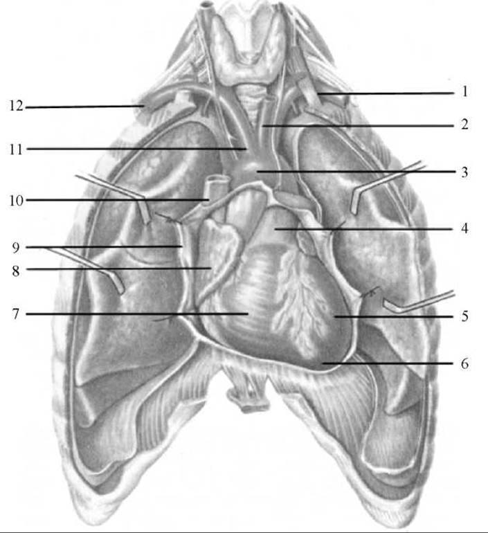
Fig. 190. Position of the heart in the thoracic cage (Pericardium opened)
1 — left Subclavian Artery; 2 — left common carotid artery; 3 — aortic arch; 4 — pulmonary trunk; 5 — left ventricle; 6 — apex of the heart; 7 — right ventricle; 8 — right atrium; 9 — pericardium; 10 — SUPERIOR VENA CAVA; 11 — brachiocephalic trunk; 12 — right subclavian artery
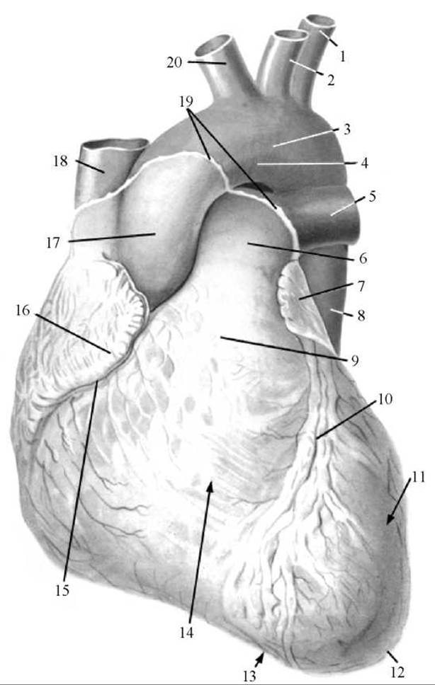
Fig. 191. Heart (anterior view)
1 — left subclavian artery; 2 — left common carotid artery; 3 — aortic arch; 4 — isthmus of aorta; 5 — left pulmonary artery; 6 — pulmonary trunk; 7 — left auricle; 8 — descending part of aorta; 9 — conus arteriosus; 10 — anterior interventricular sulcus; 11 — left ventricle; 12 — apex of the heart; 13 — notch of the apex of the heart; 14 — right ventricle; 15 — coronary sulcus; 16 — right auricle; 17 — ascending part of aorta; 18 — superior vena cava; 19 — site of transition of pericardium into epicardium; 20 — brachiocephalic trunk
In adults, the length of the heart is 10–15 cm, the greatest transverse dimension is 9–11 cm, and the anteroposterior dimension is 6–8 cm. The thickness of the atrial wall is 2–3 mm, that of the right ventricle is 5–8 mm, and that of the left ventricle is 12–15 mm. The average mass of the heart in an adult is about 300 g in men and about 250 g in women. It is generally considered that the size of the heart is roughly equal to the individual's clenched fist.
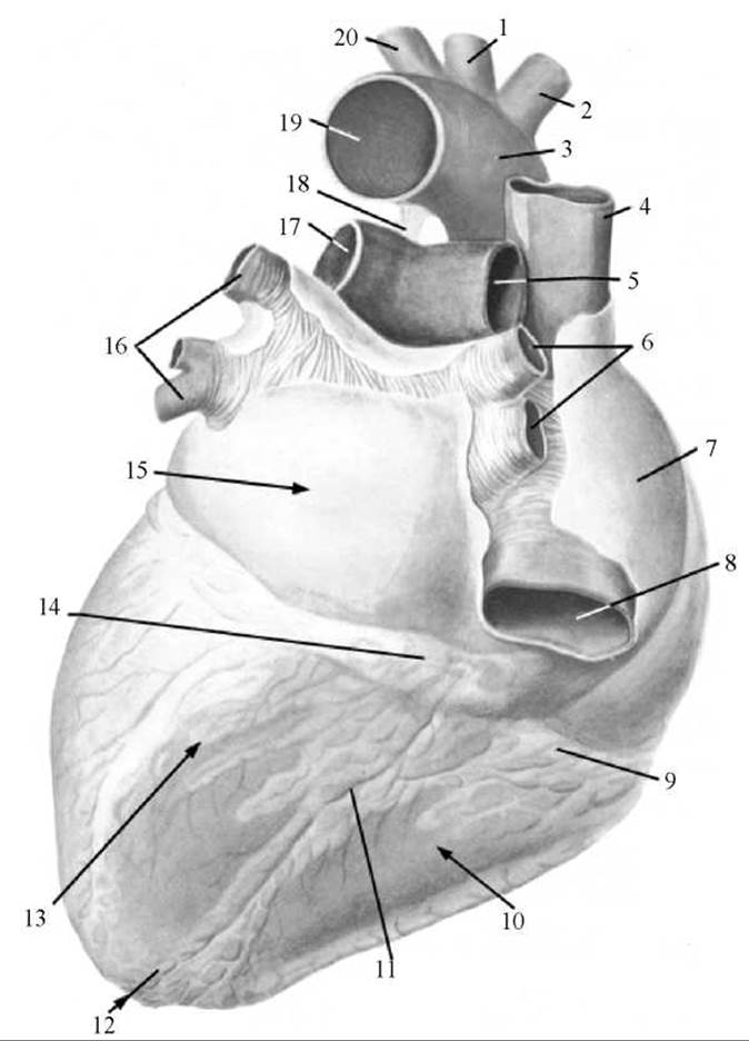
Fig. 192. Heart (posterior view)
1 — left common carotid artery; 2 — brachiocephalic trunk; 3 — aortic arch; 4 — superior vena cava; 5 — right pulmonary artery; 6 — superior and inferior right Pulmonary Veins; 7 — right atrium; 8 — INFERIOR VENA CAVA; 9 — coronary sulcus; 10 — right ventricle; 11 — posterior interventricular sulcus; 12 — apex of the heart; 13 — left ventricle; 14 — coronary sinus; 15 — left atrium; 16 — superior and inferior left pulmonary veins; 17 — left pulmonary artery; 18 — arterial ligament; 19 — aorta; 20 — left subclavian artery
The shape of the heart resembles a slightly flattened cone. The broad base of the heart (basis cordis) is directed upward, backward, and to the right, whereas the narrowed apex of the heart (apex cordis) is directed downward, to the left, and forward (Figs. 191, 192).
The surfaces of the heart include the convex sternocostal (anterior) surface (facies sternocostalis (anterior)), which faces the sternum and costal cartilages; the flat diaphragmatic (inferior) surface (facies diaphragmatica (inferior)), by which the heart rests on the Diaphragm; and two pulmonary (lateral) surfaces (facies pulmonales (laterales)), which face the mediastinal pleurae covering the Lungs. On radiographs, the pulmonary surfaces appear as the contours of the heart borders: the right border is sharper and the left is more blunt.
Three sulci are distinguished on the surfaces of the heart: a transversely positioned coronary sulcus (sulcus coronarius), which forms the external boundary between the atria and ventricles, and two longitudinal sulci — the anterior and posterior interventricular sulci (sulcus interventricularis anterior et sulcus interventricularis posterior) — which separate the ventricles from each other. The blood Vessels and nerves of the heart lie within these sulci. The anterior and posterior interventricular sulci merge near the apex of the heart to form a small notch — the notch of the apex of the heart (incisura apicis cordis).
The heart cavity consists of four chambers: two atria (right and left) and two ventricles (right and left) (Fig. 193).
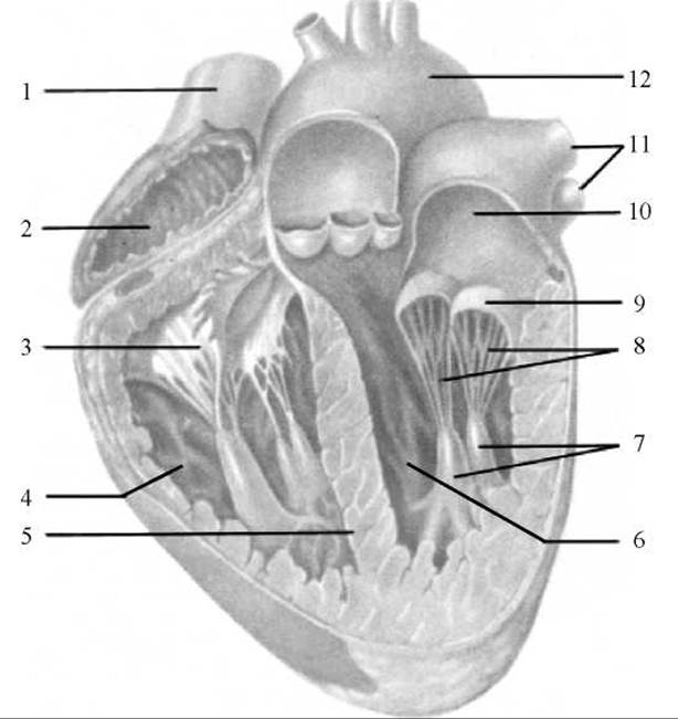
Fig. 193. Heart (longitudinal section)
1 — superior vena cava; 2 — right atrium; 3 — right atrioventricular valve; 4 — right ventricle; 5 — interventricular septum; 6 — left ventricle; 7 — papillary Muscles; 8 — tendinous cords; 9 — left atrioventricular valve; 10 — left atrium; 11 — pulmonary veins; 12 — aortic arch
The atria receive blood from the veins and force it into the ventricles; the ventricles pump blood into the Arteries: the right ventricle through the pulmonary trunk into the pulmonary arteries, and the left ventricle into the aorta, from which numerous arteries branch out to supply the body Organs and walls. The right half of the heart contains venous blood, and the left half contains arterial blood. Normally, the left and right halves of the heart are separated by a septum.
Last update: 08/08/2026
Editorial and Educational Adaptation: This material has been compiled based on the primary/original source text. The project team performed an editorial review, corrected technical inaccuracies, structured sections, and adapted the content for an educational format.
What was processed:
- elimination of formatting defects (OCR errors, structural breaks, corrupted characters);
- editorial organization of content;
- standardization of terminology in accordance with academic sources;
- verification of factual statements against the original source text.
All mentions of the author, publication year, and origin of the primary text have been preserved in accordance with the source.