Human Anatomy - Kotsan I. Ya. 2009
Cardiovascular System
Circulatory System
Structure and Functions of Blood Vessels
The Circulatory system (systema sanguineum) consists of A large number of vessels of various structures and sizes, the Blood circulating within them, and a central organ that drives the blood through the vessels — The Heart.
Blood Vessels are represented by Arteries, Veins, and the microvasculature, which comprises arterioles, precapillaries, capillaries, postcapillaries, venules, and arteriovenous anastomoses.
Arteries (arteriae) are vessels through which blood flows under pressure from the heart to Organs and Tissues. The arterial wall consists of three tunics: internal, middle, and external (Fig. 187, a).
The internal tunic (tunica intima) is formed by the endothelium, basement membrane, and subendothelial layer. Endothelial Cells line the vessel lumen; they rest on the basement membrane and are elongated along the longitudinal axis of the vessel. The subendothelial layer consists of fine elastic and Collagen fibers along with poorly differentiated Connective Tissue cells. External to the subendothelial layer lies the internal elastic membrane (membrana elastica interna), a terminal elastic lamina that separates the internal tunic from the middle one.
The middle tunic (tunica media) is formed primarily by circularly (spirally) oriented Muscle cells, as well as elastic and collagen fibers. It is separated from the external tunic by the external elastic membrane (membrana elastica externa).
Class="center">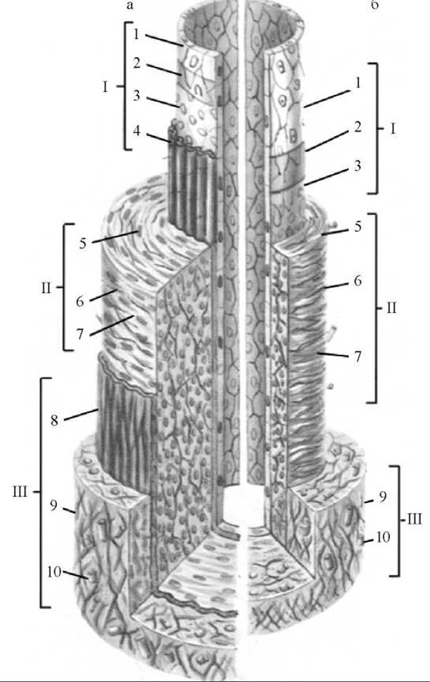
Fig. 187. Cytology/cytology/92.html">SCHEMATIC Structure OF the wall of a medium-sized muscular-type artery (a) and vein (b)
I — internal tunic: 1 — endothelium; 2 — basement membrane; 3 — subendothelial layer; 4 — internal elastic membrane; II — middle tunic containing: 5 — myocytes; 6 — elastic fibers; 7 — collagen fibers; III — external tunic containing: 8 — external elastic membrane; 9 — loose Fibrous connective tissue; 10 — blood vessels
The external tunic (adventitia) (tunica externa) is formed of loose fibrous irregular connective tissue containing elastic and collagen fibers. It transmits blood vessels — the vasa vasorum, which supply the arterial wall, and nerve fibers — the nervi vasorum, which innervate the arterial wall.
Depending on the degree of development of muscular or elastic elements in the middle tunic, arteries are classified into elastic, muscular, and mixed types.
Large arteries in which elastic fibers predominate over muscle cells in the middle tunic are called elastic-type arteries (aorta, pulmonary trunk). Elastic fibers provide the arterial wall with high tensile strength while also buffering, through their elasticity, the significant pressure pulses with which the heart propels blood into the vessels.
In some medium-sized and all small arteries, where the inertia of the cardiac pulse wanes and requires appropriate contraction of the vessel wall to maintain blood flow, contractile function predominates. This is ensured by a significant development of muscular elements in the vessel wall. Such vessels are called muscular-type arteries.
The third type of arteries—mixed (musculo-elastic) type arteries—are those in which the middle tunic contains approximately equal numbers of muscle cells and elastic fibers. These include most medium-sized arteries (e.g., carotid, subclavian, femoral, etc.)
Depending on their thickness (diameter), arteries are conventionally divided into large, medium, and small. Each artery has a main trunk and its branches.
Based on their branching patterns, arteries are classified into trunk (main) and dispersive types. In the trunk branching pattern, There is a main stem—the trunk artery—and lateral branches that arise from it gradually. As lateral branches depart from the trunk artery, its diameter progressively decreases. The dispersive branching pattern is characterized by the immediate division of the main stem into two or more terminal arteries, the overall branching layout of which resembles the crown of a deciduous tree.
A vessel that ensures blood flow bypassing the primary pathway is called a collateral vessel. When passage through the main (trunk) artery is obstructed, blood can flow via collateral bypass vessels that originate either from the same source as the main vessel or from different sources and terminate in a shared vascular network. Collateral vessels that anastomose (connect) with branches of other arteries function as arterial anastomoses. Anastomoses are of great importance when an artery is compressed or injured. An arterial injury cuts off Blood supply to a specific body area, in which case the area is supplied via the anastomotic vessel. Anastomotic vessels are typically thin and play almost no role in Regional Circulation. However, when Blood flow through the main vessel ceases, blood begins to flow through the anastomosis. Driven by enhanced blood flow, the anastomotic vessel gradually widens, its walls thicken, and it transforms into a vessel adequate for supplying the body region that ceased receiving blood through the primary artery.
Arteries that supply the body walls are called parietal arteries, whereas Arteries of the Internal Organs are called visceral arteries.
In relation to an organ, a distinction is made between extraorgan arteries, which carry blood to the organ, and intraorgan arteries, which branch within the organ and supply its individual parts (lobes, segments, lobules).
Arteries are also named after the organ they supply (e.g., renal artery, splenic artery). Some arteries are named According to the level of their origin from a larger vessel (e.g., superior mesenteric artery, inferior mesenteric artery), the bone adjacent to the vessel (radial, ulnar), the course of the vessel (medial artery, circumflex femoral artery), as well as their depth (superficial or deep). Small vessels without specific names are designated as branches.
Veins (venae) are vessels that carry blood in the direction opposite to that of arteries, i.e., from organs to the heart. The walls of veins, like those of arteries, consist of three layers, but they are significantly thinner and contain less elastic and muscular tissue; therefore, veins are less elastic and resilient than arteries (Fig. 187, b). On cross-section, arterial openings gape, whereas venous lumens collapse.
There are two types of veins: non-muscular and muscular.
In non-muscular veins, the basement membrane lies directly external to the endothelium, followed by a thin layer of loose fibrous connective tissue. Non-muscular veins include the VEINS OF THE dura mater and pia mater, the retina of the eye, bones, Spleen, and Placenta. They are tightly fused to the organ walls and therefore do not collapse.
Muscular-type veins are subdivided into veins with weak, moderate, and strong development of muscular elements. Veins with weakly developed muscular elements (generally up to 1-2 mm in diameter) are located mainly in the upper trunk, neck, face, and upper limbs. Large veins such as the SUPERIOR VENA CAVA also belong to this group. The subendothelial layer of the internal tunic in such veins is poorly developed, the middle tunic contains a small number of myocyte bundles, and myocytes are absent in the other tunics. In veins with moderately developed muscular elements, the internal elastic membrane is absent, and a network of elastic fibers lies at the boundary between the internal and middle tunics. The middle tunic consists of circularly arranged myocyte bundles separated by layers of fibrous connective tissue. The external elastic membrane is absent, while the external connective tissue tunic is well developed. Veins with strongly developed muscular elements are large veins of the lower half of the trunk and lower limbs; they are characterized by the presence of myocyte bundles in all three tunics.
Depending on their topography and position within The Human Body and organs, veins are divided into superficial and deep. Superficial (subcutaneous) veins (venae superficiales) generally run independently. Deep veins (venae profundae) accompany the corresponding arteries of the limbs in pairs and are therefore called companion veins (venae comitantes). The names of deep veins correspond to the arteries they accompany (e.g., ulnar artery — ulnar veins). Unpaired deep veins include the Internal jugular vein, subclavian, axillary, iliac (common, external, internal), femoral, and certain other veins.
Superficial veins connect to deep veins via so-called perforating veins, which function as anastomoses.
Adjacent veins frequently interconnect with each other through numerous anastomoses, collectively forming venous plexuses (plexus venosus) that are well-developed On the surface or within the walls of certain internal organs (such as the Urinary Bladder and rectum).
The largest Veins of the systemic circulation are the superior and inferior venae cavae. The hepatic veins along with their tributaries empty into the INFERIOR VENA CAVA. Collateral venous outflow provides an alternative pathway through which venous blood bypasses the main route. The tributaries of a single large (trunk) vessel interconnect via intrasystemic venous anastomoses. Intersystemic venous anastomoses (caval-caval, cavoportal, and cavo-caval-portal) are present between the tributaries of different large veins (the superior and inferior venae cavae, and the portal vein), serving as collateral pathways for venous drainage that bypass the primary veins.
The total number of veins exceeds that of arteries, and the overall capacity of the venous bed surpasses that of the arterial bed.
Blood flow velocity in veins is lower than in arteries. Blood movement through veins is facilitated by the suction effect of the thoracic cavity, which creates negative pressure during inspiration, as well as by the contraction of skeletal and visceral musculature of organs, among other factors. The contraction of the venous muscular coat also plays a significant role; it is more strongly developed in the veins of the lower half of the body—where conditions for venous return are more challenging—than in the veins of the upper body. A Specific Adaptation facilitating the return of blood to the heart is the presence of venous Valves, which are found in most small and medium-sized veins, as well as in certain large-caliber veins. Venous valves (valves)
(valvulae venosae) are crescent-shaped folds of the vessel's tunica intima, composed of fibrous connective tissue covered on both sides by endothelial cells (Fig. 188). Venous valves generally occur in pairs. They allow blood to flow exclusively toward the heart, prevent its backflow, and spare the heart from wasting excess energy on overcoming the oscillatory blood movements that constantly occur in veins. Valves are particularly abundant in the veins of the lower extremities, where blood flows against gravity, creating a predisposition for stasis and venous reflux. Numerous valves are also found in the upper extremities, whereas they are fewer in the trunk and neck veins. Only the two venae cavae, HEAD veins, renal veins, portal vein, and Pulmonary veins lack valves entirely.
Situated between the arteries and veins is the distal part of The Cardiovascular system—the microvasculature (microcirculatory bed)—which serves as the local circulatory pathway where the exchange between blood and tissues takes place. The microvasculature consists of arterioles, precapillaries, capillaries, postcapillaries, venules, and arteriovenous anastomoses.
As they distance themselves from the heart, the caliber of arteries progressively diminishes down to their smallest branches—the arterioles (arteriolae). Similar to arteries, the wall of an arteriole consists of three tunics, though they are poorly developed. An arteriole differs from an artery in that the Middle layer of its wall contains only a single layer of smooth muscle cells, through which it regulates blood flow. The arteriole continues directly into the precapillary, whose middle tunic is formed by scattered muscle cells that occasionally aggregate to form a precapillary sphincter, which regulates blood perfusion in the capillaries and the organ or body region as a whole. Numerous capillaries branch off from the precapillary. The wall of a capillary vessel (vas capillare) is extremely thin, consisting of just a single layer of endothelial cells resting on a basal lamina. This structure facilitates the free passage of dissolved substances and gases from the vessel into the surrounding tissue and vice versa. In living tissues, capillary vessels exhibit various shapes and diameters that constantly change depending on the functional state of The Vascular System and the organ in which they are located. Freely anastomosing with one another, capillaries form networks (capillary beds) that transition into postcapillaries. Postcapillaries are structurally similar to precapillaries; they possess a thin adventitial layer, and their walls are highly distensible and permeable. By converging, postcapillaries form venules (venulae), the caliber of which varies widely, typically measuring 25–50 µm under normal conditions. Postcapillaries and venules represent the initial Components of the Venous system and the terminal Links of the microvasculature. Venules empty into veins.
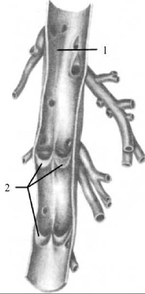
Fig. 188. Venous valves
A vein opened longitudinally and spread out
1 — lumen of the vein; 2 — cusps of the venous valves
Within the microvasculature, there are vessels that provide a direct pathway for blood to pass from an arteriole to a venule—namely, arteriovenous anastomoses. Their walls contain myocytes that regulate blood flow into the capillary bed. Under normal conditions, arteriovenous anastomoses remain closed, and blood flows through the capillary bed. However, when these anastomoses open, a portion of the blood can bypass the capillaries and flow directly into the venules. Thus, arteriovenous anastomoses function as shunts that regulate capillary circulation. An example of this regulatory function is the alteration of cutaneous capillary BLOOD FLOW IN response to ambient Temperature changes.
Typically, a capillary network is supplied by an arterial-type vessel (an arteriole) and drained by a venule. Certain organs (such as the Kidney and Liver) present exceptions to this rule. For instance, the renal corpuscle's glomerulus is supplied by an arteriole (the afferent vessel) and drained by another arteriole (the efferent vessel). In the liver, the capillary network is interposed between the afferent (interlobular) and efferent (central) veins. A capillary bed inserted between two Vessels of the same type (either arteries or veins) is referred to as a "rete mirabile" (admirable net).
The walls of blood vessels possess a rich sensory (afferent) and motor (efferent) innervation. The walls of certain large vessels (such as the ascending aorta and aortic arch, the bifurcation site of the common carotid artery into the internal and external carotid arteries, the superior vena cava, and the jugular veins, among others) contain particularly dense sensory nerve endings. Consequently, these regions are designated as reflexogenic zones, stimulation of which alters Cardiac Activity and blood pressure. Virtually all blood vessels feature extensive innervation, which plays a crucial role in regulating vascular tone and blood flow.
General Principles of vascular pathways and branching. The course of arteries and the blood supply to various organs depend on their structure, function, and development, conforming to a series of regularities studied and formulated by P. F. Lesgaft and B. A. Dolgo-Saburov. Thus, the architecture of the Arterial System corresponds to the General structural plan of the human body, which is characterized by the presence of an Axial Skeleton, a centralized Nervous system, bilateral body Symmetry, paired limbs, and the asymmetric arrangement of internal organs.
Principal arteries always travel to organs via the shortest possible path, thereby conserving the energy expended by the heart in pumping blood and accelerating its delivery. Large vessels lie along the flexor aspect of the trunk or limbs, where they are better shielded and protected. In these locations, vessels are less vulnerable to injury—a critical factor, given that arterial bleeding can be fatal. Furthermore, the flexor side provides a shorter pathway than the extensor side. On the digits, it is not the flexor surfaces that are best protected, but rather the lateral surfaces—precisely where the digital arteries run.
In accordance with the principles of bilateral symmetry and human body segmentation, most arteries are paired, and many arteries supplying the trunk are segmental. Segmentation is preserved with particular clarity in the Topography of the intercostal and lumbar arteries.
Most commonly, vessels branch off from major central arteries at the level of the organs they supply. If an organ shifts from its original embryonic site during ontogenesis—such as the Gonads—its supplying vessel does not alter its point of origin from the main artery, but instead elongates to follow the migrating organ.
A definite correspondence exists between the skeletal structural plan and the number of trunk arteries. THE Vertebral Column is accompanied by the aorta, and the clavicle by a single Subclavian Artery. The arm (containing one bone) has a single brachial artery, while the forearm (containing two bones—the radius and ulna) has two arteries corresponding to these bones. In the FOOT and hand, corresponding to the five digits, There are five pairs of arteries.
Along the path to joints, collateral arteries branch off from major trunk vessels, while running to meet them—from the distal segments of the trunk arteries—are recurrent arteries. By anastomosing with one another around the circumference of the joints, they form articular arterial networks that ensure uninterrupted blood supply during movement.
Arteries enter organs through the hilum, located on their concave medial or internal surface facing the source of blood supply. The number of arteries entering an organ and their diameter depend not only on the organ's size, but also on its functional activity.
The patterns of arterial branching are determined by the structural plan of the organ and the arrangement and orientation of connective tissue within it. In lobular organs (such as the Lungs, liver, and Kidneys), the artery enters the hilum and subsequently branches in accordance with lobes, segments, and lobules. In hollow, tube-like organs (such as the intestines, Uterus, and uterine tubes), the nourishing arteries approach from one side, while their branches assume a circular, longitudinal, or radial course. In fibrous organs (such as Muscles, ligaments, and nerves), arteries enter at multiple points and branch out along the course of the fibers.
It should be emphasized that the Nutrition of a given organ is sustained not solely by its own arteries, but also by neighboring vessels supplying blood through anastomoses. An anastomosis (connection or junction) is any tertiary vessel that connects two other vessels. Collateral circulation is of paramount importance for organ blood supply. A collateral (from the Latin *collateralis* — lateral) is a side vessel that provides an alternative (bypass) blood flow. Collateral vessels occur both within the arterial system (arterial collaterals) and the venous system (venous collaterals).
Considering the structural and Functional Characteristics of The Heart and blood vessels, the human body features two circulatory loops: the systemic (greater) and pulmonary (lesser) circulations (Fig. 189).
The systemic (greater) circulation serves to deliver nutrients and oxygen to all organs and Tissues of the body. It originates in the left ventricle of the heart, which receives oxygenated blood from the left atrium. Emerging from the left ventricle is the human body's largest artery—the aorta—which gives rise to arteries supplying all bodily organs and tissues, branching within them into increasingly smaller vessels down to arterioles and capillaries. As blood passes through the capillaries, oxygenated blood releases essential substances (oxygen, glucose, Proteins, Vitamins, Hormones, etc.) to the cells and removes Metabolic waste products and carbon dioxide, thereby transforming from arterial into venous blood. Venous blood flows from the capillaries into venules and subsequently into veins. Small veins converge into larger ones, and ultimately all blood drains into two major trunk vessels known as the superior and inferior venae cavae. The venae cavae empty into the right atrium, marking the Termination of the systemic circulation.
The Pulmonary Circulation (lesser circulation) begins at the right ventricle with the pulmonary trunk, which emerges from the heart and branches into the right and left pulmonary arteries. Upon entering the respective lung, each pulmonary artery repeatedly branches into smaller arteries that gradually transition into capillaries. These capillaries densely entwine the alveoli. Oxygen passes from the alveoli through the delicate alveolocapillary membrane into the blood, while carbon dioxide moves from the blood into the alveolar cavity. This is how gas exchange takes place, turning deoxygenated blood into oxygenated blood. Small venules originate from the capillaries and merge into larger veins, eventually forming four pulmonary veins (two from each lung) that empty into the left atrium, marking the end of the pulmonary circulation.
Some morphologists also distinguish a third (cardiac) circulation, which serves as a Supplement to the systemic circulation and supplies blood directly to the heart. The Coronary Circulation begins with two coronary arteries branching off from the aortic bulb and ends with the cardiac veins, through which blood flows into the coronary sinus that empties into the right atrium. Some of the smaller veins open directly into the cavities of the right atrium and right ventricle.
The arteries of the systemic and coronary circulations always carry oxygenated blood, while their veins carry deoxygenated blood. In the pulmonary circulation, the situation is reversed: the arteries carry deoxygenated blood, and the veins carry oxygenated blood.
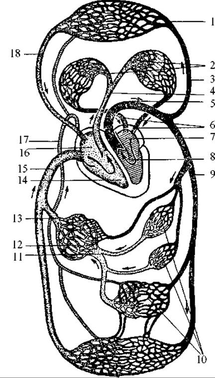
Fig. 189. Diagram of Blood Circulation
I — capillaries of the upper body; 2 — lung capillaries; 3 — arteries of the upper body; 4 — pulmonary trunk; 5 — aortic arch; 6 — pulmonary veins; 7 — left atrium; 8 — left ventricle; 9 — descending aorta; 10 — capillaries of the lower body and abdominal organs;
II — portal vein; 12 — liver capillaries; 13 — hepatic veins; 14 — right ventricle; 15 — inferior vena cava; 16 — right atrium; 17 — Thoracic duct; 18 — superior vena cava
Last update: 08/08/2026
Editorial and Educational Adaptation: This material has been compiled based on the primary/original source text. The project team performed an editorial review, corrected technical inaccuracies, structured sections, and adapted the content for an educational format.
What was processed:
- elimination of formatting defects (OCR errors, structural breaks, corrupted characters);
- editorial organization of content;
- standardization of terminology in accordance with academic sources;
- verification of factual statements against the original source text.
All mentions of the author, publication year, and origin of the primary text have been preserved in accordance with the source.