Human Anatomy, Part 1 - K. A. Dyubenko, A. K. Kolomiysev, Yu. B. Chaykovsky 2002
Special Section
Splanchnology, splanchnologia [the study of viscera] — General Overview
General structural plan of the digestive tract wall
1. The mucous membrane, tunica mucosa, is a permanent structural component of the digestive tract wall, extending from the Oral Cavity to the anus, where it gradually transitions into the Skin. It is so named because it remains constantly moist and covered with mucus. Its color depends on vascular Blood filling and ranges from pink to deep red. It consists of three lamina:
- epithelium, epitelium;
- lamina propria, lamina propria mucosae;
- muscular layer of the mucous membrane, lamina muscularis mucosae.
Depending on its functional purpose and Cell shape, the epithelium can be:
- simple squamous (Peritoneum, Pleura, Pericardium);
- simple columnar (Stomach, intestines);
- simple prismatic (Ductus deferens);
- simple cuboidal (renal tubules, gland excretory ducts, small Bronchi);
- pseudostratified ciliated (Nasal cavity, Larynx, Trachea, bronchi).
The epithelium of certain Organs contains goblet Cells that produce mucus, while others contain glands.
The lamina propria mucosae is formed by loose Fibrous Connective Tissue. It contains solitary and aggregated lymphatic nodules (follicles), capillary networks of Blood and Lymphatic vessels, glands, nerve fibers, and their endings.
Class="center">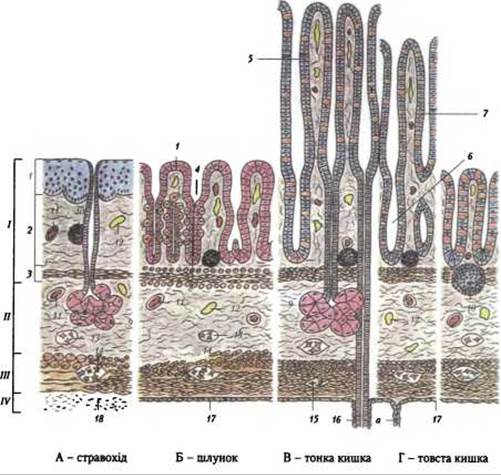
Fig. 191. General structural plan of the digestive tube wall (according to O. V. Volkova and Yu. K. Yeletsky):
I - mucous membrane; II - submucosa; III - muscular layer; IV - outer layer; 1 - epithelium; 2 - lamina propria mucosae; 3 - muscularis mucosae; 4 - gastric pits; 5 - intestinal villi; 6 - intestinal glands (crypts); 7 - unicellular intraepithelial glands; 8 - gastric glands; 9 - submucosal glands; 10 - lymphoid nodules; 11 - Blood Vessels; 12 - Lymphatic vessels; 13 - submucosal nerve plexus; 14 - myenteric nerve plexus; 15 - subserosal nerve plexus; 16 - extraintestinal gland duct; 17 - serosa (a - mesentery); 18 - adventitia
The muscular layer of the mucous membrane, lamina muscularis mucosae, lies beneath the lamina propria. It is formed by Cytology/cytology/32.html">Smooth Muscle tissue, the contraction of which determines its mobility and folding.
2. The submucosal layer, tela submucosa, is formed by loose fibrous connective tissue. It allows the mucous membrane to move relative to other layers. The submucosa is characterized by dense networks of blood and lymphatic vessels, as well as the submucosal nerve plexus. The Structure of the mucous membrane and submucosa in Internal Organs is determined by their function. Thus, its relief is heterogeneous: folded in The Stomach, and smooth in the excretory ducts of glands, bronchi, and trachea. In certain areas (Small Intestine), the mucous membrane forms finger-like projections (villi) and depressions (crypts), which increase the surface area of contact, facilitating the absorption of digested food products. The color of the mucous membrane is light pink and occasionally red, depending on the depth of the blood vessel walls within it.
3. The muscular coat, tunica muscularis, consists of layers of smooth muscle tissue—an inner (circular) and an outer (longitudinal) layer. Near the entrance and exit orifices, the muscular coat is formed by striated muscle fibers. The walls of hollow organs have a rich blood supply. The muscular coat contains blood and lymphatic networks, as well as the myenteric nerve plexus.
4. The outer coat, tunica adventitia, consists of a connective tissue membrane and covers hollow organs externally only in certain sections (Esophagus, rectum). Organs located in the serous cavities are covered by a serous membrane, tunica serosa, which is formed by a simple squamous epithelium (mesothelium) and constitutes the visceral layer of the peritoneum.
Glands, glandulae, are structures composed of epithelial cells, with connective tissue, blood vessels, and nerves participating in their structure. Depending on their origin, The Nature of secretion, the presence of excretory ducts, and their structure, glands are divided into groups. Ectodermal, mesodermal, and entodermal glands are distinguished. Ectodermal glands include sebaceous, sweat, and Mammary Glands; mesodermal glands include the interstitial Cells of the Testis; and entodermal glands include the Glands of the digestive and respiratory tracts.
Depending on the secreted substance, glands are divided into those that produce incretions and those that produce excretions. Glands with excretory ducts are called exocrine glands. Glands lacking excretory ducts, whose products enter the blood directly, are called Endocrine glands.
According to their structure and shape, glands are classified into:
- alveolar, consisting of a single spherical vesicle or pouch (simple alveolar), several alveoli (branched alveolar), or A large number of alveoli that, in turn, form lobules (compound alveolar);
- tubular, which may consist of a single tube (simple tubular) or a large number of branched tubes (branched tubular and compound tubular);
- mixed, formed simultaneously by alveoli and tubules. They can be simple, branched, and compound. For the structure and shape of glands, see Fig. 6.
Last update: 08/08/2026
Editorial and Educational Adaptation: This material has been compiled based on the primary/original source text. The project team performed an editorial review, corrected technical inaccuracies, structured sections, and adapted the content for an educational format.
What was processed:
- elimination of formatting defects (OCR errors, structural breaks, corrupted characters);
- editorial organization of content;
- standardization of terminology in accordance with academic sources;
- verification of factual statements against the original source text.
All mentions of the author, publication year, and origin of the primary text have been preserved in accordance with the source.