Human Anatomy - Kotsan I. Y. 2009
Cardiovascular System
Circulatory System
Blood and lymphatic vessels of the heart
Unlike other Organs, The Heart has its own Circulatory system, which forms what appears to be a distinct third circle of Circulation—the cardiac circulation—as part of the systemic circulation.
The cardiac Arteries branch off from the aortic bulb and encircle the heart like a crown, hence their name: the coronary arteries (Figs. 198, 199). The right and left coronary arteries originate below the free edges of the semilunar aortic cusps. During ventricular systole (contraction), the cusps close the openings of the coronary arteries, protecting them from excessive systolic Blood pressure. Blood enters the coronary arteries exclusively during ventricular diastole (relaxation), when the semilunar cusps snap shut and the entryway to the coronary vessels opens.
The right coronary artery (arteria coronaria dextra) emerges from the aorta opposite the right semilunar cusp, runs to the right beneath the right auricle, enters the coronary sulcus, curves around the right border of the heart, and extends to its posterior surface. Here, it continues as the posterior interventricular branch (r. interventricularis posterior). The latter descends along the posterior interventricular sulcus toward the apex of the heart, where it anastomoses with a branch of the left coronary artery. Branches of the right coronary artery supply blood to the walls of the right atrium and ventricle, the posterior portion of the interventricular septum, the papillary Muscles of the right ventricle, the posterior papillary Muscle of the left ventricle, and the sinuatrial and atrioventricular nodes of the heart's conduction system.
Class="center">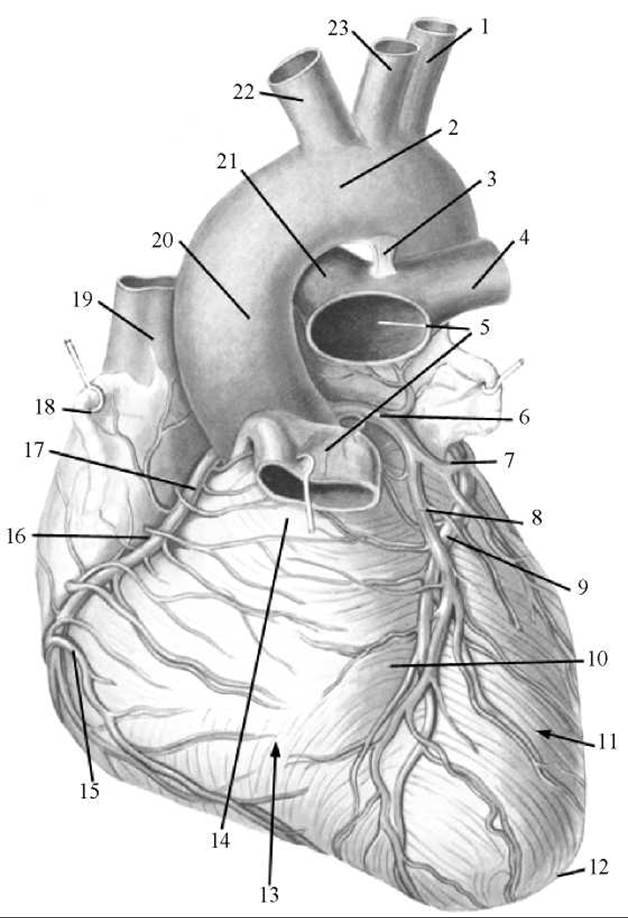
Fig. 198. Arteries and Veins OF THE Heart (anterior view)
1 — left Subclavian Artery; 2 — aortic arch; 3 — ligamentum arteriosum; 4 — left pulmonary artery; 5 — pulmonary trunk; 6 — left coronary artery; 7 — circumflex branch of the left coronary artery; 8 — anterior interventricular branch of the left coronary artery; 9 — great cardiac vein; 10 — anterior interventricular sulcus; 11 — left ventricle; 12 — apex of the heart; 13 — right ventricle; 14 — conus arteriosus; 15 — anterior cardiac vein; 16 — coronary sulcus; 17 — right coronary artery; 18 — right auricle; 19 — SUPERIOR VENA CAVA; 20 — ascending aorta; 21 — right pulmonary artery; 22 — brachiocephalic trunk; 23 — left common carotid artery
The left coronary artery (arteria coronaria sinistra) arises from the aorta opposite the left semilunar cusp, courses to the left, and lies beneath the left auricle of the heart. Beneath this auricle, the artery divides into a larger circumflex branch (r. circumflexus) and a smaller anterior interventricular branch (r. interventricularis anterior). The circumflex branch, continuing the main trunk of the left coronary artery, circles the heart to the left within its coronary sulcus, where, on the posterior surface of the organ, it anastomoses with the right coronary artery. The anterior interventricular branch descends along the sulcus of the same name toward the apex of the heart. In the region of the cardiac notch, it occasionally crosses onto the diaphragmatic surface of the heart, anastomosing there with the terminal section of the posterior interventricular branch of the right coronary artery. Branches of the left coronary artery supply blood to the wall of the left ventricle—including the papillary muscles—the majority of the interventricular septum, the anterior wall of the right ventricle, and the wall of the left atrium.
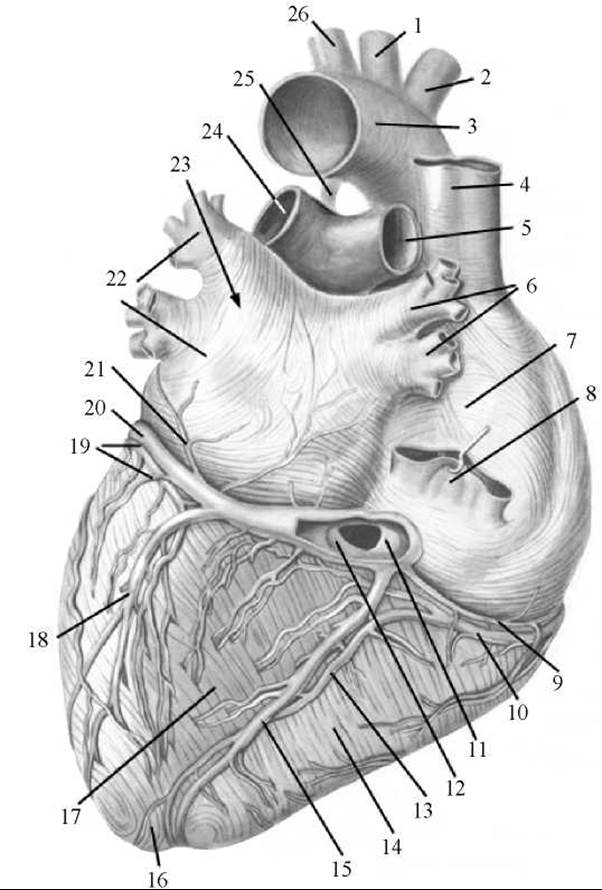
Fig. 199. Arteries and veins of the heart (posterior view)
1 — left common carotid artery; 2 — brachiocephalic trunk; 3 — aortic arch; 4 — superior vena cava; 5 — right pulmonary artery; 6 — right Pulmonary veins; 7 — right atrium; 8 — INFERIOR VENA CAVA; 9 — small cardiac vein; 10 — right coronary artery; 11 — valve of the coronary sinus; 12 — coronary sinus of the heart; 13 — posterior interventricular branch of the right coronary artery; 14 — right ventricle; 15 — middle cardiac vein; 16 — apex of the heart; 17 — left ventricle; 18 — posterior vein of the left ventricle; 19 — circumflex branch of the left coronary artery; 20 — great cardiac vein; 21 — oblique vein of the left atrium; 22 — left pulmonary veins; 23 — left atrium; 24 — left pulmonary artery; 25 — ligamentum arteriosum; 26 — left subclavian artery
Anastomosing with one another, the branches of the right and left coronary arteries form two arterial rings within the heart: a transverse ring located in the coronary sulcus, and a longitudinal ring whose vessels lie within the anterior and posterior interventricular sulci.
Various branching patterns of the coronary arteries exist, known as types of coronary blood supply. The main types are: right-coronary dominant, where the greater part of the heart is supplied by branches of the right coronary artery; left-coronary dominant, where most regions receive blood from branches of the left coronary artery; and balanced (or intermediate), in which both coronary arteries contribute equally to the blood supply of the heart walls. Transitional types of coronary supply—mid-right and mid-left—are also distinguished. It is widely accepted that the overwhelming majority of people exhibit a mid-right dominant pattern of Coronary Circulation.
Various anatomical variations and anomalies in the position and branching of the coronary arteries are also possible. These manifest as Changes in the origin site and number of coronary arteries. For instance, a coronary artery may be single (unpaired), or there may be 3 to 4 coronary arteries instead of two: two arteries arising from the right and left sides of the aorta respectively, or two from the aorta and two from the left subclavian artery.
In addition to the coronary arteries, the heart—and particularly the Pericardium—is supplied by accessory arteries that branch off from the concave surface of the aortic arch, as well as from the internal thoracic and bronchial arteries.
Veins of the heart. Most of the large cardiac veins merge into a single common, wide venous vessel—the coronary sinus (sinus coronarius)—which empties into the right atrium. The coronary sinus lies within the coronary sulcus on the posterior (diaphragmatic) surface of The Heart and opens into the right atrium inferior and anterior to the opening of the inferior vena cava.
The tributaries of the coronary sinus are 5 veins:
1) the great cardiac vein (v. cordis magna), which begins near the apex of the heart on its anterior surface, runs within the anterior interventricular sulcus alongside the anterior interventricular branch of the left coronary artery, then turns left at the level of the coronary sulcus, passes beneath the circumflex branch of the left coronary artery, enters the coronary sulcus, and continues into the coronary sinus. This vein collects blood from the veins of the anterior surface of both ventricles and the interventricular septum;
2) the middle cardiac vein (v. cordis media), which originates in the region of the posterior surface of the heart's apex, ascends along the posterior interventricular sulcus adjacent to the posterior interventricular branch of the right coronary artery, and empties into the coronary sinus. Along its path, the middle cardiac vein receives branches from the posterior walls of both ventricles;
3) the small cardiac vein (v. cordis parva), which begins on the right pulmonary surface of the right ventricle, ascends, occupies the posterior part of the coronary sulcus, and drains into the coronary sinus. This vein collects blood primarily from the right half of the heart;
4) the posterior vein of the left ventricle (v. posterior ventriculi sinistri), which is formed by several veins on the posterior surface of the left ventricle and empties into either the coronary sinus or the great cardiac vein;
5) the oblique vein of the left atrium (v. obliqua atrii sinistri), which originates on the posterior surface of the left atrium, runs superior-to-inferior, and empties into the coronary sinus.
In addition to the veins that drain into the coronary sinus, the heart features veins that open directly into the right atrium. These include the anterior cardiac veins (vv. cordis anteriores), which collect blood from the anterior wall of the right ventricle, and the smallest cardiac veins (vv. cordis minimae)—numbering 20 to 30—which originate within the substance of the heart walls and empty directly into the right atrium, as well as partially into the ventricles and left atrium via the foramina of the smallest veins (foramina venarum minimarum).
Lymphatic vessels of the heart. The lymphatic bed of the heart walls consists of lymphatic capillaries arranged in networks within the endocardium, myocardium, and epicardium. Lymph from the endocardium and myocardium drains into a superficial network of lymphatic capillaries and lymphatic vessel plexuses located in the epicardium. Anastomosing with one another, these lymphatic vessels form two main cardiac lymphatic trunks: the right and left collectors, through which lymph flows toward regional Lymph Nodes. The right cardiac lymphatic collector is formed by lymphatic vessels from the anterior and posterior surfaces of the right ventricle and empties into one of the anterior mediastinal lymph nodes. The left cardiac lymphatic collector is formed by the convergence of lymphatic vessels from the anterior surfaces of the right and left ventricles, the left pulmonary surface, and the posterior surface of the left ventricle, and drains into one of the inferior tracheobronchial lymph nodes. Small lymphatic vessels draining lymph from the atrial walls empty into the nearest anterior mediastinal lymph nodes.
Innervation of the heart. The heart receives sympathetic, parasympathetic, and sensory innervation. Sympathetic fibers, running within the cardiac nerves, carry impulses that accelerate heart rate and dilate the lumen of the coronary arteries, whereas parasympathetic fibers, traveling within the cardiac branches of the vagus nerves, transmit impulses that slow down the heart rate and constrict the coronary arterial lumen. Sensory fibers from receptors in the walls of the heart and its Blood Vessels travel within the cardiac nerves and cardiac branches to the corresponding centers in the Spinal Cord and Brain.
V. P. Vorobyov's scheme of cardiac innervation can be represented as follows: sources of cardiac innervation include the cardiac nerves and branches directed toward the heart; extraorgan cardiac plexuses (superficial and deep) located near the aortic arch and pulmonary trunk; and intraorgan cardiac plexuses situated within the heart wall across all of its layers.
The cardiac nerves (superior, middle, and inferior cervical, as well as thoracic) originate from the cervical and upper thoracic (II–V) ganglia of the Sympathetic trunk. The cardiac branches arise from the Vagus nerve.
The extraorgan cardiac plexuses are located in the region of the Base of the heart. The superficial extraorgan cardiac plexus lies on the anterior surface of the pulmonary trunk and on the concave surface of the aortic arch, while the deep one lies behind the aortic arch (anterior to the tracheal bifurcation). The superficial extraorgan cardiac plexus is formed by the superior left cervical cardiac nerve (from the left superior cervical sympathetic ganglion) and the superior left cardiac branch (from the left vagus nerve). All other aforementioned cardiac nerves and branches enter the deep extraorgan cardiac plexus.
The branches of the extraorgan cardiac plexuses extend into a single intraorgan cardiac plexus. Depending on which layer of the heart wall it is located in, this single intraorgan cardiac plexus is conventionally subdivided into closely interconnected subepicardial, intramuscular, and subendocardial plexuses. The intraorgan cardiac plexus contains Nerve Cells and their clusters that form small cardiac nerve ganglia. Nerve cells are particularly abundant in the subepicardial cardiac plexus.
Last update: 08/08/2026
Editorial and Educational Adaptation: This material has been compiled based on the primary/original source text. The project team performed an editorial review, corrected technical inaccuracies, structured sections, and adapted the content for an educational format.
What was processed:
- elimination of formatting defects (OCR errors, structural breaks, corrupted characters);
- editorial organization of content;
- standardization of terminology in accordance with academic sources;
- verification of factual statements against the original source text.
All mentions of the author, publication year, and origin of the primary text have been preserved in accordance with the source.