Cytology, General Histology and Embryology - V. K. Napkhanyuk 2002
Tissues
Muscle Tissues
Striated Cardiac Muscle Tissue
Cytology/practical/59.html">Striated cardiac Muscle tissue (textus muscularis striatus cardiacus) develops from symmetrical areas of the visceral layer of the splanchnotome (coelomic lining) in the cervical region of the embryo, known as the myoepicardial plate.
Most Cells of this tissue differentiate into cardiac myocytes (cardiomyocytes), while the remainder differentiate into epicardial mesothelial cells.
During histogenesis, cardiomyocytes differentiate into several types: contractile, conducting, transitional (intermediate), and secretory.
Contractile cardiomyocytes are elongated, nearly cylindrical cells measuring 100-150 µm in length. They are interconnected in such a way that their chains form so-called functional fibers 10-20 µm in thickness, with their contact areas forming intercalated discs.
Cardiomyocytes are capable of branching, forming a spatial network. Their lateral surface is covered by a basal lamina, into which fine reticular and Collagen fibers weave from the outside.
Cardiomyocytes contain one or two oval-shaped nuclei located in the central part of The Cell. The nuclei are mostly polyploid.
Near the nuclear poles, the Cytoplasm contains general-purpose Organelles, including the cell center, Golgi apparatus, poorly developed granular Endoplasmic reticulum, and occasional Lysosomes. The agranular endoplasmic reticulum is well developed, forming subsarcolemmal cisternae adjacent to T-tubules, as well as anastomosing tubules of the L-system. The cytoplasm contains Glycogen and lipid inclusions, Myoglobin, and A large number of Mitochondria forming chains around specialized organelles—myofibrils.
Myofibrils (myofibrillae) consist of orderly arranged Actin and Myosin filaments, which represent contractile Proteins.
The anchoring of actin and myosin filaments within the cardiomyocyte is accomplished by specialized structures—telophragmata and mesophragmata—constructed from other proteins.
The telophragm (telophragma) is a network of protein molecules stretched across the cell and attached to the cytolemma. In longitudinal sections of cardiomyocytes, telophragmata appear as lines approximately 100 nm thick (Z-lines). The region of a myofibril between two telophragmata is called a sarcomere.
In the middle of the sarcomere lies the mesophragm (mesophragma), which in a longitudinal section appears as a line (M-line). Myosin filaments extend from the mesophragm toward the telophragm, while actin filaments extend from the telophragm to meet them. They overlap and run parallel for some distance, with each myosin (thick) filament being accompanied by six actin (thin) myofilaments.
The region of the sarcomere occupied by the M-line and adjacent zones containing only myosin filaments is designated as the H-band (light zone), whereas the region containing both myosin and partial actin filaments is designated as the A-band (A-disc).
The regions of two adjacent sarcomeres separated by the Z-line, which contains only actin filaments, together form the I-band (I-disc). The bands received this name due to their different MOLECULAR Organization AND light refraction: light refraction is isotropic in the I-band region and anisotropic in the A-band region.
Mitochondria and the agranular endoplasmic reticulum are located between the myofibrils. The mitochondria are very large and form a three-dimensional network with a very dense arrangement in the I-disc region.
At the level of the telophragm, the cytolemma forms deep invaginations known as transverse tubules (T-tubules), the shape of which is replicated by the basal lamina. This system ensures the rapid conduction of the Action Potential to each myofibril. The tubules of the agranular endoplasmic reticulum in the cytoplasm are arranged longitudinally, anastomose with one another, reach the T-tubules, and run parallel to them for some distance.
The opposite ends of cardiomyocytes terminate at different levels within the region of the intercalated discs. The protruding part of one cardiomyocyte is fitted into the recessed part of the next. The cell contact surface forms finger-like junctions—interdigitation—and numerous desmosomes into which the actin myofilaments of the nearest myofibril sarcomere are woven. The lateral surfaces of the protrusions also Touch each other and are united by numerous Gap Junctions (nexuses). Through interdigitations and desmosomes, cardiomyocytes are firmly bound to one another, which ensures the generation of a unified force during the contraction of many neighboring cells. Ionic and chemical interactions, which synchronize cardiomyocyte contraction, are mediated by nexuses.
Conducting cardiomyocytes are cells whose dimensions are significantly larger than those of working cardiomyocytes (length about 100 µm, thickness about 50 µm), which distinguishes them in histological preparations.
The cytoplasm contains all general-purpose organelles; the mitochondria are small and evenly distributed throughout the cytoplasm. Myofibrils are few in number and located at the periphery of the cell. The cytolemma does not form T-systems. Conducting cardiomyocytes connect to each other into fibers not only by their ends but also by their lateral surfaces.
Intercalated discs are formed between cardiomyocytes, which are structurally simpler than those between contractile cardiomyocytes. Fibers composed of conducting cardiomyocytes branch among the contractile working cardiomyocytes, transmitting the excitatory impulse to them.
The function of conducting cardiomyocytes is to receive controlling signals from pacemaker elements and transmit information to the contractile cardiomyocytes.
Slides for Study
Slide 31. Striated Cardiac Muscle tissue (Fig. 95).
Low power. Locate a muscle fiber in longitudinal section.
High power. The preparation clearly shows that the muscle fiber consists of cells—cardiomyocytes—of nearly rectangular shape, with one or two nuclei located in the center. Intercalated discs in the form of dark strips are located perpendicularly to the long axis of the fiber. At this magnification, the junctions of cardiac muscle fibers—anastomoses—can be observed. At the light-optical level, the cross-striation of myofibrils is visible as the striation of the entire fiber.
Draw the preparation. Label on the drawing: 1) cardiomyocyte; 2) cardiomyocyte Nucleus; 3) intercalated disc; 4) Connective Tissue septa with Blood Vessels; 5) anastomosis between two muscle fibers.
To deepen your understanding of The Structure of skeletal and cardiac Striated muscle tissue, examine the electron micrographs and diagrams (Figs. 96–100).
Class="center">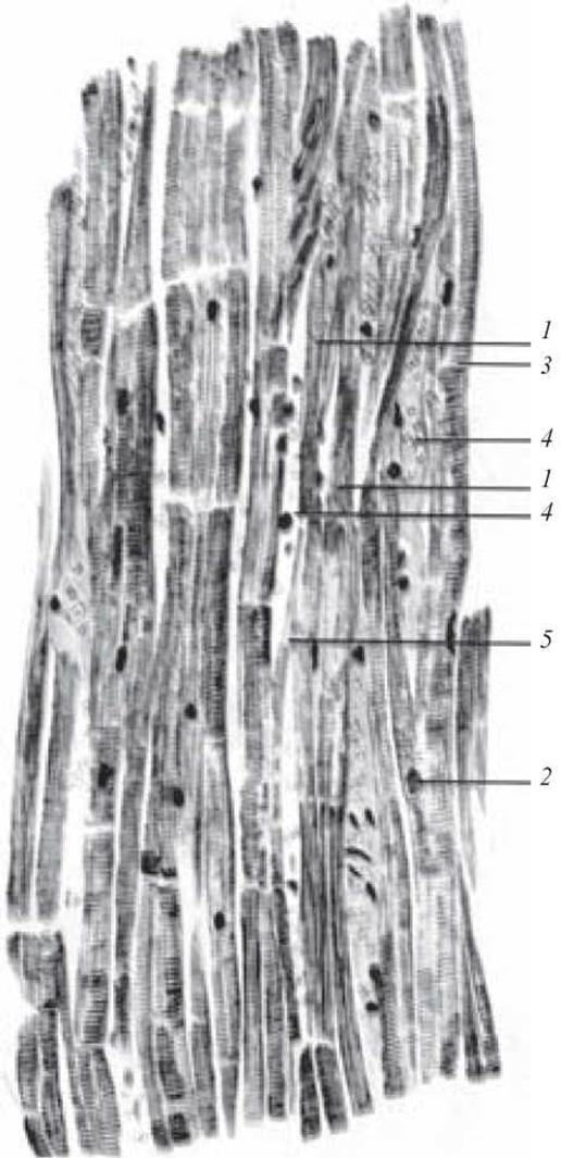
Fig. 95. Cardiac striated muscle tissue. Iron hematoxylin stain. x 320:
1 — cardiomyocyte; 2 — cardiomyocyte nucleus; 3 — intercalated disc; 4 — connective tissue layers containing blood vessels; 5 — anastomosis between two muscle fibers
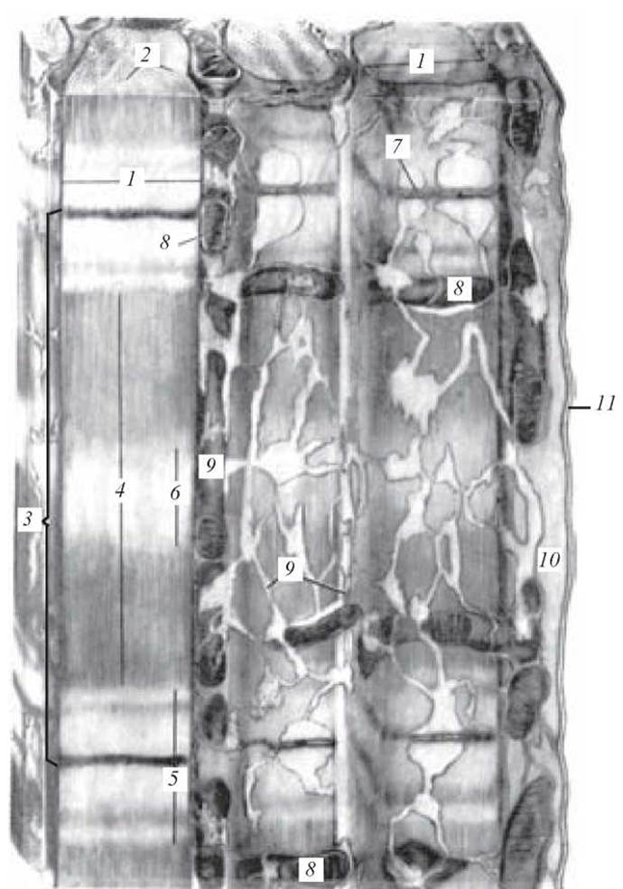
Fig. 96. Ultramicroscopic structure of a striated muscle fiber. Electron micrograph:
1 — striated myofibrils; 2 — myoprotoprism/myofilaments; 3 — sarcomere; 4 — dark anisotropic disc (A band); 5 — light isotropic disc (I band); 6 — M line; 7 — Z line/disc (T line); 8 — mitochondria-sarcosomes; 9 — sarcoplasmic reticulum of the transverse muscle fiber; 10 — sarcoplasm; 11 — sarcolemma (after Bennett)
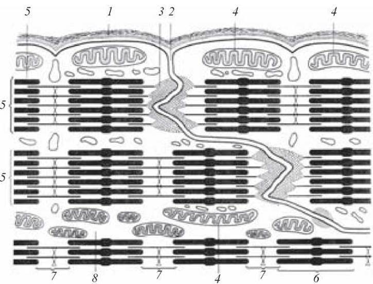
Fig. 97. Intercalated discs between cardiac muscle cells (diagram):
1 — muscle fiber sheaths; 2 — intercalated disc; 3 — endings of myofilaments on the cytolemma; 4 — mitochondria; 5 — myofilaments; 6 — A disc (anisotropic disc); 7 — I disc (isotropic disc); 8 — sarcoplasm
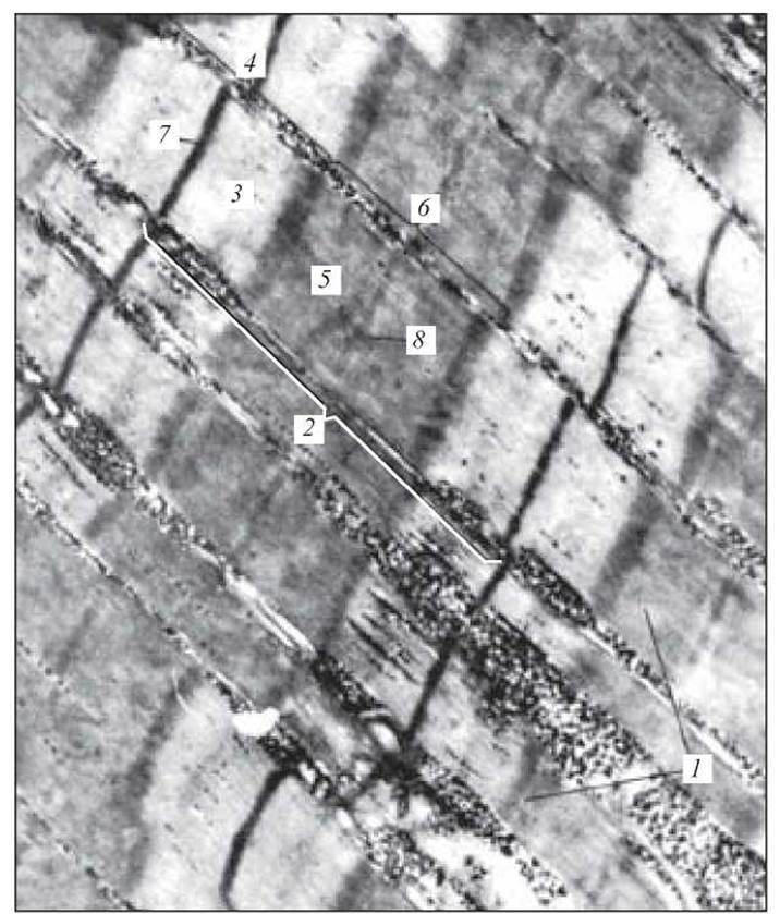
Fig. 98. Striated muscle fiber from an axolotl Skeletal Muscle. Electron micrograph. x 27,000:
1 — striated myofibrils; 2 — sarcomere; 3 — 1/2 of I disc; 4 — I disc; 5 — 1/2 of A disc; 6 — A disc; 7 — Z line (T line); 8 — M line (after V. P. Hilyov)
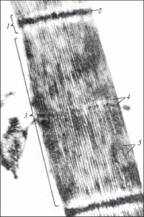
Fig. 99. Striated myofibril from a strongly contracted mouse skeletal muscle fiber. Electron micrograph. x 43,200:
1 — I disc; 2 — Z line (T line); 3 — A disc; 4 — M line; 5 — myofilaments (after Szent-Györgyi)
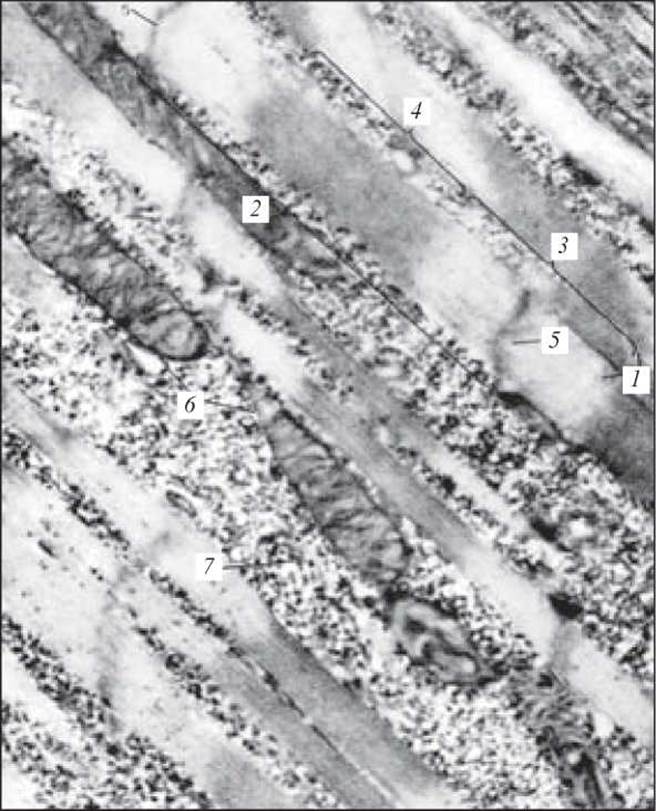
Fig. 100. Striated myofibrils from a relaxed axolotl skeletal muscle fiber. Electron micrograph. x 27,000: 1 — striated myofibrils; 2 — sarcomere; 3 — I disc; 4 — A disc; 5 — Z line (T line); 6 — mitochondria-sarcosomes; 7 — Ribosomes (after V. P. Hilyov)
1. General morphofunctional characteristics of Muscle Tissues.
2. Classification of muscle tissues.
3. Sources of muscle tissue development.
4. Structural and functional features of smooth muscle myocytes.
5. Skeletal muscle tissue: localization and functional features.
6. The muscle fiber as the structural unit of skeletal muscle tissue.
7. STRUCTURE OF THE myofibril. The sarcomere.
8. STRUCTURE AND Functions of myosatellite cells.
9. Sarcoplasmic reticulum. T-systems.
10. Histophysiology of contraction.
11. Types of muscle fibers.
12. Structure of the muscle as an organ.
13. Morphofunctional characteristics of cardiac muscle. Contractile cardiomyocyte.
14. Structural Features of atrial myocytes.
15. Conducting cardiomyocytes. Types and structural features.
16. Mechanisms of regeneration in various types of muscle tissue.
Situational problems
1. A preparation of muscle tissue shows muscle fibers containing numerous peripherally located nuclei. Cross-striations are visible. What type of muscle tissue is this?
2. Under a Microscope, a preparation of muscle tissue reveals spindle-shaped cells with an elongated, rod-shaped nucleus in the center of each cell. What type of muscle tissue is this?
3. Preparations of skeletal and cardiac muscle tissues are provided. By what structural features can they be distinguished?
Sample exam questions
1. General morphofunctional characteristics of muscle tissues. Histogenesis. Classification.
2. Morphofunctional characteristics of Smooth muscle tissue.
3. Morphofunctional characteristics of Striated skeletal muscle tissue.
4. Morphofunctional characteristics of cardiac muscle tissue.
Last update: 10/08/2026
Editorial and Educational Adaptation: This material has been compiled based on the primary/original source text. The project team performed an editorial review, corrected technical inaccuracies, structured sections, and adapted the content for an educational format.
What was processed:
- elimination of formatting defects (OCR errors, structural breaks, corrupted characters);
- editorial organization of content;
- standardization of terminology in accordance with academic sources;
- verification of factual statements against the original source text.
All mentions of the author, publication year, and origin of the primary text have been preserved in accordance with the source.