Cytology, General Histology and Embryology - V. K. Napkhanyuk 2002
Tissues
Muscle Tissues
Smooth Muscle Tissues
Muscle Tissues (textus muscularis) represent a group of specialized tissues that vary in origin and Structure, yet share a common functional feature: the ability of their structural elements to contract, which is accompanied by A change in Membrane Potential.
All of these groups are characterized by the elongated shape of their structural components and specialized Organelles known as myofibrils. Myofibrils can be unstriated (smooth) or striated (cross-striated). Accordingly, one distinguishes between unstriated and Striated Muscle tissue. Based on their origin, muscle tissues are subdivided into five histogenetic types (Fig. 92).
Unstriated (smooth) muscle tissue (textus muscularis nonstriatus) is classified into three types depending on its origin.
Cytology/cytology/32.html">Smooth muscle tissue of mesenchymal origin is a tissue whose stem Cells and progenitor cells are presumably related to Connective Tissue fibroblast precursors and reside, in a determined state, within the mesenchyme. Much like fibroblasts, they synthesize glycosaminoglycans and Collagen molecules, which subsequently assemble extracellularly to form the basement membrane matrix and fibers.
The Structural and functional unit of smooth muscle tissue is the smooth myocyte (myocytus nonstriatus). This is a spindle-shaped Cell ranging from 20 to 500 µm in length and 2 to 20 µm in diameter. In the Uterus, endocardium, aorta, and Urinary Bladder, branched myocytes can be found.
Class="center">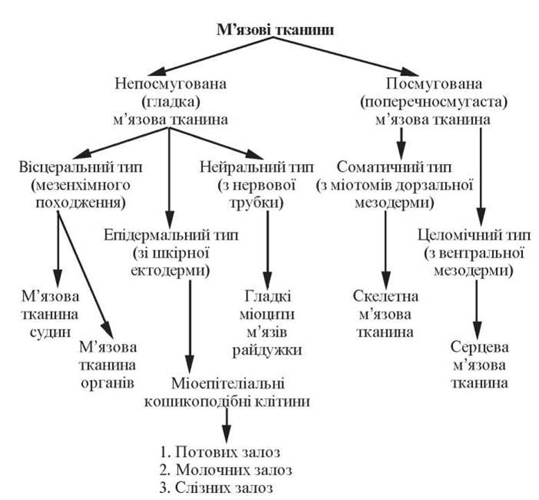
Fig. 92. Classification of muscle tissues
During contraction, a myocyte can bend or even twist.
The Cytoplasm of a smooth myocyte forms numerous invaginations, pinocytotic vesicles, and caveolae, through which Calcium Ions enter the cytoplasm. General-purpose organelles are located near the nuclear poles. The Golgi apparatus, Endoplasmic reticulum, and especially the granular reticulum are poorly developed, indicating reduced synthetic activity. Free Ribosomes, lipid and carbohydrate inclusions, as well as pigment granules are present.
Myofilaments are subdivided into thin Actin filaments (myofilamentum tenue) and thick Myosin filaments (myofilamentum crassum).
Actin myofilaments lie longitudinally or at an angle to the long axis of The Cell in the cytoplasm, forming a three-dimensional network. In addition to the protein actin, they contain Tropomyosin and caldesmon.
Myosin myofilaments are located predominantly longitudinally in the cytoplasm and are thicker than actin filaments. Contraction involves the sliding of Actin and myosin filaments relative to each other. The phosphorylation of myosin, which depends on the concentration of calcium ions, plays a significant role in the contraction mechanism of smooth myocytes.
Each myocyte is surrounded by a basement membrane containing fenestrations; in the region of these openings, Gap Junctions (nexus) are formed between adjacent myocytes. Reticular, elastic, and fine collagen fibers form a network around the muscle cells called the endomysium, which connects neighboring myocytes. Muscle groups of 10–12 myocytes unite into a muscle layer, between which lies loose connective tissue containing Blood Vessels and nerves.
Smooth muscle tissue of epidermal origin is a tissue whose cells develop from the surface ectoderm. They have a stellate or basket-like shape and embrace the terminal portions and small excretory ducts of glands with their processes. The processes of these cells contain a contractile apparatus structured similarly to that of unstriated myocytes of mesenchymal origin. The Nucleus and organelles are located in the central part of the cell.
These cells are localized in sweat, mammary, salivary, and lacrimal glands. Differentiating simultaneously with their secretory epithelial cells from common precursors, myoepithelial cells lie directly adjacent to the epithelial cells in such a way that a thin shared basement membrane separates them from the connective tissue.
By contracting, the processes of these cells facilitate the expulsion of glandular secretions.
Myocytes of neural-origin smooth muscle tissue develop from neural rudiment cells within the wall of the optic cup. Structurally, these are unstriated myocytes with a corresponding contractile apparatus. They are constituents of the iris Muscles of the eye, which dilate and constrict the pupil.
Smooth muscle tissues within Organs
Within organs, smooth unstriated myocytes are aggregated into bundles separated by thin layers of loose Fibrous connective tissue (perimysium). The totality of these bundles forms a muscle, which is surrounded by thicker layers of fibrous connective tissue (epimysium). These layers carry Blood Vessels supplying nutrients to the smooth muscle, as well as nerve fibers terminating among the myocytes.
Smooth muscles are present in the walls of many Internal Organs, forming layers and tunics within them (respiratory organs, gastrointestinal tract, reproductive organs, blood vessels).
Slides for study
Slide 29. Smooth muscle tissue of the urinary bladder (Fig. 93).
Low magnification. Under this magnification, locate the muscular tunic of the organ.
High magnification. Smooth myocytes, which appear spindle-shaped in longitudinal section, are clearly visible. The cytoplasm is oxyphilic. A rod-shaped purple nucleus is located in the center of the cell. Layers of loose fibrous connective tissue are visible between the longitudinal and circular muscle layers. Make a drawing of the slide.
Label on the drawing: 1) smooth muscle cells in longitudinal section; 2) smooth muscle cells in transverse section;
3) connective tissue layers with blood vessels;
4) nucleus of a smooth muscle cell.
Striated Muscle Tissues
Striated (skeletal) muscle tissue (textus muscularis striatus) is a tissue whose structural elements are characterized by the presence of striated myofibrils.
Depending on its origin, structural features, and functional purpose, striated muscle tissue is divided into two types: skeletal and cardiac.
Striated Skeletal Muscle Tissue
Striated skeletal muscle tissue (textus muscularis striatus skeletalis) develops from the myotome Cells of the dorsal mesoderm. Its structural and functional unit is the muscle fiber, which is a multinucleated syncytium (symplast). This tissue contains specialized cells known as myosatellite cells, which serve as cambial elements. They are located between the Plasmalemma of the muscle fiber and its basal lamina.
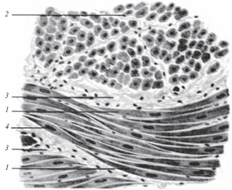
Fig. 93. Smooth muscle tissue of the urinary bladder. Hematoxylin and eosin staining, ×400:
1 — smooth muscle cells in longitudinal section; 2 — smooth muscle cells in transverse section; 3 — connective tissue layers with blood vessels; 4 — nucleus of a smooth muscle cell
The muscle fiber (myofibrillae), formed by the myosymplast and myosatellite cells, is cylindrical in shape with rounded, oblique, or jagged ends. It ranges from 9-150 µm in diameter and can reach a length equal to that of the entire muscle.
Externally, the muscle fiber is covered by the sarcolemma, which consists of a basal lamina intertwined with reticular and fine collagen fibers of the surrounding connective tissue, and the plasmalemma of the myosymplast, forming its inner layer.
The plasmalemma covers the myosymplast externally, conducting the Action Potential that spreads to the membrane of the T-tubules.
The myosymplast contains A large number of nuclei (up to several tens of thousands) located beneath the plasmalemma; these nuclei are elongated in shape and contain nucleoli along with a small amount of heterochromatin.
The cytoplasm of the symplast (sarcoplasm) contains three groups of organized structures:
— general-purpose organelles;
— inclusions — lipid, pigment, and carbohydrate;
— special-purpose organelles — myofibrils.
General-purpose organelles are represented by the Golgi
apparatus, areas of agranular endoplasmic reticulum, and Mitochondria located near the poles of the nuclei.
Inclusions are predominantly represented by Glycogen, Lipids, and Myoglobin.
Special-purpose organelles (myofibrils) run parallel to the length of the muscle fiber; their length matches that of the fiber, and their thickness is 1-2 µm.
The myofibrils of striated muscle fibers consist of thick myofilaments, which contain the protein myosin, and thin myofilaments, which contain the protein actin.
Thick myofilaments form the anisotropic disc or A band (discus anizotropicus, stria A), which exhibits birefringence.
Thin myofilaments form the isotropic disc or I band (discus izotropicus, stria I), which does not exhibit birefringence. The Z line, or telophragma (telophragma, linea Z), passes through the middle of the I disc, while the H band, within which the M line, or mesophragma (mesophragma, linea M), is located, runs through the center of the A disc.
The segment between two Z lines is called a sarcomere (myomere). The sarcomere serves as the structural and functional unit of a myofibril, with a length of 2-3 nm.
Myosatellitocytes (myosatellitocyti) are mononuclear cells that lie adjacent to The surface of the symplast in such a way that their cytolemmas are in contact.
A considerable number of myosatellitocytes are associated with a single symplast.
The nucleus of a myosatellitocyte is smaller than that of the symplast and has a more rounded shape.
The cytoplasm contains evenly distributed mitochondria and endoplasmic reticulum. The Golgi apparatus and cell center are located near the nucleus, while specialized organelles are absent.
Myosatellitocytes function as cambial elements of skeletal muscle tissue.
MOLECULAR MECHANISMS OF muscle fiber contraction
In the relaxed state of a myocyte, calcium ions accumulate within the tubules of its agranular endoplasmic reticulum. The action potential, propagating along the cytolemma and T-tubules, triggers the release of calcium ions, which reach the myofibrils and interact with the regulatory Proteins troponin and tropomyosin. As a result, tropomyosin molecules shift and expose the actin sites, enabling them to interact with myosin and slide toward each other. This inward movement brings the actin and myosin filaments, as well as the telophragms, closer together; since they are attached to the cytolemma, the entire fiber shortens.
Energy required for Muscle contraction is utilized in the form of ATP. Myosin protein heads capable of binding ATP molecules also exhibit ATPase activity (i.e., The ability to hydrolyze ATP). The actin-myosin-ATP complex is unstable and rapidly dissociates into actin and myosin-ATP. Cross-bridges presumably detach at the moment when the myosin HEAD binds ATP molecules. This cycle repeats at a rate of 50-100 times per second. Following death, ATP synthesis ceases, and actin fails to detach from myosin, leaving the Actomyosin complex stable for several hours. Consequently, the filament remains fixed in a bound state—a process known as rigor mortis.
Types of muscle fibers
At the light-optical level, several types of muscle fibers are distinguished: red muscle fibers (type I); white muscle fibers (type II); and transitional forms.
Each of these fiber types exhibits distinct ultrastructural and metabolic features.
Ultrastructural features are manifested by the degree of Development of the sarcoplasmic reticulum, the length of contact between T-tubules and this reticulum, The structure of telophragms, the arrangement of actin and myosin filaments within myofibrils, the number of mitochondria, and varying contents of myoglobin, glycogen, and lipid inclusions.
Metabolic features are evident in the varying activity of Enzymes such as adenosine triphosphatase and succinate dehydrogenase.
For instance, type I fibers contain slow-type adenosine triphosphatase and exhibit high succinate dehydrogenase activity, along with a high content of myoglobin and glycogen. Type II fibers contain fast-type ATPase, lower SDH activity, a higher amount of glycogen inclusions, and less myoglobin.
Muscle as an organ
Muscle fibers, bound together by connective tissue, form an organ known as a muscle (musculus).
Individual muscle fibers are separated by connective tissue layers called the endomysium.
Reticular and collagen fibers of the endomysium intertwine with the fibers of the sarcolemma. On each muscle fiber, the plasmalemma forms narrow, deep invaginations into which reticular and collagen fibers penetrate. They pierce the basal lamina and form a loop that attaches to the plasmalemma at sites where actin filaments of the sarcomeres contact it from the inside. Extending beyond the basal lamina, reticular fibers intertwine with collagen fibers, which continue into the tendon.
Each muscle fiber possesses its own innervation and is surrounded by a network of blood capillaries.
The complex formed by a fiber and its surrounding loose connective tissue elements constitutes the structural and functional unit of skeletal muscle (the myon).
Muscle fibers of various types are grouped in specific combinations into bundles, separated by thicker layers of loose fibrous connective tissue known as the perimysium. The perimysium also contains elastic fibers. The connective tissue surrounding the entire muscle is called the epimysium.
Slides for study
Slide 30. Striated muscle tissue of the Tongue (Fig. 94).
Low magnification. At this magnification, locate bundles of striated muscle fibers cut in various directions; they are basophilic, multinucleated with dark purple nuclei lying beneath the sarcolemma (in both longitudinal and cross sections).
High magnification. The longitudinal section clearly shows the alternation of dark and light basophilic bands. Intercellular layers of loose connective tissue (endomysium) are visible between the muscle fibers.
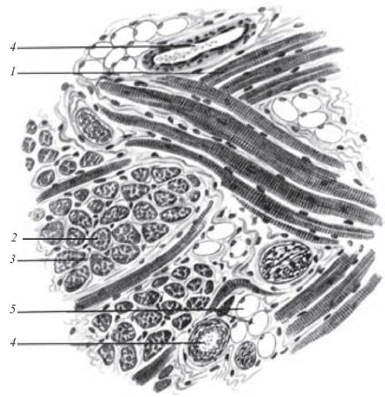
Fig. 94. Striated muscle tissue of the tongue. Stained with iron hematoxylin. x 400:
1 — muscle fibers in longitudinal section; 2 — muscle fibers in cross section; 3 — endomysium; 4 — blood vessels; 5 — fat cells
Make a drawing of the slide. Indicate the following on the drawing: 1) muscle fibers in longitudinal section; 2) muscle fibers in cross section; 3) endomysium; 4) blood vessels; 5) fat cells.
Last update: 10/08/2026
Editorial and Educational Adaptation: This material has been compiled based on the primary/original source text. The project team performed an editorial review, corrected technical inaccuracies, structured sections, and adapted the content for an educational format.
What was processed:
- elimination of formatting defects (OCR errors, structural breaks, corrupted characters);
- editorial organization of content;
- standardization of terminology in accordance with academic sources;
- verification of factual statements against the original source text.
All mentions of the author, publication year, and origin of the primary text have been preserved in accordance with the source.