Protein Chemistry - Part 2 - Selected Topics in Special Protein Chemistry - Ashmarin I. P. 1968
Contractile proteins of muscle tissue
Actomyosin
An essential property of both Myosin and Actin is their ability to interact with each other to form a stable complex known as actomyosin. When solutions of myosin and actin are mixed in 0.35–1.5 M potassium chloride, the viscosity increases sharply to values significantly exceeding the sum of the viscosities of the individual components. This viscosity surge is particularly pronounced within the concentration range of 0.35–0.5 M potassium chloride (Fig. 9). At lower salt concentrations, the precipitation zone expands, covering an interval from 0 to 0.25–0.3 M potassium chloride. The linear proportionality between viscosity and protein concentration, typical of myosin, is replaced by a curvilinear relationship—viscosity rises faster than concentration. Thixotropy also appears, and Flow Birefringence increases. All of this points to The formation of large, elongated molecules prone to interacting with one another.
The most stable complexes of myosin and actin are formed at quite specific quantitative ratios of these Proteins—from 2.5 : 1 to 3.7 : 1 by weight. This corresponds to approximately two to three actin monomers per myosin molecule, which is roughly close to their 3:1 ratio in the myofibril.
Unlike myosin, actomyosin gel has The ability to drastically alter its properties upon interacting with ATP. Even upon adding very low concentrations of ATP (0.5–1.0∙10-4 M) to a suspension of actomyosin in 0.02–0.15 M potassium chloride, a rapid and distinct shrinkage and compaction of the suspension particles is observed. Translucent, rather voluminous, and slowly settling in the initial suspension, they become opaque, sharply contoured, decrease in size, and settle rapidly after the Introduction of ATP. This phenomenon is often referred to as actomyosin superprecipitation. The particles of the actomyosin gel undergo significant dehydration in the process. Whereas before The addition of ATP they contain 98% Water or even more, after superprecipitation they retain no more than 50% water. It is easy to calculate that superprecipitation squeezes out at least 97% of the water from the gel.
Even such comparatively superficial observations of actomyosin gel particles naturally draw analogies to the phenomena of Muscle contraction. Even more associations of this kind arise when observing actomyosin threads obtained by extruding concentrated actomyosin solutions (1–2% by weight) through a capillary (d ≈ 0.2 mm) into a 0.05–0.15 M potassium chloride solution. Actomyosin molecules in such threads are arranged less randomly than in ordinary suspension particles; in this case, orientation along the thread axis predominates. If ATP is added to the solution surrounding the thread, its contraction to a third of its initial length can be observed within a few dozen seconds.
However, despite the greater similarity between the contraction of a thread and a muscle compared to a suspension, A number of significant differences can also be noted. The primary one is that it is very difficult to observe the ability of such a thread to perform mechanical work. Moreover, actomyosin threads with a small weight suspended from them not only fail to contract under METABOLISM/18.html">The Influence of ATP, but, on the contrary, stretch. Only in experiments where metallic powder was applied to one end of the thread was it possible to reveal some mechanical performance. Obviously, the inadequacy of such actomyosin threads as a molecular model of the elementary contraction mechanism is due to the insufficient orientation of the actomyosin molecules along the thread axis. To enhance their orientation, a technique for producing so-called film threads has recently been developed. They are prepared from thin actomyosin films spread On the surface of a 0.05 M potassium chloride solution. A thread is formed by compressing the film between two barriers. Upon the addition of ATP, such a thread proves capable of performing quite significant mechanical work, namely lifting a 20 mg weight to a height of up to 50% of the initial thread length. It is also telling that actin-free myosin threads prepared by the same method are incapable of such contraction with weight-lifting.
Class="center">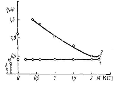
Fig. 9. Dependence of actomyosin viscosity on potassium chloride concentration (Szent-Györgyi, 1947).
1 — in the presence of ATP, 2 — without ATP.
In all experiments of this kind, the presence of 0.001–0.002 M magnesium chloride in the solution promotes the processes of superprecipitation and contraction. Calcium Ions in this case also act as antagonists to magnesium ions, suppressing these processes. Note that The Effect of magnesium and calcium ions on actomyosin contraction is the reverse of their action on the ATPase activity of myosin. As indicated earlier, the latter is enhanced by calcium ions and inhibited by magnesium ions. However, it should be especially noted that the relationship between the enzymatic activity of myosin and the effect of magnesium undergoes important changes upon complex formation with actin. Thus, in the presence of low potassium chloride concentrations (0.01–0.03 M), magnesium ions not only do not suppress but even enhance the ATPase activity of actomyosin. Admittedly, at higher potassium chloride concentrations (0.1 M), the inhibitory effect of magnesium ions reappears. In all other respects, the enzymatic activity of actomyosin and myosin has no substantial differences.
Actomyosin reacts quite uniquely to the addition of ATP not only in gels, but also in its solutions in 0.35–2 M potassium chloride. In the latter case, a rapid drop in solution viscosity is observed, down to values close to the total viscosity of the myosin and actin that make up the given actomyosin preparation. Further, as ATP is cleaved by myosin, the viscosity gradually returns to its initial level. This phenomenon is apparently explained by the dissociation of actomyosin into myosin and actin, which occurs As a result of ATP binding to the same myosin groups required for interaction with actin, which appear to be specialized SH-groups.
The superprecipitation of actomyosin gel Suspensions, the contraction of actomyosin threads, and The phenomenon of decreased viscosity in actomyosin solutions upon the addition of ATP are Characteristic Properties of actomyosin.
The intimate mechanism of actomyosin interaction with ATP leading to the effects described above is currently a realm of hypotheses, sometimes contradictory. Researchers' attention has naturally been drawn to the question of whether the configuration and packing of actomyosin polypeptide chains change during superprecipitation and contraction.
The attempt to explain these phenomena by the transition of an extended polypeptide chain—the β-Structure—into a helical α-structure seemed very attractive. However, X-Ray Diffraction Analysis of the changes occurring in actomyosin gels under the action of ATP led to the opposite Conclusion. Instead of the 5.1 Å meridional reflection characteristic of the α-structure, a 4.65 Å meridional reflection appears, which is characteristic of super-contracted keratin (see Chapter I). Apparently, a transition from the α-structure to the β-structure takes place, with the latter additionally forming transverse folds that ultimately cause particle shortening. The sections of The polypeptide chains within the folds are linked by Hydrogen Bonds. Naturally, such transverse folds are formed specifically by β- and not α-structures, since the latter possess greater resistance to bending, and most of the hydrogen bonds in them close not between distant sections of the polypeptide chains, but between closely neighboring components. A schematic diagram of such changes is presented in Fig. 10.
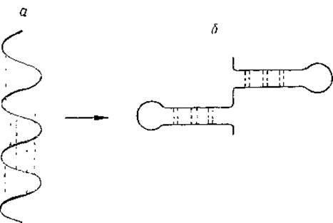
Fig. 10. Diagram of presumed polypeptide chain configuration changes during actomyosin contraction.
a — α-structure, b — variant of the transverse β-structure.
What are the factors that can induce such changes in polypeptide chain configuration? Particular attention among researchers is drawn to The concepts of charge alteration on various sections of the chain under the influence of ATP. For instance, it has been suggested that the initial extended state of actomyosin is maintained by the mutual repulsion of positively charged side radicals located along the polypeptide chains. The introduced ATP, by contrast, is negatively charged and can neutralize these charges, triggering the transition of the chain into a folded conformation. Enzymatic Cleavage of ATP releases the positively charged groups once again, and the chain returns to its initial state.
In actomyosin molecules at physiological concentrations of hydrogen, potassium, magnesium, and calcium ions, positively charged groups do indeed predominate. However, this hypothesis implies that the energy liberated during ATP breakdown is utilized to stretch the molecule, i.e., at the stage analogous to muscle relaxation. Contraction, in turn, must be powered by the energy preliminarily accumulated within the stretched molecule. There is a series of indications that ATP breakdown is actually directly linked to the contraction process rather than relaxation. In particular, actomyosin that has contracted under the influence of low ATP concentrations remains in this state even after the complete Cleavage of the latter. If the hypothesis cited above were correct, one would expect a rapid spontaneous Swelling of the gel and elongation of the threads. Furthermore, many factors that suppress ATPase activity (such as a drop in Temperature, certain SH-Reagents, etc.) also inhibit the contraction process. True, the influence of calcium and magnesium ions does not entirely fit this proposition, although it does not refute it either, since under conditions particularly favorable for contraction—at low potassium chloride concentrations—magnesium ions, as already mentioned, enhance the ATPase activity of actomyosin.
When considering the relationship between the ATPase activity of myosin and actomyosin contraction, it must be emphasized that the transformation of the energy bound in the high-energy bonds of ATP into contraction energy is a very complex, multi-stage process. The cleavage of ATP into ADP and inorganic phosphate is merely the final, cumulative result. There is considerable evidence, gathered thus far through various indirect Methods, indicating that the initial outcome of ATP interaction with myosin consists in The transfer of an orthophosphate residue to one of the Functional groups of the protein, once again forming a new high-energy bond that exists for a very brief interval. Myosin is thus modified, forming what is known as phosphorylmyosin. The chemical energy confined within this bond is expended to alter the configuration of the actomyosin polypeptide chains or to shift the relative positions of the contractile complex components. Crucially, this energy migration occurs within a single molecule. Simultaneously, the release of inorganic phosphate takes place.
The remarkable transformations of actomyosin during its reaction with ATP, characterized above, have served as the foundation for a number of hypotheses regarding the mechanisms of muscle contraction. However, it must be emphasized that despite the temptation of direct analogies between muscle contraction and the changes in actomyosin induced by ATP, extreme caution is necessary here. Undoubtedly, the interaction of actomyosin with ATP is the defining, key reaction of muscle contraction. Yet between this reaction and muscle contraction as a whole lies a very complex mechanism, the concepts of which are examined in the final section of this chapter.
Last update: 06/08/2026
Editorial and Educational Adaptation: This material has been compiled based on the primary/original source text. The project team performed an editorial review, corrected technical inaccuracies, structured sections, and adapted the content for an educational format.
What was processed:
- elimination of formatting defects (OCR errors, structural breaks, corrupted characters);
- editorial organization of content;
- standardization of terminology in accordance with academic sources;
- verification of factual statements against the original source text.
All mentions of the author, publication year, and origin of the primary text have been preserved in accordance with the source.