Cytology, General Histology and Embryology - V. K. Napkhanyuk 2002
Tissues
Skeletal Tissues
Osteohistogenesis
Osteohistogenesis is The process of initiation, development, formation, and regeneration of Bone tissue.
There are two types of osteohistogenesis: embryonic and postembryonic.
During the Embryonic period, The Development of bone tissue can occur in two ways: direct or indirect osteohistogenesis.
Direct osteohistogenesis, also known as membranous osteohistogenesis (osteogenesis membranacea), develops directly from mesenchyme and is characteristic of coarse-fibered bone tissue formation in flat bones, such as the cranial vault bones. This process is observed primarily During the first month of intrauterine development and consists of the following stages.
The First stage is The formation of the osteogenic (skeletogenic) island. In the areas where the future bone will form, mesenchymal Cells proliferate and the osteogenic island becomes vascularized.
The Second Stage (osteoid stage) is characterized by the differentiation of cells within the osteogenic islands into osteoblasts, which begin to produce the organic matrix of bone tissue. As a result, they surround themselves with intercellular substance and transform into osteocytes located within lacunae. The proliferating fibers push the cells apart; however, the cells retain their processes and remain interconnected. The primary osteoid matrix located between the fibers thickens and becomes enriched with osteomucoid.
The Third Stage is the calcification (impregnation with salts) of the intercellular substance. Osteoblasts produce the enzyme alkaline phosphatase, which breaks down glycerophosphates found in peripheral Blood into sugar and phosphoric acid. Phosphoric acid reacts with calcium salts to initially form amorphous substances in the form of [Ca3(PO4)2], which subsequently give rise to hydroxyapatite crystals. The fibers grow outward from the ends of the bone model, forming long bone trabeculae, or beams. Afterward, the network of trabeculae branches out, and the spaces between them are filled with Connective Tissue containing Blood Vessels.
Indirect osteohistogenesis, or cartilaginous osteohistogenesis (osteogenesis cartilaginea), is the process of bone development replacing Cartilage.
In the second month of embryonic development, a cartilaginous precursor forms from the mesenchyme at the sites of future tubular bones, rapidly acquiring the shape of the future bone. This primordium consists of embryonic hyaline cartilage covered by perichondrium.
The replacement of cartilage tissue with bone tissue involves perichondral and endochondral ossification.
Perichondral ossification (ossificatio perichondrialis) begins when osteoblasts appear in the perichondrium of the mid-diaphysis, forming coarse-fibered bone tissue. This newly formed tissue surrounds the diaphysis of the cartilage model like a collar, thereby disrupting the Blood supply to the diaphyseal cartilage. Consequently, dystrophic changes appear in the cartilage of the diaphysis, cartilage cells undergo vacuolization, and their nuclei become condensed (pyknosis). Mineral deposits are detected between the cells.
The process of cartilage destruction is accelerated by The activity of specialized cells called osteoclasts, which break down the cartilage. Blood vessels grow into the resulting cavities.
Endochondral ossification (osseficatio endochondrialis) is the process of Bone Formation inside the cartilage model.
Osteoblasts form from the mesenchyme accompanied by blood vessels, initiating the construction of endochondral bone upon the trabeculae of uncalcified cartilage. Endochondral bone differs from perichondral bone by retaining remnants of uncalcified cartilage intercellular substance.
During the development of endochondral bone, osteoblasts gradually break it down, creating large cavities that merge to form the medullary cavity. The mesenchyme invading this space differentiates into the Bone Marrow stroma, which is then colonized by stem cells. Simultaneously, new trabeculae of bone tissue continue to grow along the periphery of the diaphysis from the periosteum. Expanding longitudinally toward the epiphyses and increasing in thickness, they form a dense layer of bone.
Ossification of the epiphyses. Cartilage tissue persists in the epiphyses for a long time, featuring several distinct zones:
— the zone of cartilage resorption (zona resorbeus), which lies directly adjacent to the diaphysis;
— the hypertrophic zone (zona hypertrophica), where chondrocytes destined for destruction swell and resemble vesicles;
— the proliferation zone (zona proliferativa), which serves as the future metaepiphyseal growth plate, where chondrocytes are arranged in vertical columns; and
— the reserve zone (zona reservata).
Subsequently, secondary centers of endochondral ossification appear in the epiphyses as well, although the cartilage proliferation zone persists in humans until approximately 20 years of age.
Growth, Regeneration, and Age-Related Changes of Bone Tissue
Bone tissue growth occurs exclusively through apposition—the deposition of newly formed bone tissue onto existing bone. Growth in bone thickness is driven by the periosteum through proliferation and the synthetic activity of osteoblasts in its deep osteogenic layer.
Longitudinal growth is ensured by The Cell proliferation within the zone of proliferation of the metaepiphyseal plate.
Physiological regeneration of bone tissue is accomplished through the continuous replacement of old bone lamellae with newly formed ones, and the creation of new osteons in place of destroyed ones. These opposing processes are driven by the coordinated activity of osteoclasts and osteoblasts.
Age-related changes in bone tissue involve the gradual loss of the inorganic matrix beginning after the age of twenty.
A characteristic feature is that in men, mineral loss occurs at a steady rate of approximately 0.4% of body mass annually.
In women, following the onset of menopause—likely due to estrogen deficiency in the body—demineralization processes accelerate, reaching 1–1.5% annually.
Slides for Study
Slide 27. Development of bone tissue from mesenchyme (Fig. 89).
Low magnification. Locate areas of coarse-fibered bone tissue (osteoid) stained homogeneously bright pink. The islets are surrounded by mesenchymal cells, which are stellate in shape with weakly basophilic Cytoplasm. Osteoblasts are located On the surface of the osteoid; these are polygonal cells with intensely basophilic cytoplasm. Deep within the osteoid lie osteocytes—cells with a distinct cytoplasmic rim. Osteoclasts are situated at the periphery of the osteoid and appear as large, multinucleated cells.
High magnification. A ruffled border is visible on the side of the osteoclast facing the bone. Blood vessels appear as cross and oblique sections of thin-walled tubules containing Blood Cells. Draw the slide.
Label the following on the drawing: 1) skeletogenic islet; 2) mesenchyme; 3) blood vessel; 4) bone trabecula: a) osteocyte, b) unmineralized ground substance (osteoid); 5) osteoblasts; 6) osteoclast.
Slide 28. Development of bone in place of cartilage (Fig. 90).
Low magnification. Find the diaphyseal zone of the cartilaginous model in the slide. In this zone, beneath the perichondrium, the perichondral bone collar (bone cuff) is visible. Its intercellular substance is homogeneously pink, while the osteoblasts and osteocyte nuclei are basophilic. In the central zone of the diaphysis, where endochondral ossification takes place, endochondral bone forms around blue and light-blue areas of unmineralized cartilage matrix. Accumulations of Cytology/practical/86.html">Red bone marrow cells (round-shaped cells, often with hyperchromatic nuclei) are visible within the bone cavity. At the border with the epiphysis is the zone of cartilage resorption, where unmineralized cartilage is degraded and replaced by bone tissue. Further along is the zone of hypertrophy, where chondrocytes appear as clear vesicles. Beyond this lies the zone of proliferation, where chondrocytes actively divide and arrange themselves linearly in coin-like stacks. Most of the epiphysis is occupied by the zone of unchanged hyaline cartilage. Draw the slide.
Label the following on the drawing: 1) epiphyseal hyaline cartilage; 2) perichondrium; 3) Column layer; 4) vesicular layer; 5) perichondral bone collar; 6) zone of calcified cartilage; 7) endochondral bone; 8) blood vessel; 9) primary osteon canal; 10) bone marrow; 11) periosteum.
Class="center">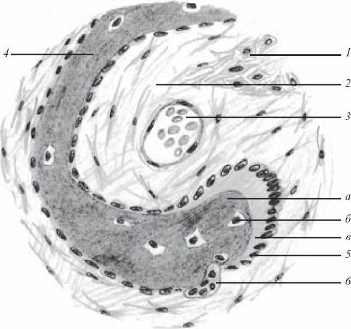
Fig. 89. Development of bone tissue from mesenchyme. Hematoxylin and eosin staining, ×400:
1 — skeletogenic islet; 2 — mesenchyme; 3 — blood vessel; 4 — bone trabecula (a — calcified intercellular substance; b — osteocyte; c — unmineralized ground substance, or osteoid); 5 — osteoblasts; 6 — osteoclast
For a better understanding of calcification processes, examine the electron micrograph (Fig. 91).
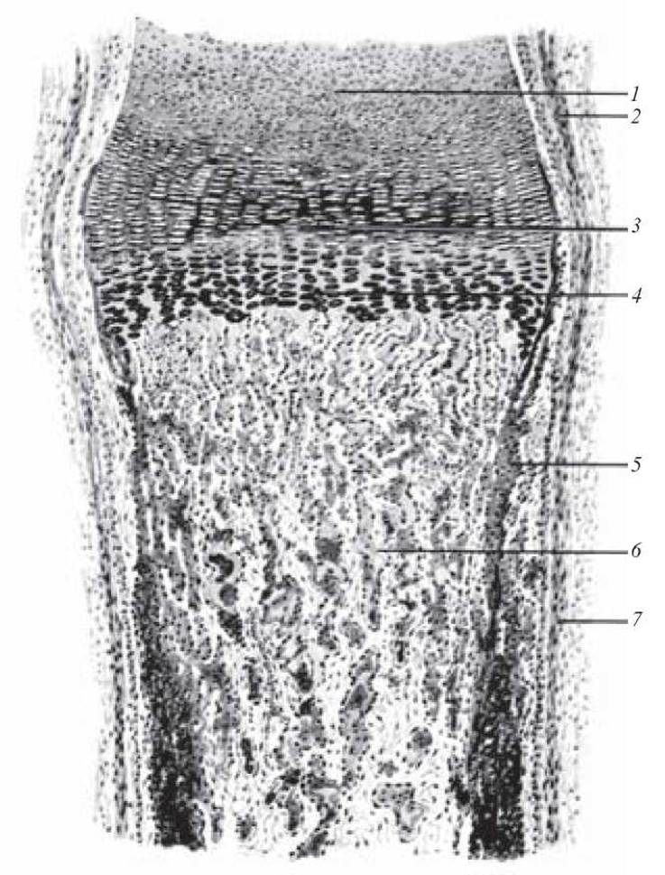
Fig. 90. Development of bone replacing cartilage. Hematoxylin and eosin staining, ×56:
1 — epiphyseal hyaline cartilage; 2 — perichondrium; 3 — layer of columnar cartilage; 4 — layer of vesicular cartilage; 5 — perichondral bone collar; 6 — endochondral bone; 7 — periosteum
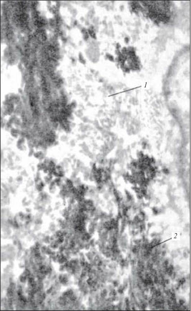
Fig. 91. Calcification of regenerating bone tissue. Electron micrograph of the calcification zone in regenerating bone, ×30,000:
1 — osein (Collagen) protofibrils; 2 — hydroxyapatite crystals in the intercellular substance (after L. N. Mikhailova)
Control Questions
1. General characteristics of osteohistogenesis.
2. Stages of bone tissue development directly from mesenchyme (direct osteohistogenesis).
3. Stages of bone tissue development replacing cartilage (indirect osteohistogenesis).
4. Bone tissue regeneration and its age-related changes.
Case studies
1. In a tubular bone, bone lamellae that do not form osteons are located between the osteons. What is THE ORIGIN OF these lamellae?
Sample exam questions
1. Characteristics of direct osteohistogenesis. Stages.
2. Development of bone tissue from cartilage.
Last update: 10/08/2026
Editorial and Educational Adaptation: This material has been compiled based on the primary/original source text. The project team performed an editorial review, corrected technical inaccuracies, structured sections, and adapted the content for an educational format.
What was processed:
- elimination of formatting defects (OCR errors, structural breaks, corrupted characters);
- editorial organization of content;
- standardization of terminology in accordance with academic sources;
- verification of factual statements against the original source text.
All mentions of the author, publication year, and origin of the primary text have been preserved in accordance with the source.