Biological Membranes - A. N. Ogurtsov 2012
Structure and Functions of Biomembranes
Passive Transmembrane Transport
Pores in the Lipid Bilayer
Cell/29.html">The Lipid Bilayer forms the foundation of any cell membrane. Its continuity determines the barrier and mechanical Properties of the cell. During cellular activity, this bilayer continuity can be disrupted, leading to the Formation of Structural defects known as trans-membrane hydrophilic pores. The formation of such pores affects all Functions of The Cell membrane related to its permeability.
Furthermore, lipid pores play an active role in the membrane mechanisms through which Cells respond to external stress factors.
Just like any real crystal, a phospholipid film may contain defects, which serve as focal points for structural rearrangements. While defects vary in type, we will examine only a single bilayer defect: the trans-membrane hydrophilic pore (Figure 57).
Class="center">
Figure 57 - Lipid Bilayer Membrane with lipid pores
In the lipid bilayer film of a cell membrane, pores emerge (barring purely mechanical damage) As a result of:
1) thermal fluctuations of the bilayer surface;
2) electrical breakdown;
3) film freezing;
4) the action of Surfactants;
5) osmotic pressure;
One of the most typical and well-studied Examples of biological membrane destabilization is erythrocyte hemolysis. This phenomenon begins with cell Swelling in a hypotonic medium driven by osmotic pressure. As the cell swells, the membrane stretches, leading to an increase in membrane tension.
At a certain threshold tension level, hydrophilic lipid pores appear. These pores are large enough to allow the escape of Hemoglobin molecules and low-molecular-weight substances. This results in a drop in the osmotic pressure gradient, which in turn reduces membrane tension, allowing the pores to reseal. Cytoskeletal Proteins help the erythrocyte maintain its shape, producing what is known as an erythrocyte ghost. The ghost retains its osmotic activity, making the destabilization process cyclical in nature.
In the absence of a Cytoskeleton, or if it is insufficiently developed, the mechanical strength of the cell is entirely determined by The Fate of the lipid pores. If a pore is smaller than a critical size, it reseals. Otherwise, the uncontrolled growth of the pore leads to membrane rupture.
Let us consider a model of a lipid pore (Figure 58). We assume that the lateral surface of the pore is shaped like a circular cylinder. Suppose the cylindrical side surface is curved with a radius of curvature of h/2. The pore radius is denoted as r. While the lipid bilayer as a whole is flat, the pore possesses two radii of curvature: h/2 and r.
Surface curvature at the lipid-Water interface gives rise to an additional pressure known as the Laplace pressure, which is expressed as

where a is the interfacial (surface) tension inside the pore, and r is the radius of curvature.
In the model under consideration, There are two such radii (h/2 and r) and, consequently, two pressures. One of them, p(h/2), promotes pore expansion, while the other, p(r), drives pore constriction. The ultimate fate of the pore depends on the balance between these two pressures. If p(h/2) > p(r), the pore will expand; if p(h/2) < p(r), the pore will reseal.

Figure 58 - Model of a hydrophilic lipid pore: h is the thickness of the lipid bilayer; h/2 is the radius of curvature of the wall inside the pore; r is the pore radius
Thus, two opposing forces act at the pore boundary: one is the edge line tension of the pore perimeter, which promotes pore growth, and the other is the surface tension of the bilayer, which causes pore constriction.
The energy of the pore rim is proportional to the first power of the radius and increases the total energy, whereas the surface tension energy is proportional to the square of the radius and decreases the total energy. As a result, the total energy E(r) is equal to

where the first term is determined by the pore rim energy with line tension y, and the second term is determined by the surface tension energy σ.
The shape of the curve in Figure 56 indicates the existence of unstable equilibrium at the maximum point with critical values of energy (E*) and radius (r*).

Figure 59 — Dependence of pore energy on its radius at various values of the Membrane Potential
At the equilibrium point (the extremum of the function)  and the equation turns into an identity: 0 = 2πy-2πσr*, from which the critical pore radius can be determined
Substituting r* into the equation E(r) = 2πrу-πr2σ, the height of the energy barrier will be equal to 
Given the instability of equilibrium, it can be stated that the appearance of pores with r > r* will be accompanied by membrane rupture as a result of unrestricted pore growth.
Conversely, at r < r*, the pore will reseal, and membrane stability will be preserved.
Such is the quantitative criterion for the stability of a lipid bilayer membrane.
Lipid pores under stress. Introduction/36.html">Biological Membranes are subjected to a high-intensity electric field generated by ion diffusion across the membrane and electrogenic ion pumps. Since the potential difference between the Cytoplasm and the extracellular environment reaches about 0,1 V, and the membrane thickness does not exceed 10 nm, the field strength is equal to 107 V/m.
Thus, the membrane is a more efficient electrical insulator than many liquid insulators used in engineering. In some cases, the membrane potential in a living cell can be higher and reach 0,2 V (freshwater Algae, Bacteria, energized Mitochondria).
In excitable nerve and Muscle cells, a brief membrane repolarization occurs with an increase in potential amplitude. However, breakdown of the cell membrane by its own membrane potential is unlikely.
At the same time, the increase in membrane potential resulting from the application of an external electric field can reach a value exceeding the threshold for electrical breakdown. In this case, structural defects of the transmembrane lipid pore type appear.
The developed technique of cell membrane electrical breakdown is called electroporation and is widely used in biotechnology.
In physics, electrical breakdown is understood as a sharp increase in electric current in an initially weakly conducting medium. In a living cell, this medium is the lipid bilayer.
In this case, the formula for the dependence of pore energy on its radius must be modified by introducing an additional term reflecting THE CONTRIBUTION OF the electric field,


εводы and εмембраны are the dielectric constants of water and the membrane, respectively; φ is the membrane potential; C0 is the capacitance per unit area of the defect-free membrane.
The dependence of pore energy on its radius for this case is shown in Figure 59, which presents a family of curves plotted According to the derived equation for various values of the membrane potential.
The higher the membrane potential, the lower the pore energy value and the more the curve maximum shifts toward the origin.
As the radius increases, the pore energy should, on the one hand, increase because the pore perimeter grows, and, on the other hand, simultaneously decrease in proportion to the increase in the surface tension of the membrane and the membrane potential. As a result (Figure 59), a curve with a maximum appears, which makes it possible to quantitatively estimate the critical parameters of the membrane: the critical pore radius and the height of the energy barrier.
Taking the field into account, the height of the energy barrier is equal to
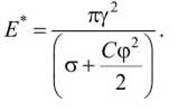
It can be seen that with an increase in the membrane potential and surface tension, the barrier height decreases.
The critical pore radius can be calculated using the formula

The critical radius also decreases as both a and φ increase. It follows from the formula that the dependence of critical pore parameters on the membrane potential becomes significant only when the electrical component vastly exceeds the surface tension.
Calculations show that for a lipid bilayer in the liquid-crystalline state, the critical membrane potential cannot be less than 0.23 V.
The stability of bilayer membranes is determined by the probability of critical-radius pores appearing. Naturally, any factor that lowers the energy barrier will increase this probability. Such factors include:
1) a decrease in the pore edge energy;
2) an increase in surface tension;
3) an increase in the membrane potential.
As seen in Figure 59, an increase in the breakdown voltage up to 1 V shifts the critical radius to values below 0.5 nm, which is close to the radii of natural Ion Channels in cell membranes. This implies that electrical breakdown is accompanied by The Emergence of a wide spectrum of lipid pores of various radii, including pores with dimensions matching ion-selective protein channels.
Currently, external electric field application is one of the primary Methods in modern biotechnology. Its Applications include enhancing membrane porosity (electroporation), introducing DNA (electrotransfection), releasing large molecules from cells (electropermeabilization), and Cell Fusion (electrofusion).
Thermal Processing of bilayer lipid membranes significantly affects the energetics of pore formation, as the phase transition is accompanied by a substantial change in surface tension. For instance, upon freezing hydrogenated egg lecithin, the surface tension a increased from 1.1∙1 (Г3 to 5.6 ∙ 10-3 N/m.
Taking this into account, the dependence of pore energy on its radius in liquid and solid membranes was calculated using the formula E(r) = 2πrу-πr2σ (Figure 60).

Figure 60 - Dependence of pore energy on its radius in the liquid-crystalline state (A) and gel state (B) of Membrane Lipids
As Figure 60 demonstrates, the critical pore radius in the gel state is significantly smaller compared to the liquid-crystalline state, not exceeding 2 nm in absolute value. The long-term Stability of the lipid bilayer in the gel state indicates that existing pores, as well as those arising during the phase transition (liquid-crystalline state) – (gel state), have dimensions of less than 2 nm.
A comparison of Figures 59 and 60 demonstrates the high efficiency of thermal processing of bilayer lipid membranes for obtaining lipid pores with parameters similar to those produced by electrical breakdown.
Indeed, freezing membrane lipids during a phase transition (which occurs at room Temperature for many saturated lipids) is equivalent to the electrical breakdown of the membrane by an external electric field with a voltage of 0.5 V. At the same time, electrical stimulation is clearly more convenient in terms of calibrating the intensity and duration of exposure.
In terms of permeability, lipid pores differ fundamentally from protein channels due to their origin and exceptional dynamicity.
While protein channels have strictly defined dimensions that remain constant throughout the cell's lifespan, the dimensions of lipid pores vary over a wide range during closure. However, this Variability is not limitless. If the pore radius is less than the critical value, the pore must traverse all intermediate radii during closure until it reaches its minimum size.
The question of whether lipid pores can close completely remains open. It is hypothesized that complete pore closure is prevented by strong Hydration forces that manifest when the walls of hydrophilic pores come into close proximity.
Unlike protein ion channels, lipid pores lack pronounced selectivity, which correlates with their relatively large initial dimensions.
Nevertheless, it is clear that during closure, lipid pores can reach arbitrarily small sizes, including those comparable to the dimensions of protein ion channels, which may lead to the redistribution of ionic currents across the membrane, for example, during excitation.
Furthermore, it is known that after the stress factor is removed, the bilayer lipid membrane can return to a low-conductivity state, implying that the pores have shrunk below the size required for hydrated ions to pass through.
Thus, hydrophilic lipid pores are versatile in the sense that they can be utilized by the cell to transport macromolecular substances, ions, and water molecules.
Research into lipid pore permeability currently follows two main directions: the first investigates the largest possible pores, while the second focuses, conversely, on lipid pores of the minimum radius.
The first case concerns electrotransfection, a method used to introduce DNA molecules into living cells or Liposomes for the transfer and intracellular delivery of foreign genetic material. It has been shown that a high-intensity external electric field facilitates the penetration of the giant DNA molecule through the membrane envelope.
The maximum size of the critical pore corresponds to the liquid-crystalline state of the lipid bilayer in the absence of an external electric field and equals 9 nm. Applying an external electric field with a strength of 100 kV/m reduces the critical pore radius to 1 nm within 0.2 s. Since the membranes remain undamaged in this process, the size of the lipid pores evidently does not fall below this lower limit.
The paradox lies in the fact that the effective diameter of the statistical DNA coil that must enter the particle reaches 2000 nm. Therefore, it is clear that the DNA molecule must cross the membrane as an unwound single strand. It is known that the end of the strand has a diameter of 2 nm and can enter the pore. However, free diffusion of the DNA strand within the pore is unlikely to be possible. Currently, The Mechanism of this process is not fully understood.
It is hypothesized, in particular, that:
1) the DNA molecule is capable of expanding the pore and thus slipping through the membrane;
2) DNA penetration may be facilitated by additional electrophoretic and electroosmotic forces, taking into account the net negative charge of the DNA molecule;
3) it is also possible that pores with the ends of the DNA molecule anchored within them act as anchors holding the molecule at a specific site near the vesicle on the membrane surface, and the transfer process itself represents a form of pinocytosis.
METABOLISM/35.html">Selection/41.html">Review Questions and Exercises
1. What Three types of processes are supported by the selective permeability of biological membranes?
2. What is the difference between Electrochemical Potential and chemical potential?
3. Write down the Teorell and Nernst-Planck equations.
4. List the Types of Passive transmembrane transport.
5. Which substances can diffuse across the lipid bilayer via passive (simple) diffusion?
6. How do small polar molecules (e.g., water molecules) diffuse across the biological membrane?
7. How do kinks transport water molecules across the membrane?
8. What Types of Membrane transport proteins are distinguished?
9. What are the three types of ion pumps?
10. What is the characteristic rate of Ion transport across the membrane by an ion pump?
11. What is the characteristic rate of molecule and ion Transport Across the membrane via a protein channel?
12. What are the three schemes of transmembrane transport mediated by carrier transporters?
13. Which membrane transporters are referred to as cotransporters?
14. What is the difference in function between primary active transporters (ion pumps) and secondary active transporters?
15. What is the characteristic rate of molecular transport across the membrane by secondary active transporters?
16. What are the Similarities and differences between simple diffusion and Facilitated Diffusion?
17. What are the Two Types of facilitated diffusion?
18. List the main differences between facilitated and simple diffusion.
19. Give examples of GLUT family glucose transporter proteins.
20. What two primary methods are used to study the function of transporter proteins?
21. How does the presence of lipid pores affect the stability of Biomembranes?
22. As a result of what external influences can lipid pores form in the membrane?
23. What is The Role of lipid pores in erythrocyte hemolysis?
24. List the Main parameters of the hydrophilic lipid pore model.
25. What is the quantitative criterion for the stability of a lipid bilayer membrane?
26. Write down the expression for the height of the energy barrier of a lipid pore.
27. Write down the expression for the critical pore radius.
28. Under what stress conditions can the size of lipid pores exceed the critical value?
29. What is meant by biomembrane electroporation?
30. How does the critical pore radius change during the transition from the liquid-crystalline to the gel state?
31. What is the fundamental difference in permeability between lipid pores and protein channels?
32. What is meant by biomembrane electrotransfection?
33. What are the three distinct types of mechanisms for DNA transfection across a biomembrane?
Last update: 13/08/2026
Editorial and Educational Adaptation: This material has been compiled based on the primary/original source text. The project team performed an editorial review, corrected technical inaccuracies, structured sections, and adapted the content for an educational format.
What was processed:
- elimination of formatting defects (OCR errors, structural breaks, corrupted characters);
- editorial organization of content;
- standardization of terminology in accordance with academic sources;
- verification of factual statements against the original source text.
All mentions of the author, publication year, and origin of the primary text have been preserved in accordance with the source.