MEDICAL BIOLOGY, ANATOMY, HUMAN PHYSIOLOGY AND PATHOLOGY - Ya.I. Fedoniuk 2010
BIOLOGY
CHAPTER 1. BIOLOGICAL FOUNDATIONS OF HUMAN VITAL ACTIVITY
1.2. CELL. TISSUES. CONCEPT OF ORGAN, ORGAN SYSTEM, ORGANISM
CONNECTIVE TISSUE
3.Connective Tissue with Special Properties
Connective tissue with special properties is subdivided into reticular, adipose, mucous, and pigment Tissues. This group of tissues is characterized by the predominant development of a specific type of Cells, as well as distinct Features of the Extracellular matrix.
Reticular tissue forms The system of hemopoietic Organs (Bone Marrow, Spleen, Lymph Nodes) and constitutes the microenvironment for Blood Cells. It is composed of reticular cells and reticular fibers that intertwine to form a meshwork (Fig. 1.14).
Class="center">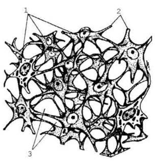
Fig. 1.14. Reticular tissue: 1 - reticular Cell; 2 - processes of reticular cells; 3 - reticular fibers.
Adipose tissue is represented by adipocytes (Fig. 1.15). This tissue has a lobular Structure—adipocytes are arranged in groups, between which it performs the function of lipid storage and METABOLISM. White and brown adipose tissue are distinguished. Cytology/practical/49.html">White adipose tissue is located subcutaneously, in the greater omentum, and in other fat depots. A significant portion of the fat from white adipose tissue is easily mobilized during starvation, utilized to cover the body's Energy Expenditure, and performs a Shock-absorbing function. Brown adipose tissue is found in newborns; its function is participation in thermoregulation. White adipose tissue is composed of adipocytes containing a single large lipid droplet that pushes The Nucleus to the periphery. Brown adipose tissue is composed of adipocytes containing numerous small fat inclusions in the Cytoplasm, and is located between the shoulder blades, on the back of the neck, and in the axillary fossae. The nucleus in these adipocytes occupies a central position.
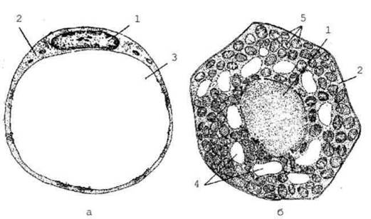
Fig. 1.15 Structure of adipocytes in white and brown adipose tissues: a - white adipose tissue adipocyte; b - brown adipose tissue adipocyte: 1 - nucleus; 2 - cytoplasm; 3 - large lipid droplet; 4 - fat inclusions; 5 - Mitochondria.
Pigment tissue consists of accumulations of pigment cells, known as melanocytes. It is found in areas of Skin around the nipples, in the vascular and iris Tunics of the eye, and in birthmarks (nevi). Due to its high melanin content, which can absorb ultraviolet rays, pigment cells play a protective role against the damaging effects of solar radiation.
Mucous tissue is a modified loose Fibrous connective tissue characterized by a marked quantitative predominance of the extracellular matrix, which is virtually devoid of fibrous structures. Mucous tissue has a gelatinous consistency and lacks Blood Vessels, Lymphatic vessels, and nerves. In the fetus, it is found within the umbilical cord, where it is known as "Wharton's jelly" and, due to its high elasticity, prevents the compression of umbilical vessels. In adults, a similar structure is found in the tissue forming the vitreous body of the Eyeball.
The source of origin for the cells of all Connective Tissues is embryonic connective tissue, or mesenchyme (Fig. 1.16). Mesenchymal cells have an irregular (stellate or spindle-shaped) Morphology with branched processes that form a network; the gelatinous extracellular material consists almost exclusively of ground substance with a minimal amount of reticular fibers. It is well developed in the fetus.
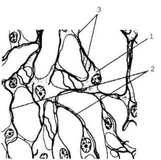
Fig. 1.16. Mesenchyme: 1 - mesenchymal cells; 2 - processes of mesenchymal cells; 3 - extracellular matrix.
Last update: 08/08/2026
Editorial and Educational Adaptation: This material has been compiled based on the primary/original source text. The project team performed an editorial review, corrected technical inaccuracies, structured sections, and adapted the content for an educational format.
What was processed:
- elimination of formatting defects (OCR errors, structural breaks, corrupted characters);
- editorial organization of content;
- standardization of terminology in accordance with academic sources;
- verification of factual statements against the original source text.
All mentions of the author, publication year, and origin of the primary text have been preserved in accordance with the source.