Human Histology - O.D. Lutsyk 2003
Systemic Histology
Endocrine System
The Endocrine System includes A number of glands and individual Cells of the body (Fig. 4.23), whose common and defining feature is The ability to produce BIOLOGICALLY ACTIVE SUBSTANCES called Hormones. The latter act as mediators in regulating the Functions of Organs and their systems. Several classes of hormones are distinguished: Peptides (oligopeptides, Polypeptides, glycopeptides), Amino Acid Derivatives (neuroamines), and Steroids (Sex Hormones, corticosteroids). All these biologically active substances are produced in extremely small quantities. Entering the Blood or Lymph, they bind specifically to receptors On the surface of cells within target organs. This mediates the distant action of the endocrine organs on the body. In addition to endocrine secretion proper, where hormones are released into the blood or lymph, there is also paracrine secretion, where a hormone binds to target cells immediately adjacent to the endocrine Cell, and autocrine secretion, in which a hormone released from one part of a cell binds to receptors on another part of the same cell.
Class="center">
Fig. 4.23. Major endocrine organs and hormone-producing cells of The Human Body
The Mechanism of hormone Action can be described as follows. A hormone molecule circulating in the bloodstream or lymph "finds" its receptor on The surface of the Plasmalemma, in the Cytoplasm, or in The Nucleus of a particular target cell. The stereochemical complementarity between the Active Site of the hormone molecule and the configuration of its receptor plays a decisive role in this highly specific recognition. The binding of a hormone to its receptor induces conformational (three-dimensional) Changes in the receptor molecule, which, in turn, affects cellular enzyme systems, particularly the adenylate cyclase system. The Mechanism of hormone action is discussed in greater detail in biochemistry and physiology textbooks. The Effect of hormones can manifest not only as stimulation but also as inhibition of The activity of cells and their systems.
The basis of interaction between individual Components of the endocrine system, as well as between endocrine cells and target cells, is the feedback principle (Fig. 4.24). The effect of a particular hormone on a target cell leads to an increase in its production of certain chemical substances. An increase in the concentration of these substances in the body's internal environment serves as a signal to inhibit the activity of the endocrine cell. Conversely, a decrease in hormone concentration in the blood or lymph stimulates the synthetic activity of the endocrine cell. The feedback principle also holds true for the inhibitory effect of a hormone on a target organ.
All Endocrine glands share several structural features. They lack excretory ducts. All of them have a well-developed vascular network, especially the microcirculatory bed. Cells of endocrine organs form characteristic clusters in the form of follicles (sacs) or trabeculae (cords) (Fig. 3.5). Specific granules containing the accumulated biologically active substance can usually be found in endocrine cells (hormone-producing cells). Unlike exocrine cells, endocrine cells accumulate secretory granules in the basal part of The Cell adjacent to the Vessels of the microcirculatory bed, into which the hormones are released.
The components of the body's endocrine system can be broadly divided into four groups. The first group—the central Organs of the endocrine system—includes the Hypothalamus, Pituitary Gland (hypophysis), and Pineal Gland (epiphysis). These organs are closely linked to the Central Nervous system and coordinate the activity of all other PARTS OF THE endocrine system. The second group—Peripheral endocrine organs—includes the thyroid, parathyroid, and Adrenal Glands. These are purely endocrine glands that exert a multi-vector influence on the body, enhancing or weakening metabolic processes. The third group includes organs that combine endocrine function with several others, such as the Pancreas, Gonads (Testis, Ovary), Kidneys, Placenta, etc. The human body also contains a large group of cells known as the Cytology/cytology/65.html">Diffuse endocrine system, which make up the fourth group of endocrine elements.
The hypothalamus (Fig. 4.23, 4.24) is a central neuroendocrine organ that integrates neural and humoral (hormonal) Regulation of the body's major visceral systems. It consists of about 30 pairs of nuclei (clusters of Nerve Cells) located at the Base of the Brain, in the floor of the Third ventricle. It is conventionally divided into the anterior, middle (mediobasal), and posterior hypothalamus. The endocrine function of the hypothalamus is associated with the activity of neurosecretory cells in the anterior and middle hypothalamus. Neurons of the posterior and, to a lesser extent, middle and anterior hypothalamus send their processes within sympathetic and parasympathetic nerve trunks to the respective organs, thereby providing neural regulation of their activity.

Fig. 4.24. The hypothalamo-hypophyseal axis of NEUROENDOCRINE REGULATION OF body functions and THE PRINCIPLE OF negative feedback between central and peripheral endocrine organs
In the anterior hypothalamus, There are two pairs of nuclei composed of large peptidocholinergic neurosecretory cells: the supraoptic and paraventricular nuclei. Cells of the supraoptic and, to a lesser extent, paraventricular nuclei produce the hormone vasopressin, which causes contraction of smooth Muscle cells in the vascular wall, thereby increasing blood pressure. The second effect of vasopressin is to reduce urine output by enhancing Water reabsorption in the kidneys. Given this effect, vasopressin is also called antidiuretic hormone. In recent years, an important role of vasopressin has also been shown in regulating BODY Temperature AND cardiovascular activity; this hormone is also essential for normal brain development. Cells of the paraventricular nuclei synthesize oxytocin, which causes contraction of uterine smooth myocytes and mammary gland myoepithelial cells. The Hormones of the supraoptic and paraventricular nuclei descend along the axons of neurosecretory cells into the posterior pituitary, where they are released into the bloodstream via axovasicular synapses.
The middle (mediobasal) hypothalamus includes the arcuate, dorsomedial, ventromedial, and suprachiasmatic nuclei, as well as the preoptic area. Small peptidoadrenergic neurosecretory cells of the middle hypothalamic nuclei produce two groups of biologically active substances—liberins (releasing hormones) and statins (release-inhibiting hormones)—which act on the cells of the anterior pituitary gland.
Hypothalamic statins as a type of releasing hormones should not be confused with statins—pharmacological drugs used to inhibit Cholesterol synthesis in the Liver.
Liberins and statins are collectively known as releasing factors (from the English "to release"). Liberins and statins are physiological antagonists: the former stimulate, while the latter inhibit the production and release of Pituitary Hormones into the blood. Liberins and statins are transported to the pituitary gland via its portal system. The following types of liberins are known: folliberin, luliberin, somatoliberin, prolactoliberin, thyroliberin, melanoliberin, and corticoliberin; among the statins, Somatostatin, prolactostatin, and melanostatin are currently known. The names of the hormones of the Middle group of hypothalamic nuclei are composed of two parts: the first part corresponds to the name of the pituitary hormone produced by the target cell (e.g., follitropin, lutropin, somatotropin), and the second part is the word "liberin" or "statin," depending on the physiological action of the hormone. For their discovery of hypothalamic releasing hormones, American scientists R. Guillemin and A. Schally were awarded the Nobel Prize in 1977.
Hypothalamic nuclei are composed of small or large multipolar neurons with well-developed elements of the Golgi complex and granular Endoplasmic reticulum. These Organelles ensure the Synthesis and Secretion of hormones, which are chemically oligopeptides. Specific granules containing biologically active substances prepared for release can be detected in the cytoplasm of all neurosecretory cells. Currently, immunohistochemical Methods (using Antibodies against the hormones they produce) are used for the selective detection of specific neurosecretory cells of the hypothalamus, as there are no clear morphological criteria for differentiating these cells. Some hypothalamic cells produce Neuropeptides responsible for the sensation of pain, hunger, thirst, etc. (Table 25).
Table 25. Some hypothalamic neuropeptides
|
Substance P |
Pain |
|
Angiotensin II |
Thirst |
|
Luliberin |
Sexual desire |
|
Cholecystokinin-8 |
Hunger |
|
ß-endorphin |
Pleasure |
Development of the hypothalamus. The hypothalamus begins to form at the fourth to fifth weeks of Embryogenesis in the basal part of the diencephalic vesicle of the brain.
The pituitary gland (hypophysis, glandula pituitaria) is a central endocrine organ whose function is to regulate the activity of peripheral components of the endocrine system (so-called pituitary-dependent organs), as well as to exert a direct effect on a number of non-endocrine cells in the body. Pituitary-dependent elements of the endocrine system include The Thyroid Gland, adrenal cortex, and endocrine cells of the gonads. Among non-endocrine cells, the pituitary gland affects mammary gland lactocytes, melanocytes, adipocytes, chondrocytes, testicular spermatogonia, etc. Oxytocin and vasopressin—hormones that cause contraction of smooth myocytes of the Uterus and vascular wall—are stored in the pituitary gland.
The pituitary gland is located at the base of the brain, in the hypophyseal fossa of the sella turcica of the cranial base. It is a spherical, pea-sized organ weighing 500–600 mg. It consists of four lobes: distal (anterior), intermediate (middle), tuberal, and posterior (Figs. 4.25, 4.26). The latter forms the so-called pituitary stalk, which connects the pituitary gland to the brain Tissues. The distal, intermediate, and tuberal lobes are collectively called the adenohypophysis, as they are composed of cells that synthesize and release biologically active substances into the blood. The posterior lobe is called the neurohypophysis; it stores and releases into the blood the hormones oxytocin and vasopressin, synthesized by the neurosecretory cells of the anterior hypothalamus.
Two groups of cells are distinguished among the endocrine cells of the distal lobe of the pituitary gland: chromophilic and chromophobic. Chromophilic cells contain granules in their cytoplasm that intensely bind histological stains. They account for about 40% of the cell mass of the distal lobe of the pituitary gland. Chromophobic cells are more numerous, making up about 60%. Their cytoplasm lacks granules, and these cells stain weakly in histological preparations. Chromophobic and chromophilic endocrine cells form elongated multicellular clusters—trabeculae (cords)—in the distal lobe of the pituitary gland. In this arrangement, chromophobic cells occupy a central position, while chromophilic cells are located at the periphery of the trabeculae.

Fig. 4.25. The hypothalamo-hypophyseal system, illustrating its vascularization, hormone production, and release

Fig. 4.26. The pituitary gland: A — semi-schematic representation of a midsagittal section of the human pituitary gland, x 15; B — light Microscopy of a pituitary gland specimen demonstrating its three parts, x 340; C — transmission Electron microscopy of a somatotropic cell of the pituitary gland, x 7500
The group of chromophilic endocrinocytes includes two cell types: basophils and acidophils. Basophilic endocrinocytes of the pituitary gland contain granules that stain with basic Dyes. Among them, gonadotropic, thyrotropic, and corticotropic cells are distinguished. Acidophilic endocrinocytes of the pituitary gland contain large, dense granules in their cytoplasm that stain with acidic dyes. Among acidophilic adenocytes, lactotropic and somatotropic cells are distinguished. Table 26 presents the types of chromophilic cells of the adenohypophysis, the hormones they synthesize, and their physiological actions, along with the Morphological Characteristics of their specific granules and the presence of corresponding regulatory hypothalamic releasing hormones.
Table 26. Secretory cells of the pars distalis of the pituitary gland
|
Cell type |
Staining |
Hormones |
Main functions |
Secretory granules in humans |
Hypothalamic liberins |
Hypothalamic statins |
|
Somatotroph |
Acidophil |
Somatotropin |
Stimulates longitudinal bone growth |
Numerous round; 300-400 nm |
Somatoliberin |
Somatostatin |
|
Lactotroph |
Acidophil |
Prolactin |
Stimulates milk secretion |
Prolactoliberin |
Prolactostatin |
|
|
Gonadotroph |
Basophil |
Follitropin |
Stimulates follicular cell proliferation and estrogen secretion in females; stimulates Spermatogenesis in males |
250-400 nm |
Folliberin |
|
|
Lutropin |
Stimulates progesterone secretion in females and testosterone secretion in males |
250-400 nm |
Luliberin |
|||
|
Thyrotroph |
Basophil |
Thyrotropin |
Stimulates synthesis and secretion of THYROID HORMONES |
120-200 nm |
Thyroliberin |
|
|
Corticotroph |
Basophil |
Adrenocorticotropin |
Stimulates secretion of Adrenal Cortex Hormones |
400-550 nm |
Corticoliberin |
All hormones of the pars distalis of the pituitary gland are proteinaceous in nature. It is generally accepted to distinguish among adenohypophyseal hormones between glycoprotein hormones, which are produced by basophils, and polypeptide hormones, which are produced by acidophilic endocrinocytes.
For the synthesis and secretion of biologically active substances, the cytoplasm of pituitary endocrinocytes contains a well-developed rough Endoplasmic reticulum and Golgi complex elements. Although there are methods to identify various types of hormone-producing pituitary cells based on the shape, size, and tinctorial properties of their granules, structural features and localization of organelles, and the shape and size of cells and nuclei, immunohistochemical methods (using specific antibodies against particular hormones) are considered the most specific for detecting individual cell types of the adenohypophysis.
Chromophobe endocrinocytes of the pars distalis of the pituitary gland represent a rather heterogeneous cell population. These are poorly differentiated cambial cells that serve as a reserve to replace endocrinocytes that have completed their life cycle. A significant portion of chromophobe endocrinocytes consists of cells that have entered the differentiation stage but have not yet accumulated specific hormone-containing granules in their cytoplasm. Chromophobe endocrinocytes may also include cells that had discharged their secretory granules from the cytoplasm at the moment the pituitary gland was sampled for histological examination. Chromophobes also include folliculostellate cells, whose function remains unclear. Accumulations of folliculostellate cells can form microfollicular structures with the deposition of secretory products in the follicular lumen.
The pars intermedia of the pituitary gland is separated from the pars distalis by a layer of loose Connective Tissue. It is composed of two cell types: melanotropic and lipotropic cells. Melanotrophs secrete melanotropic hormone into the blood, which affects pigment METABOLISM. Lipotropic endocrinocytes, mediated by lipotropin, stimulate lipid Metabolism in the Body. Melanotropic, lipotropic, and adrenocorticotropic hormones are formed by the Cleavage of a large precursor molecule, proopiomelanocortin (POMC).
The pars tuberalis of the adenohypophysis is located between the pituitary stalk and the median eminence of the hypothalamus. It is formed by cords of cuboidal epithelial cells with moderately basophilic cytoplasm; individual cells of the tuberal cords contain basophilic granules in their cytoplasm. The function of the pars tuberalis cells remains unclear.
The adenohypophysis is connected to the hypothalamus by the portal Vascular System. The afferent hypophyseal Arteries branch in the median eminence of the hypothalamus into a primary capillary plexus, which receives releasing hormones from the neurosecretory cells of the medial hypothalamus. The capillaries of this primary plexus merge into portal Veins that run along the pituitary stalk to the adenohypophysis, where they branch into a secondary capillary plexus. In the latter, the blood delivers the corresponding liberins or statins to the pituitary endocrinocytes and is enriched with pituitary hormones. Recently, it has been discovered that thyroliberin, gonadoliberin, neurotensin, angiotensin, gastrin, and secretin are also produced in the pituitary gland. Obviously, today we are still far from knowing all the hormones and, consequently, Functions of the adenohypophysis.
The posterior lobe of the pituitary gland (neurohypophysis) contains Herring bodies—terminal expansions of the axons of neurosecretory cells from the anterior hypothalamus, where secretory granules containing oxytocin and vasopressin accumulate. The supportive and trophic apparatus of the neurohypophysis is formed by pituicytes—spindle-shaped or irregularly star-shaped ependymal glial cells.
Development. The pituitary gland begins to develop during the fourth week of embryogenesis from epithelial and neural primordia. The epithelium of the upper part of the embryonic stomodeum forms the hypophyseal pouch, which deepens toward the developing brain and gives rise to the structures of the adenohypophysis. The pars distalis is formed by the proliferation of the anterior wall epithelium of the hypophyseal pouch, while the pars intermedia develops from its posterior wall. An outgrowth from the diencephalic vesicle of the developing brain moves toward the hypophyseal pouch, eventually transforming into the infundibulum of the third ventricle. The neuroglia of the distal end of the infundibulum proliferates to form the neurohypophysis, while the proximal part of the infundibulum becomes the pituitary stalk. Adrenocorticotropic cells are first detected in the human pituitary gland during the fifth week of embryogenesis, and cells producing other pituitary hormones appear during the thirteenth week. By birth, the Differentiation of the pituitary gland is generally complete. In the postnatal period, a phased activation of adenohypophyseal endocrinocytes is observed: in the early postnatal period, somatotropic and thyrotropic cells are predominantly activated, whereas the activation of gonadotropic adenocytes prevails during Puberty.
Pituitary insufficiency in early childhood causes dwarfism, known as Pituitary dwarfism. Pituitary dwarfs are not mentally retarded, but their Reproductive System development is delayed, rendering them incapable of reproduction. Hyperfunction of somatotropic cells in children leads to gigantism. In adults, overproduction of Growth Hormone results in acromegaly, characterized by disproportionate enlargement of the limbs, Tongue, brow ridges, lower jaw, etc.
The pineal gland (epiphysis cerebri, corpus pineale) is a central organ of the endocrine system that regulates the photoperiodicity of body organs and systems, primarily its circadian rhythms (fluctuations in cell activity associated with the cycle of day and night), as well as the activity of the reproductive system. The mechanism of the pineal gland's response to changes in illumination is linked to its perception of stimuli from the retina via sympathetic nerve trunks. The pineal gland is located at the base of the Diencephalon, in the dorsal part of the roof of the third ventricle. In an adult, its mass is 120–130 mg, and its shape resembles a pine cone, 0.5–1 cm in length. Externally, it is covered by a connective tissue capsule, from which septa extend into the organ, dividing it into lobules (Fig. 4.27). Each lobule of the pineal gland consists of two cell types: neurosecretory pinealocytes and gliocytes (astrocytic Glia). Pinealocytes are localized mainly in the central parts, while astrocytes are found at the periphery of the lobules. The function of pineal gliocytes is primarily supportive and mechanical: their processes interweave with the connective tissue stroma of the organ.

Fig. 4.27. Pineal gland: A — semi-schematic representation of a median section of the pineal gland of a newborn, x 20; B — micrograph of the pineal gland of a 69-year-old woman with specific concretions, known as brain sand, x 160
Pinealocytes are large polygonal cells with branched processes. Their cytoplasm contains a well-developed smooth and rough endoplasmic reticulum, Golgi complex elements, Mitochondria, and Lysosomes. The endings of the processes form club-shaped expansions near hemocapillaries, containing secretory granules and mitochondria. Depending on the functional state of these cells, a variety poor in secretory inclusions (so-called light cells) and dark pinealocytes, which accumulate acidophilic or basophilic granules in their cytoplasm, are distinguished. Based on the composition of their secretory products, pinealocytes represent a rather heterogeneous cell population: they synthesize about 40 types of regulatory peptides, as well as biologically active amines—serotonin and melatonin. The synthesis and release of melatonin depend on the level of illumination: it increases in the dark and is inhibited in the light. Conversely, the release of serotonin, which is a metabolic precursor of melatonin, occurs intensively during daylight hours and slows down when light is lacking. Melatonin has the ability to inhibit the secretion of gonadotropin-releasing hormone by the hypothalamus, thereby delaying premature puberty. In adults, melatonin controls pigment metabolism, sexual functions, diurnal and seasonal rhythms, Cell Division and differentiation, and exhibits antitumor activity. A lack of serotonin in brain tissue is the pathogenetic basis of depression; conversely, an increase in serotonin concentration leads to emotional elevation. Among the regulatory Peptides of the pineal gland, the following are distinguished: luliberin and thyroliberin (with these hormones, the pineal gland complements the hypothalamus); thyrotropic hormone (analogous to pituitary TSH); and hormones regulating Mineral Metabolism, particularly potassium metabolism in the body.
Development. The pineal gland begins to develop during the fifth week of embryogenesis from the neuroectoderm as an outgrowth (pouch) in the region of the future roof of the third ventricle. After birth, the pineal gland loses its afferent and efferent connections with the brain. It reaches its maximum development by the seventh year of life, after which age-related involution is observed. Some pinealocytes atrophy, and stromal components proliferate. In the latter, spherical microscopic layers of carbonate and phosphate salts accumulate, which are called brain sand.
The thyroid gland (glandula thyroidea) is a peripheral organ of the endocrine system that regulates the body's basal metabolic rate and ensures blood Calcium Homeostasis. It is located on the anterior surface of the thyroid and cricoid cartilages of the Larynx, as well as the second and third tracheal rings. The mass of the gland is 20–30 g; it consists of two polygonal lobes connected by an isthmus. The dimensions of each lobe are 7x3x2 cm.
The thyroid gland is covered by a connective tissue capsule, from which septa extend into the organ. The Structural and functional unit of the thyroid gland is the follicle—a microscopic vesicle whose wall is formed by a single layer of thyrocytes (Figs. 4.28, 4.29, 4.30, A). Inside the follicle, colloid accumulates—a gelatinous substance consisting of the protein thyroglobulin. In the thyroglobulin molecule, thyroxine (the thyroid hormone) is bound to a polypeptide chain (globulin). Externally, each follicle is surrounded by a basement membrane, which serves as the base for the thyrocytes. In addition to follicles, clusters of thyrocytes without an internal cavity, known as interfollicular islets, can be seen in histological sections of the thyroid gland. Their presence is due to the potential for budding—the Separation of poorly differentiated cells and The formation of new follicles. The detection of some interfollicular islets is likely due to the sectioning plane passing through the edge of mature follicles without capturing their colloid during histological preparation.
Follicular thyrocytes are the primary cellular component of the thyroid gland. The shape of these cells is related to their functional activity: normally in adults they are cuboidal; in cases of hyperfunction and in children, they become columnar; under conditions of hypofunction and in old age, they become squamous. On the apical surface (facing the follicular lumen) of the thyrocyte, there are microvilli involved in the release of secretory products into the follicular lumen. The lateral surfaces of adjacent cells form desmosomal junctions. The plasmalemma of the basal surface of the thyrocyte forms numerous invaginations. An increase in the functional activity of thyrocytes is accompanied by an increase in the number and height of microvilli, as well as an increased number of invaginations.
The cytoplasm of thyrocytes contains a well-developed rough endoplasmic reticulum and Golgi complex elements (Fig. 4.28, B). Thyrocytes have the ability to absorb iodide ions and The amino acid Tyrosine from the bloodstream, which is incorporated into the polypeptide component of thyroglobulin synthesized by the thyrocyte. From the apical part of the thyrocyte, thyroglobulin enters the follicle via exocytosis. Thyrocytes are capable of converting iodide ions into atomic iodine, which, after being released into the follicular space, binds to tyrosine within the polypeptide chain of thyroglobulin. Iodinated thyroglobulin accumulates as colloid inside the follicle. Two Types of iodinated tyrosine possess hormonal activity: triiodothyronine (T3) and tetraiodothyronine (T4), or thyroxine. Approximately 90–95% of the hormones produced by thyrocytes consist of T4, and only 5–10% of T3. However, the latter possesses significantly higher physiological activity compared to T4. When the body requires thyroxine, colloid droplets are phagocytosed, and the process proceeds in reverse: the polypeptide chain is hydrolyzed by lysosomal Enzymes of the thyrocyte, and the released thyroxine is discharged through the basal surface of the cell into the capillary network surrounding the follicle. Thyroid hormones in Blood Plasma bind to thyroid-binding globulin; however, only T3 or T4 molecules that have dissociated from thyroid-binding globulin possess physiological activity. During Circulation in the body, T4 is converted into T3 in various organ tissues by the removal of one iodine atom. By affecting The rate of oxygen consumption and the overall level of metabolic processes in the cell, thyroxine regulates the body's basal metabolic rate.

Fig. 4.28. Thyroid gland: A — semi-schematic representation of the microstructure, x 180; B — scheme of thyroglobulin synthesis and accumulation (left), its cleavage and release of thyroid hormones into the bloodstream (T3, triiodothyronine; T4, thyroxine)
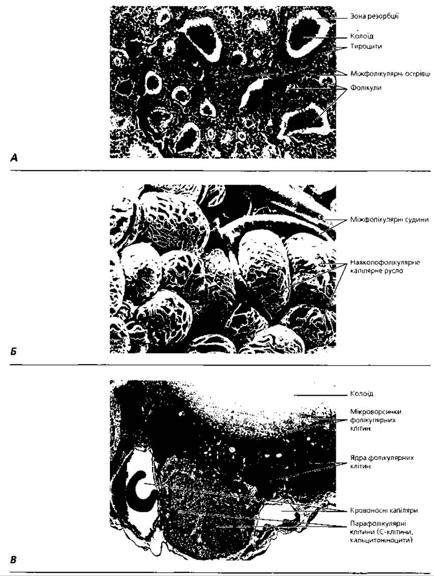
Fig. 4.29. Thyroid gland: A — light microscopy, x 300; B — scanning electron micrograph of the microvasculature of the thyroid gland, x 400. C — transmission electron microscopy of a segment of the thyroid follicle wall with two parafollicular cells, x 3600

Fig. 4.30. Parathyroid glands: A - topography and vascularization; B - light microscopy, x 220
The second type of thyroid cells are the so-called parafollicular cells, calcitoninocytes, or C-cells. They lie singly between the base of thyrocytes and the basement membrane of the follicles. These are large cells of irregular round or polygonal shape, containing A large number of secretory granules in their cytoplasm (Fig. 4.29, B). A characteristic feature of parafollicular cells is their ability to reduce heavy metal oxides, which accounts for their so-called argyrophilia or osmiophilia. The cytoplasm contains a well-developed rough endoplasmic reticulum and elements of the Golgi complex. Parafollicular cells synthesize the hormone Calcitonin. Calcitonin reduces blood calcium levels by depositing it in Bone tissue.
Development. The thyroid gland primordium appears during the fourth week of embryonic development as an epithelial outgrowth of the pharyngeal wall between the First and Second pairs of pharyngeal (branchial) pouches. The growth of the epithelial cord is accompanied by its bifurcation at the level of the 3rd–4th pairs of pharyngeal (branchial) pouches, giving rise to the thyroid lobes. In the Early stages of embryogenesis, the thyroid gland has a trabecular (cord-like) Structure; as colloid accumulates inside the trabeculae, they transform into follicles. Note that thyrocytes and parafollicular cells have different origins. Calcitoninocytes develop from the fifth pair of pharyngeal pouches. In vertebrates, they exist as a pair of isolated glands - the so-called ultimobranchial bodies. In mammals, ultimobranchial tissue disperses around the follicles as parafollicular cells.
Parathyroid gland (glandula parathyroidea). Humans typically have four (less commonly, two) parathyroid glands. They are located on the posterior surface of the thyroid gland, beneath a common connective tissue capsule (Fig. 4.30). These are oval-shaped organs, measuring 6x3x2 mm, with a mass of 35-50 mg each. The structural and functional unit of the parathyroid gland is the trabecula. Trabeculae are composed of clusters of parathyrocytes that connect with each other, forming desmosomal junctions.
Parathyrocytes have a well-developed rough endoplasmic reticulum, Golgi complex, and mitochondria, and they accumulate secretory granules in their cytoplasm. Depending on the functional state of the parathyrocytes, their cytoplasm may stain basophilically (the so-called chief parathyrocytes) or acidophilically (acidophilic parathyrocytes). Parathyrocytes produce parathyroid hormone (parathormone), which increases blood calcium levels through bone demineralization (by stimulating osteoclast activity). Calcitonin and parathormone are antagonists; their interaction maintains calcium homeostasis in the blood. The mechanism of parathyroid cell activation is associated with the presence of receptors on their plasmalemma capable of directly sensing Calcium Ions.
Development. The Formation of the parathyroid glands begins during the fifth week of embryogenesis from epithelial outgrowths of the 3rd and 4th pairs of pharyngeal (branchial) pouches. Gradually, these outgrowths detach, and each transforms into an independent parathyroid gland. In newborns and young children, the gland parenchyma consists only of chief cells; acidophilic cells appear during the fifth to seventh years of life. After 20-25 years of age, an accumulation of adipocytes is observed in the parathyroid glands.
In the event of a decrease or complete loss of parathyroid gland function (for example, if they are accidentally removed during thyroid surgery), tetany develops, characterized by spasms of striated Muscles. If urgent measures are not taken, this condition leads to death.
The Adrenal gland (glandula suprarenalis) is a paired endocrine organ located above the superior pole of the Kidney. The mass of each adrenal gland is 6-7 g, its shape is triangular or semilunar with a concave base, and its dimensions are 5x3x1 cm. Externally, the adrenal glands are covered by a connective tissue capsule. Its parenchyma consists of two parts that differ in origin, structure, and function: the superficial cortex and the central medulla (Figs. 4.31, 4.32). Cortical endocrine cells form cords oriented perpendicular to the surface of the adrenal gland. The spaces between the cords are filled with layers of loose connective tissue.
The adrenal cortex contains three morphologically and functionally distinct zones: the superficial zona glomerulosa, the middle zona fasciculata, and the deep zona reticularis. The ratio of the width of these zones in the adrenal cortex of a normal mature Organism is 1:9:3, respectively. Small polygonal cells of the zona glomerulosa form rounded clusters (glomeruli). Endocrine cells of this zone produce mineralocorticoid hormones, mainly aldosterone, which regulates sodium levels in the body (Fig. 4.33). This hormone also has the property of enhancing inflammatory processes. Large cells of the zona fasciculata are arranged in parallel rows (bundles). Depending on their functional state, these cells can have light or dark cytoplasm, and a cuboidal or columnar shape. Endocrine cells of the zona fasciculata synthesize glucocorticoid hormones (cortisol, corticosterone), which regulate the metabolism of CARBOHYDRATES, Proteins, and Lipids, stimulate Energy Metabolism, and suppress inflammatory processes in the body. To a lesser extent, cells of the zona fasciculata produce androgens - dehydroepiandrosterone and androstenediol. Cells of the zona reticularis are polygonal or rounded, slightly smaller than the cells of the zona fasciculata, and form branching cords that resemble a network under the microscope. Endocrine cells of the zona reticularis synthesize steroids with weak androgenic activity (resembling the action of Male Sex Hormones). To a lesser extent, cells of the zona reticularis synthesize glucocorticoids, thereby complementing the function of the endocrine cells of the zona fasciculata.
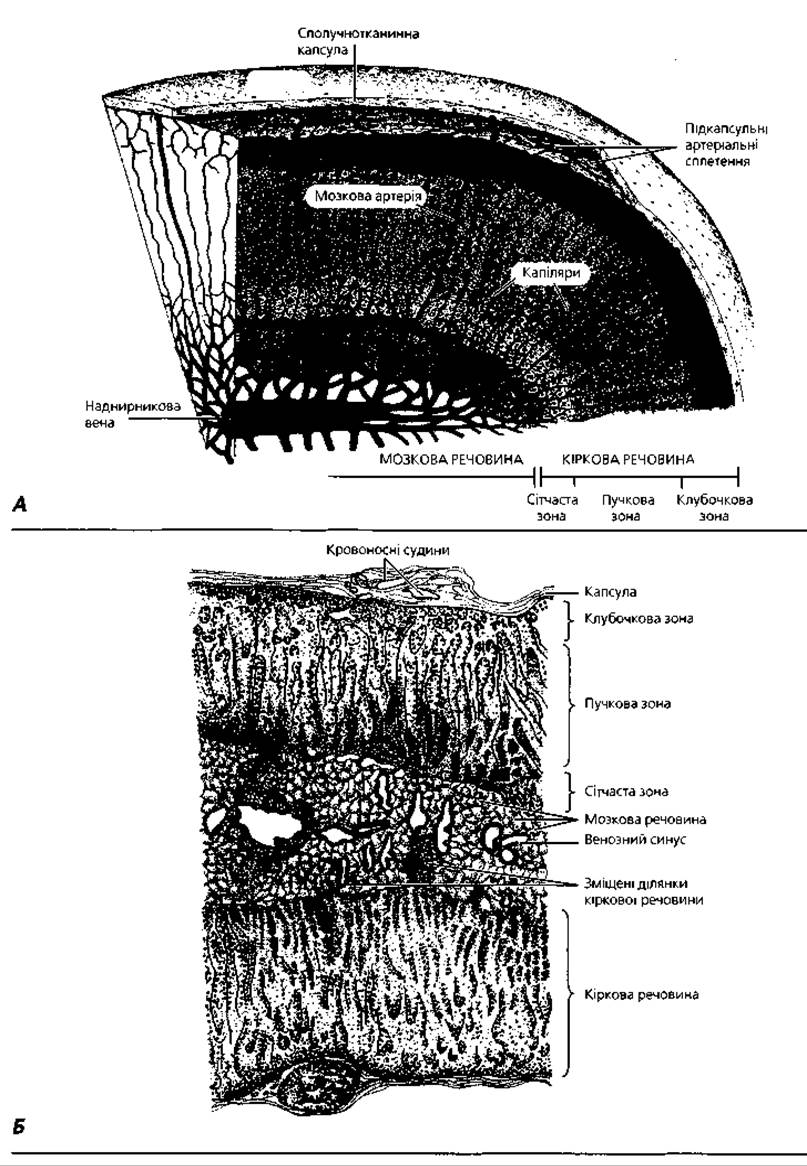
Fig. 4.31. Adrenal gland: A - diagram of histoarchitectonics and vascularization; B - semi-schematic representation of a histological specimen of the adrenal gland, median section, x30

Fig. 4.32. Light microscopy of the adrenal gland: A - general view of the cortex structure, x 65; B - capsule, glomerular region of the zona fasciculata; C - zona fasciculata: cells with numerous lipid inclusions; D - zona reticularis; E - medulla: epinephrine cells - cells with light cytoplasm; norepinephrine cells - cells with dark cytoplasm, x 270
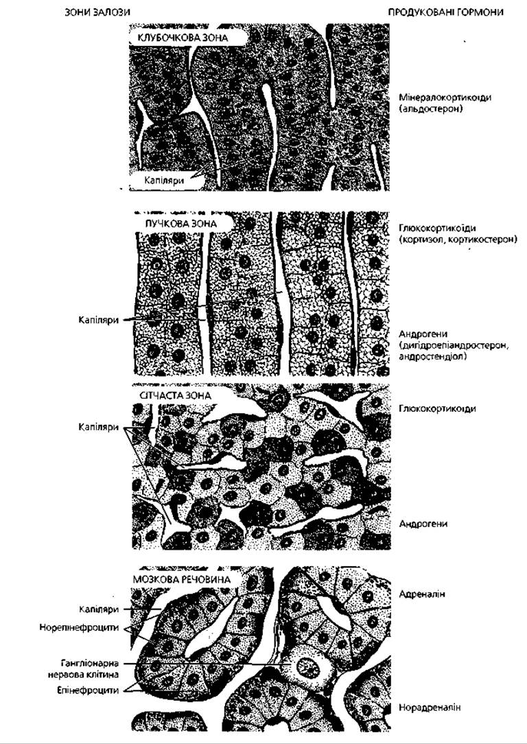
Fig. 4.33. Histophysiology of the adrenal gland: cells of the zona fasciculata and zona reticularis of the cortex are stimulated by adrenocorticotropic hormone of the pituitary gland; glucocorticoids produced by the zona fasciculata and zona reticularis inhibit the corticotropic activity of the hypothalamo-pituitary system via a negative feedback mechanism; medullary cells are stimulated by the endings of preganglionic sympathetic nerve fibers
In the cells of the adrenal cortex, the smooth endoplasmic reticulum and elements of the Golgi complex are well developed. The mitochondria of these cells contain characteristic tubular cristae and provide the synthesis of steroid hormone precursors. A feature of the cells of the zona fasciculata and zona reticularis is the presence of a large number of small lipid inclusions in their cytoplasm. Between the three main morphofunctional Zones of the cortex, there are clusters of poorly differentiated cells that serve as a source of physiological regeneration of the adrenal cortex. The first layer of such cells is localized between the capsule and the zona glomerulosa. The second germinative layer, called the sudanophobic zone, is located between the zona glomerulosa and the zona fasciculata. Between the zona reticularis and the medulla, cells with acidophilic cytoplasm are observed, forming the so-called X-zone. These are remnants of the embryonic adrenal cortex cells.
Regulation of the activity of cells in the zona fasciculata and zona reticularis is mediated by adrenocorticotropin (ACTH) of the pituitary gland, which interacts with a specific receptor on their plasmalemma. The synthesis and secretion of mineralocorticoids by the cells of the zona glomerulosa are independent of the pituitary gland and are regulated mainly by the Renin-Angiotensin System.
The adrenal medulla is demarcated from the cortex by an incomplete layer of connective tissue. It is composed of large round or polygonal cells, which are divided into epinephrine cells and norepinephrine cells based on The Nature of the substances they synthesize. Epinephrine cells have a light cytoplasm filled with secretory granules and produce epinephrine. Under an Electron microscope, the cytoplasm of norepinephrine cells appears dark and contains secretory granules of norepinephrine. Epinephrine and norepinephrine (collectively known as catecholamines) mobilize the body's defenses. An increase in the level of these hormones in the blood is a sign of the body's response to stress - the action of very strong stimuli or environmental factors that may pose a threat to life.
Development. The adrenal gland develops from two embryonic primordia: the medulla from para-aortic ganglia, and the cortex from outgrowths of the coelomic epithelium, which form the so-called interrenal body. The Development of the cortex begins during the fifth week of embryogenesis. Large acidophilic cells of the interrenal body are the source of the primary (fetal) cortex of the future adrenal glands. During the tenth week of embryonic development, the primary cortex is overgrown by small basophilic cells originating from the coelomic epithelium of the ROOT of the mesentery, which form the definitive cortex. The migration of neuroblasts from the para-aortic sympathetic ganglia into the interrenal body occurs during the sixth to seventh weeks of embryogenesis. Initially, the chromaffin cells of the medulla produce only norepinephrine; at later Selection/3.html">Stages of development, epinephrine production begins.
The adrenal gland reaches its maximum development at 20-25 years of age. Starting from 50-60 years of age, age-related involution of the zona glomerulosa and zona fasciculata is observed, with their endocrine elements being replaced by connective tissue. The CHARACTERISTICS OF THE medulla and zona reticularis do not change significantly with age.
The diffuse endocrine system consists of isolated endocrine cells scattered throughout the vast majority of the body's organs and systems. There are two types of cellular elements in the diffuse endocrine system: cells of neural origin that develop from neural crest neuroblasts, and cells of non-neural origin. Endocrine cells of the first group are grouped into the APUD system. They have the ability to take up and decarboxylate precursors of biologically active amines (serotonin, norepinephrine, epinephrine) - hence their name (Amine Precursor Uptake and Decarboxylation). The formation of neuroamines in these cells is combined with the synthesis of biologically active regulatory peptides. THE CONCEPT OF the APUD system was formulated in 1968 by the English histochemist E. Pearse. Currently, about 50 different apudocytes (cells of the APUD system) and their corresponding hormones are known; about 20 hypothetical hormones produced by cells of the APUD series are still awaiting the determination of their chemical nature.
The APUD system includes diffuse endocrine cells of the Digestive System, a number of neurosecretory cells of the brain, melatonin-synthesizing cells of the pineal gland, and cells of the adrenal medulla. Regulatory peptides of the APUD system cells provide local (paracrine) as well as distant regulation of the activity of the body's organs and systems. Their function is independent of the pituitary gland, but is closely linked to the action of nerve impulses arriving via sympathetic and parasympathetic trunks.
Diffuse cells of non-neural origin do not have the ability to take up and decarboxylate precursors of biologically active amines. This group of cells includes, in particular, endocrine cells of the testis, as well as follicular cells and luteal cells of the Ovaries. These cells produce steroid rather than protein hormones (testosterone, estrogens, progesterone), and their activity depends on the Influence of the corresponding tropic hormones of the pituitary gland.

Fig. 4.34. Schematic diagram of the interactions between the three major Regulatory Systems of the body: The Immune System influences The Nervous System via cytokines and is simultaneously regulated by Neurotransmitters; hormones mediate communication between the nervous and endocrine systems, as well as between the endocrine and immune systems. Through the coordinated action of the nervous, endocrine, and immune systems, the body maintains functional balance and the stability of its internal environment (homeostasis)
In concluding the Overview of the endocrine system, it should be noted that certain regulatory peptides (Insulin, gastrin, somatostatin, cholecystokinin, substance P) are produced by individual nerve cells, while several biologically active amines (adrenaline, serotonin) combine The properties of both a hormone and a neurotransmitter, transmitting excitation across the synaptic cleft. Endocrine mechanisms underlie many brain dysfunctions: serotonin deficiency is a pathogenetic factor in the development of depression, and serotonin and noradrenaline play an important role in the mechanisms of Schizophrenia. These facts point to a close Phylogenetic and Ontogenetic relationship between the nervous and endocrine systems as the body's primary regulatory systems, with the nervous system apparently being a later evolutionary acquisition. The function of these two systems is closely linked to the activity of yet another regulatory system—the immune system (Fig. 4.34). The integrative, regulatory, and Protective Functions of all three systems are mediated by another system that integrates the body into a single whole—The Cardiovascular system.
Terms to remember
1. Hormone. 2. Endocrinocyte. 3. Humoral regulation. 4. Hypothalamus. 5. Anterior hypothalamus. 6. Supraoptic nucleus. 7. Vasopressin (antidiuretic hormone). 8. Paraventricular nucleus. 9. Oxytocin. 10. Middle hypothalamus. 11. Liberins. 12. Statins. Releasing hormones. 13. Pituitary gland. 14. Distal (anterior) lobe of the pituitary gland. 15. Intermediate (middle) lobe of the pituitary gland. 16. Tuberal lobe of the pituitary gland. 17. Adenohypophysis. 18. Posterior lobe of the pituitary gland. 19. Pituitary stalk. 20. Neurohypophysis. 21. Chromophilic endocrinocyte. 22. Acidophil. 23. Lactotroph. 24. Prolactin. 25. Somatotropin. 26. Somatotropic hormone. 27. Basophil. 28. Gonadotroph. 29. Follitropin. 30. Lutropin. 31. Thyrotroph. 32. Thyrotropin. 33. Corticotroph. 34. Adrenocorticotropin. 35. Chromophobic endocrinocyte. 36. Melanotroph. 37. Melanotropin. 38. Lipotroph. 39. Lipotropin. 40. Herring body. 41. Pituicyte. 42. Pineal gland. 43. Pinealocyte. 44. Brain sand. 45. Thyroid gland. 46. Thyroid follicle. 47. Thyrocyte. 48. Colloid. 49. Thyroglobulin, triiodothyronine, T3. 50. Tetraiodothyronine, T4 (thyroxine). 51. Interfollicular islet. 52. Parafollicular cell. Calcitoninocyte, C-cell. 53. Calcitonin. 54. Parathyroid gland. 55. Trabecula of the parathyroid gland. 56. Chief parathyrocyte. 57. Acidophilic parathyrocyte. 58. Parathyroid hormone (parathormone). 59. Adrenal gland. 60. Adrenal cortex. 61. Adrenal medulla. 62. Zona glomerulosa. 63. Aldosterone. 64. Zona fasciculata. 65. Glucocorticoids (cortisol, corticosterone). 66. Zona reticularis. 67. Sex steroids. 70. Epinephrocyte. 71. Norepinephrocyte. 72. Catecholamines (adrenaline, noradrenaline). 73. APUD system.
Last update: 09/08/2026
Editorial and Educational Adaptation: This material has been compiled based on the primary/original source text. The project team performed an editorial review, corrected technical inaccuracies, structured sections, and adapted the content for an educational format.
What was processed:
- elimination of formatting defects (OCR errors, structural breaks, corrupted characters);
- editorial organization of content;
- standardization of terminology in accordance with academic sources;
- verification of factual statements against the original source text.
All mentions of the author, publication year, and origin of the primary text have been preserved in accordance with the source.