BIOLOGY Volume 2 - A Guide to General Biology - 2004
17. COORDINATION AND REGULATION IN ANIMALS
17.6. The Endocrine System
The Endocrine System is formed mainly by Glands of Internal Secretion, which are also called Endocrine glands (from the Greek éndon — within, krinō — separate). A gland is a Structure that secretes a specific substance (secretion) or substances. In the body of humans and animals, Two Types of glands are distinguished: exocrine and endocrine. Exocrine glands (from the Greek éxō — outside), or glands of external secretion, possess ducts. An example of such glands is Sweat Glands, located deep in the Skin and releasing their secretion (sweat) onto its surface. Endocrine glands are characterized by the following features:
1) they secrete substances called Hormones;
2) they have no ducts, and hormones are secreted directly into the Blood;
3) they are richly supplied with blood, i.e., permeated by a relatively large number of Blood Vessels.
Some glands combine endocrine and exocrine Functions, such as the Pancreas, which, In addition to the hormones Insulin and Glucagon, secretes pancreatic juice containing digestive Enzymes that enters the duodenum via a duct.
A hormone is a chemical messenger with the following properties:
1) it is transported by the bloodstream;
2) it acts on target Organs distant from its site of synthesis;
3) it binds to target receptor molecules due to its precise structural fit (like a key fitting a lock), i.e., it acts highly specifically;
4) it is a relatively small, soluble organic molecule;
5) it is effective at low concentrations.
The endocrine and nervous systems act in a coordinated manner to maintain a constant internal environment (Homeostasis) within the body. A comparison of these two systems is presented in Table 17.1. As will be shown below, the Hypothalamus, or more precisely, the hypothalamo-hypophyseal system, plays an important role in neuroendocrine interaction (see also Section 17.2.4). Despite obvious differences, both systems are similar in that they use chemical messengers for cellular communication. It is believed that both systems arose and evolved in parallel as The Need for communication between different PARTS OF THE body increased with its growing size and complexity. In both cases, The primary function of these systems is to coordinate and control vital physiological processes.
The major human endocrine glands are shown in Fig. 17.46, and their hormones and effects are listed in Table 17.9.
Class="center">
Fig. 17.46. Location OF THE major endocrine glands in The Human Body.
Table 17.9. Major human endocrine glands, their functions, and factors regulating their secretion
Gland |
Hormones |
Functions |
Factors regulating secretion |
Hypothalamus |
Releasing hormones (liberins) and inhibiting hormones (statins) Hormones of the posterior pituitary are also synthesized here |
Regulation of Hormone secretion by the anterior pituitary |
Secretion is regulated by hormone and metabolite levels via feedback mechanisms |
Posterior pituitary |
No hormone synthesis occurs; the following Hypothalamic hormones are stored and secreted here: Oxytocin |
Milk ejection by the mammary gland, uterine contractions during childbirth |
Negative feedback mechanisms involving hormones and The Nervous system |
Antidiuretic hormone (ADH, vasopressin) |
Reduction of urine secretion by the Kidneys |
Osmotic potential of the blood |
|
Anterior pituitary |
Follicle-stimulating hormone (FSH, follitropin) |
In males — stimulation of Spermatogenesis In females — follicle growth in the Ovary |
Blood levels of estrogens and testosterone; act via the Hypothalamus and Pituitary |
Luteinizing hormone (LH, lutropin) |
In males — testosterone secretion |
Blood testosterone level; acts via the hypothalamus and pituitary |
|
In females — secretion of estrogens and progesterone, ovulation, and Maintenance of the corpus luteum |
Blood estrogen levels; act via the hypothalamus and pituitary |
||
Prolactin |
Stimulation of milk production and secretion |
Hypothalamic hormones |
|
Thyroid-stimulating hormone (TSH, thyrotropin) |
Synthesis and Secretion of THYROID HORMONES and growth of The Thyroid Gland |
Blood thyroxine level; acts via the hypothalamus and pituitary |
|
Adrenocorticotropic hormone (ACTH, corticotropin) |
Synthesis and secretion of Adrenal Cortex Hormones, growth of these glands |
Blood ACTH level; acts via the hypothalamus |
|
Growth Hormone (GH, somatotropin) |
Protein Synthesis; growth, especially of limb bones |
Hypothalamic hormones |
|
Parathyroid gland |
Parathyroid hormone (PTH, parathormone) |
Increases blood calcium levels and decreases blood phosphate levels |
Blood levels of Ca2+ and PO43- |
Thyroid gland |
Triiodothyronine (T3) and thyroxine (T4) |
Regulation of basal metabolic rate, growth, and development |
TSH |
Decreases blood calcium levels |
Blood Ca2+ level |
||
Adrenal cortex |
Glucocorticoids (cortisol, etc.) |
Protein breakdown, glucose/Glycogen synthesis, adaptation to stress, anti-inflammatory/anti-allergic effects |
ACTH |
Mineralocorticoids (aldosterone, etc.) |
Na+ retention by the kidneys; maintenance of the Na+/K+ ratio in extracellular and intracellular fluids; elevation of blood pressure |
Blood levels of Na+ and K+; low blood pressure |
|
Adrenal medulla |
Adrenaline (epinephrine) |
Increase in Heart rate and force of contraction; constriction of skin and gastrointestinal capillaries. Dilation of arterioles in The Heart and skeletal Muscles, elevation of blood glucose levels |
Sympathetic nervous system |
Noradrenaline (norepinephrine) |
General constriction of small Arteries, elevating blood pressure |
Nervous system |
|
Islets of Langerhans |
Insulin (beta Cells) |
Decreases blood glucose levels; increases uptake and utilization of glucose and Amino Acids by cells |
Blood levels of glucose and amino acids |
Glucagon (alpha cells) |
Increases blood glucose levels; enhances breakdown of glycogen to glucose in the Liver |
||
Gastrin |
Secretion of gastric juice |
Presence of food in The Stomach |
|
Duodenum |
Secretin |
Secretion of pancreatic juice; inhibition of gastric juice secretion |
Presence of acidic food in the duodenum |
Cholecystokinin (pancreozymin) |
Emptying of the Gallbladder and secretion of pancreatic juice into the duodenum |
Presence of Fatty acids and amino acids in the duodenum |
|
Kidneys |
Renin |
Conversion of angiotensinogen to angiotensin |
Blood Na+ level, low blood pressure |
Ovary |
Estrogens (17β-estradiol, etc.) |
Development of female secondary sexual characteristics; Regulation of the Menstrual cycle |
FSH and LH |
Progesterone |
Pregnancy, inhibition of ovulation |
LH |
|
Corpus luteum |
Progesterone and estrogens |
GROWTH AND DEVELOPMENT of the Uterus |
LH |
Progesterone and estrogens |
Fetal development |
Developing fetus |
|
Chorionic gonadotropin |
Maintenance of the corpus luteum |
Developing fetus |
|
Placental lactogen |
Stimulation of mammary gland growth |
Developing fetus |
|
Testosterone |
Development of male secondary sexual characteristics |
LH and FSH |
17.6.1. Mechanism of hormone action
As shown in Table 17.10, Vertebrate Hormones can be divided into four groups based on their chemical nature: 1) Peptides and Proteins; 2) amine derivatives (e.g., Tyrosine); 3) Steroids; and 4) fatty acids.
Table 17.10. Chemical Nature of major human hormones
Chemical group |
Hormone |
Main source |
Peptides and proteins |
Releasing and inhibiting hormones |
Hypothalamus |
Growth hormone Follicle-stimulating hormone Luteinizing hormone Prolactin Thyroid-stimulating hormone Adrenocorticotropic hormone |
Anterior pituitary |
|
Oxytocin ADH (vasopressin) |
Posterior pituitary |
|
Parathyroid hormone |
Parathyroid gland |
|
Calcitonin |
Thyroid gland |
|
Insulin Glucagon |
Islets of Langerhans (pancreas) |
|
Gastrin |
Gastric mucosa |
|
Secretin |
Duodenal mucosa |
|
Amines |
Adrenaline |
Adrenal medulla |
Noradrenaline |
Adrenal medulla and sympathetic nervous system |
|
Thyroxine Triiodothyronine |
Thyroid gland |
|
Steroids |
Testosterone |
|
Estrogens Progesterone |
Ovaries and placenta |
|
Corticosteroids |
Adrenal cortex |
|
Fatty acids |
Many Tissues |
Hormone release
The mechanisms regulating hormone release by endocrine glands are listed below: 1. The presence of a specific metabolite in the blood. For example, an excess of glucose in the blood triggers the secretion of insulin by the pancreas, which lowers blood glucose levels. 2. The presence of another hormone in the blood. For example, many hormones secreted by the anterior Pituitary Gland are 'trophic' hormones (tropins), meaning they stimulate hormone secretion by other endocrine glands. 3. Stimulation by the sympathetic nervous system. For example, in Response to nerve impulses generated during stressful situations, The adrenal medulla secretes adrenaline and noradrenaline. Negative feedback In the first two cases described above, the timing and amount of hormone released are regulated by feedback mechanisms, usually negative ('the less, the more' and vice versa). This type of control is discussed in detail in Section 17.6.4 using thyroxine function as an example. Cascade effect Hormone release in response to the presence of another hormone in the blood is typically controlled by the hypothalamus and pituitary gland, and the final effect is often the result of the secretion of three different hormones. A good example is the ACTH-mediated release of cortisol, shown schematically in Fig. 17.47. This mechanism, known as a cascade effect, is important because the action of a small amount of 'trigger' hormone is amplified at each stage, leading to significant Changes in the final target organ. Thus, cortisol in Fig. 17.47 is an anti-stress factor secreted by the outer part (cortex) of the Adrenal Glands. It belongs to the glucocorticoids, which, among other things, raise blood sugar levels when the body requires increased Energy Expenditure (see also Section 17.6.5).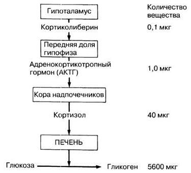
Fig. 17.47. An example of a cascade effect in the REGULATION OF GLUCOSE-to-glycogen conversion through the release of ACTH-releasing factor (corticotropin-releasing hormone). The overall 'Amplification' in this case is 56,000-fold (data from: Bradley, 1976).
Action on target cells Each hormone is highly specific and acts only on particular target cells that carry the corresponding protein receptors on their surface. Non-target cells lack these receptors, so the hormone has no effect on them. Upon binding to its receptor, a hormone can alter the function of various target Cell structures. These primarily include: 1) The Cell membrane; 2) membrane-bound enzymes (second messenger mechanism); 3) genes. Examples of these effects are given below. CELL MEMBRANE. One of the effects of insulin—increased cellular glucose uptake—occurs because the hormone, upon binding to receptors, increases the permeability of the cell membrane to glucose. Adrenaline acts on smooth Muscle cells by opening or closing their sodium, potassium, or both channels, which alters the Membrane Potential and consequently stimulates contraction or relaxation. SECOND MESSENGER MECHANISM. Adrenaline and many Peptide Hormones, upon binding to outer Membrane Receptors, cannot penetrate the cell; however, they trigger the intracellular release of a 'second messenger,' which initiates a cascade of enzymatic reactions leading to the desired effect. In many cases, the second messenger is the nucleotide cyclic adenosine monophosphate (cAMP), whose simplified MECHANISM OF ACTION is shown in Fig. 17.48. When a hormone binds to a receptor protein, this protein is activated and becomes the enzyme adenylate cyclase, which catalyzes The conversion of ATP to cAMP. In principle, cAMP can initiate a wide range of responses depending on the cell type in which it is formed.
Fig. 17.48. A simplified diagram showing how adrenaline stimulates glucose release from a liver cell. Activation of membrane-bound adenylate cyclase leads to The formation of cAMP, which activates enzyme systems that ultimately break down (phosphorolyze) glycogen into glucose. Glucose then diffuses out of the cell into the blood.
Thus, in the case of adrenaline, cAMP activates the enzyme protein kinase, which in turn activates another enzyme, phosphorylase, which converts glycogen to phosphorylated glucose. The ultimate effect of adrenaline in this scenario is the release of glucose from the cell. Amplification occurs at each step of this process: only a few molecules of adenylate cyclase are needed to activate many protein kinase molecules, and so on. This is another example of a cascade effect. Other Hormones that use cAMP as a second messenger include ADH (Sections 17.6.2 and 20.6), TSH (Section 17.6.4), ACTH (Section 17.6.5), glucagon (Section 17.6.6), LH and FSH (Sections 21.7.4 and 21.7.6), and most hypothalamic releasing hormones. GENES. Steroid Hormones (Sex Hormones and hormones secreted by the adrenal cortex) pass through the outer membrane of target cells and bind to a receptor protein in the Cytoplasm. The resulting complex enters the Cell Nucleus, where the hormone acts directly on the Chromosomes, 'switching on' specific genes by triggering their METABOLISM/31.html">Transcription—the synthesis of Messenger RNA (mRNA). This RNA moves into the cytoplasm, where it is translated, leading to the synthesis of new proteins, such as enzymes that perform specific functions. The hormone thyroxine, upon entering the cell, binds directly to receptor proteins on the chromosomes, producing a similar effect.Last update: 06/08/2026
Editorial and Educational Adaptation: This material has been compiled based on the primary/original source text. The project team performed an editorial review, corrected technical inaccuracies, structured sections, and adapted the content for an educational format.
What was processed:
- elimination of formatting defects (OCR errors, structural breaks, corrupted characters);
- editorial organization of content;
- standardization of terminology in accordance with academic sources;
- verification of factual statements against the original source text.
All mentions of the author, publication year, and origin of the primary text have been preserved in accordance with the source.