Human Biochemistry, Volume 2 - Murray R. 1993
Biochemistry of Intra- and Intercellular Communication
Pituitary and Hypothalamic Hormones
Hypothalamic Hormones
The secretion (and in some cases, synthesis) of each of the Pituitary Hormones listed in Table 45.1 is under the tonic control of at least one hypothalamic hormone. Hypothalamic hormones are released from the endings of hypothalamic nerve fibers surrounding the capillaries of the hypothalamo-pituitary system in the pituitary stalk, and reach the anterior lobe via a specialized portal vessel system connecting the Hypothalamus and this lobe. Information regarding The Structure of certain hypothalamic hormones can be found in Table 45.2.
Class="center">Table 45.1. Hypothalamo-pituitary hormones
|
Hypothalamic hormones |
Abbreviation |
Released pituitary hormone1) |
|
Corticotropin-releasing hormone (corticoliberin) |
CRH |
ACTH (LPH, MSH, endorphins) |
|
Thyrotropin-releasing hormone (thyroliberin) |
TRH |
TSH |
|
Gonadotropin-releasing hormone (gonadoliberin) |
GnRH |
LH, FSH |
|
Growth Hormone-releasing hormone (somatoliberin) |
GHRH |
GH |
|
Growth hormone-inhibiting hormone (Somatostatin) |
GHIH |
GH (TSH, FSH, ACTH) |
|
Prolactin-inhibiting hormones; dopamine and GAP |
PIH (prolactin-inhibiting hormone) |
Prolactin |
1) Parentheses indicate pituitary hormones whose release is subject to secondary or weaker effects by the given hypothalamic hormone.
Hypothalamic hormones are released in a pulsatile manner, and isolated anterior pituitary target Cells respond more effectively to the pulsatile administration of these hormones than to their continuous exposure. The release of lutropin (LH) and follitropin (FSH) is controlled by the concentration of a single releasing hormone, gonadoliberin, the concentration of which, in turn, is determined by the Blood levels of Sex Hormones reaching the hypothalamus (see the feedback loop in Fig. 43.1). Adrenocorticotropic hormone (ACTH) release is controlled primarily by corticoliberin (corticotropin-releasing hormone, CRH), although A number of Other Hormones may also be involved in regulating this process, including antidiuretic hormone (ADH), catecholamines, vasoactive intestinal peptide (VIP), and angiotensin II. Corticoliberin secretion is influenced by cortisol (a glucocorticoid hormone secreted by the Adrenal Glands). Thyrotropin (TSH) release depends mainly on thyroliberin (thyrotropin-releasing hormone, TRH), the secretion of which is regulated by THYROID HORMONES, triiodothyronine (T3) and thyroxine (T4); TSH secretion is inhibited by somatostatin (see Fig. 46.4). Growth hormone (GH) secretion and production are under the tonic control of both stimulatory and inhibitory hypothalamic hormones. In addition, a peripheral feedback loop participates in regulating growth hormone secretion. Insulin-like growth factor 1 (somatomedin C), which mediates certain effects of growth hormone, stimulates somatostatin release and inhibits somatoliberin secretion (Fig. 45.5). The regulation of prolactin (PRL) Synthesis and Secretion is primarily driven by the tonic suppression of these processes by hypothalamic agents. Its distinguishing feature is the combination of neural (nipple stimulation) and neurotransmitter/neurohormonal factors. Dopamine (Table 45.2) inhibits prolactin synthesis (by inhibiting prolactin Gene METABOLISM/31.html">Transcription) and secretion; however, evidence suggests that the suppression of prolactin secretion is not mediated by dopamine alone. A recently discovered 56-residue neuropeptide exhibits both gonadoliberin activity and prolactin-inhibiting hormone (prolactostatin) activity. It is referred to as gonadotropin hormone-releasing hormone-associated peptide (GAP). GAP, whose overall structure and specific features are shown in Fig. 45.1, is a potent inhibitor of prolactin secretion and can clearly be considered a prolactostatin. The existence of GAP may explain the intriguing link between gonadoliberin and prolactin secretion that is particularly pronounced in certain species.
Table 45.2. Structure of hypothalamic releasing hormones (liberins)
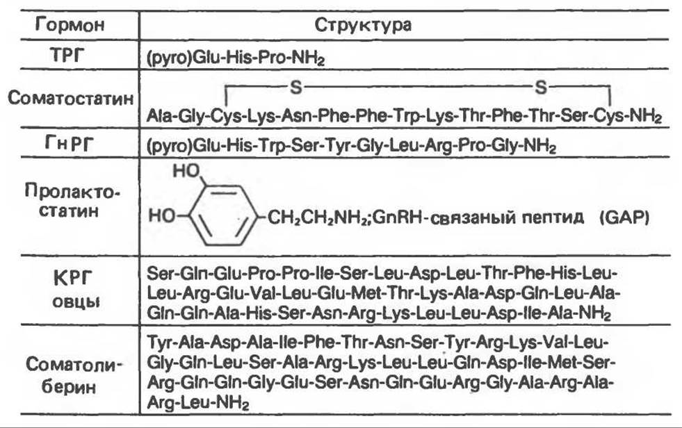

Fig. 45.1. Structure and Amino Acid Sequence of placental cDNA for prepro-gonadotropin-releasing hormone (GnRH). The protein consists of three domains: a signal peptide, GnRH, and gonadotropin-releasing hormone-associated peptide (GAP). The Amino acid sequences of GnRH and GAP are shown at the bottom of the figure. The site of enzymatic Processing leading to the Cleavage of the two molecules is indicated. Numbers denote positions within The amino acid sequences of GnRH (1–10) and GAP (1–56). (Reproduced with permission, from Nicolics K. et al. A prolactin-inhibiting factor with the precursor for human gonadotropin-releasing hormone. Nature 1986, 316, 511. Copyright 1985 by Macmillan Journals Ltd.)
Many hypothalamic hormones, particularly thyroliberin, corticoliberin, and somatostatin, are found in other regions of The Nervous system and in a number of peripheral Tissues. Somatostatin concentration in the Pancreas is higher than in the hypothalamus. It is produced by D-Cells of the islets of Langerhans and appears to regulate Glucagon and insulin secretion. Furthermore, somatostatin is among the more than 40 Peptides produced by Neurons of the central and peripheral nervous systems.
cAMP was initially thought to act as the mediator of releasing hormone action on the adenohypophysis; however, recent experiments with gonadoliberin and thyroliberin suggest the involvement of a calcium-phospholipid mechanism similar to that described above (see Fig. 44.5). Whether releasing hormones affect the synthesis of corresponding pituitary hormones In addition to their secretion remains controversial; it has recently been demonstrated that somatoliberin increases the transcription rate of the growth hormone gene, and thyroliberin exerts a similar effect on the prolactin gene.
Last update: 06/08/2026
Editorial and Educational Adaptation: This material has been compiled based on the primary/original source text. The project team performed an editorial review, corrected technical inaccuracies, structured sections, and adapted the content for an educational format.
What was processed:
- elimination of formatting defects (OCR errors, structural breaks, corrupted characters);
- editorial organization of content;
- standardization of terminology in accordance with academic sources;
- verification of factual statements against the original source text.
All mentions of the author, publication year, and origin of the primary text have been preserved in accordance with the source.