Biological Chemistry - Berezov T. T., Korovkin B. F. 1998
Hormones
Hypothalamic hormones
The Hypothalamus serves as the direct interface between the higher centers of the Central Nervous system and the Endocrine System. The Nature of the connections between the central nervous system and the endocrine system began to clear up in recent decades when the first humoral factors were isolated from the hypothalamus, which turned out to be hormonal substances with exceptionally high biological activity. It took considerable effort and experimental skill to prove that these substances* are synthesized in the hypothalamic Nerve Cells, from where they reach the Pituitary Gland via the portal capillary system and regulate the secretion of Pituitary Hormones, specifically their release (and possibly Biosynthesis as well). These substances were initially termed neurohormones, and later releasing factors (from the English word release), or liberins. Substances with the opposite effect, i.e., inhibiting the release (and possibly biosynthesis) of pituitary hormones, came to be called inhibiting factors, or statins. Thus, Hypothalamic Hormones play a key role in the physiological system of hormonal regulation across diverse biological Functions in individual Organs, Tissues, and the Organism as a whole.
* In the early 1970s, R. Guillemin and A. Schally were the first to isolate substances from hypothalamic tissue that exerted a regulatory effect on pituitary function. For their discovery of these so-called superhormones, alongside R. Yalow—who developed the radioimmunoassay METHOD FOR DETERMINING Peptide Hormones—these authors were awarded the Nobel Prize in 1977.
Class="center">
To date, 7 stimulators (liberins) and 3 inhibitors (statins) of pituitary hormone secretion have been discovered in the hypothalamus, namely: corticoliberin, thyroliberin, luliberin, folliberin, somatoliberin, prolactoliberin, melanoliberin, Somatostatin, prolactostatin, and melanostatin (Table 8.1). Five hormones have been isolated in pure form, and their primary structures have been established and confirmed by chemical synthesis.
Major difficulties in obtaining hypothalamic hormones in pure form are due to their extremely low concentration in the starting tissue. For instance, isolating just 1 mg of thyroliberin required Processing 7 tons of hypothalami obtained from 5 million sheep.
It should be noted that not all hypothalamic hormones are strictly specific to a single pituitary hormone. In particular, thyroliberin has been shown to stimulate the release of prolactin In addition to thyrotropin, while luliberin stimulates the release of follicle-stimulating hormone alongside luteinizing hormone.
Table 8.1. Hypothalamic hormones controlling the release of pituitary hormones 1
Old Name |
Accepted Abbreviation |
Recommended Name |
Corticotropin-releasing factor |
CRF |
Corticoliberin |
Thyrotropin-releasing factor |
TRF |
Thyroliberin |
Gonadotropin-releasing factor |
GRF |
Gonadoliberin |
Follicle-stimulating hormone-releasing factor |
FRF |
Folliberin |
FSH-RF |
||
Somatotropin-releasing factor |
SRF |
Somatoliberin |
Somatotropin-inhibiting factor |
SIF |
Somatostatin |
Prolactin-releasing factor |
PRF |
Prolactoliberin |
Prolactin-inhibiting factor |
PIF |
Prolactostatin |
Melanotropin-releasing factor |
MRF |
Melanoliberin |
Melanotropin-inhibiting factor |
MIF |
Melanostatin |
1 Hypothalamic hormones do not have universally established names. It is recommended to append the suffix "liberin" to the first part of the corresponding pituitary hormone name; for example, "thyroliberin" refers to the hypothalamic hormone that stimulates the release (and possibly the synthesis) of thyrotropin, the corresponding pituitary hormone. Similarly, the names of hypothalamic factors that inhibit the release (and possibly synthesis) of trophic pituitary hormones are formed by appending the suffix "statin". For instance, "somatostatin" refers to the hypothalamic peptide that inhibits the release (or synthesis) of the pituitary Growth Hormone, somatotropin.
It has been established that in terms of chemical Structure, all hypothalamic hormones are low-molecular-weight Peptides—specifically, oligopeptides of unusual structure—although their exact Amino Acid Composition and Primary Structure have not yet been elucidated for all of them. Below are the currently available data on the Chemical Nature of six of the ten known hypothalamic hormones.
1. Thyroliberin (Pyro-Glu-His-Pro-NH2):
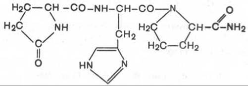
Thyroliberin is a tripeptide consisting of pyroglutamic (cyclic) acid, Histidine, and prolinamide linked by peptide bonds. Unlike classical peptides, it lacks free NH2 and COOH groups at the N- and C-terminal Amino Acids.
2. Gonadoliberin is a decapeptide consisting of 10 amino acids in the following sequence:

The C-terminal amino acid is glycinamide.
3. Somatostatin is a cyclic tetradecapeptide (consisting of 14 amino acid residues)*:

Aside from its cyclic structure, this hormone differs from the previous two in that it lacks pyroglutamic acid at the N-terminus: a disulfide bond is formed between two Cysteine residues at positions 3 and 14. It should be noted that the synthetic linear analogue of somatostatin also exhibits similar biological activity, indicating that the disulfide bridge of the natural hormone is not essential. In addition to the hypothalamus, somatostatin is produced by Neurons of the central and peripheral nervous systems, and is also synthesized in S-Cells of the pancreatic islets (islets of Langerhans) in the Pancreas as well as in intestinal cells. It exerts a wide range of biological effects; in particular, it has been shown to inhibit growth hormone synthesis in the adenohypophysis, while also directly suppressing The biosynthesis of Insulin and Glucagon in the ß- and a-cells of the islets of Langerhans.
4. Somatoliberin has recently been isolated from natural sources. It consists of 44 amino acid residues with a fully elucidated sequence. Furthermore, biological activity characteristic of somatoliberin is also exhibited by a chemically synthesized decapeptide:
H-Val-His-Leu-Ser-Ala-Glu-Gln-Lys-Glu-Ala-OH.
This decapeptide stimulates the Synthesis and Secretion of the pituitary growth hormone, somatotropin.
5. Melanoliberin, whose chemical structure is analogous to the open-ring STRUCTURE OF THE hormone oxytocin (lacking the tripeptide side chain), has the following structure:
H-Cys-Tyr-Ile-Gln-Asn-Cys-OH.
6. Melanostatin (melanotropin-inhibiting factor) is represented either by the tripeptide Pyro-Glu-Leu-Gly-NH2 or by a pentapeptide with the following sequence:
Pyro-Glu-His-Phe-Arg-Gly-NH2.
It should be noted that melanoliberin exerts a stimulating effect, whereas melanostatin, conversely, has an inhibitory effect on the synthesis and secretion of melanotropin in the anterior pituitary gland.
In addition to the aforementioned hypothalamic hormones, the chemical nature of another hormone, corticoliberin, has been extensively studied. Active preparations of this hormone have been isolated from both the hypothalamic tissue and the posterior pituitary; it is believed that the latter may serve as a storage depot for Vasopressin and Oxytocin. Recently, ovine hypothalamic corticoliberin, consisting of 41 amino acids with a elucidated sequence, has been successfully isolated.
* When writing the Amino acid sequences of Polypeptides, it is currently standard practice to denote the N-terminus with the symbol H and the C-terminus with OH.
The primary site of hypothalamic hormone synthesis is most likely nerve terminals, specifically hypothalamic synaptosomes, as they exhibit the highest concentration of both hormones and biogenic amines. The latter, alongside hormones from peripheral Endocrine glands acting via feedback loops, are considered key regulators of the secretion and synthesis of hypothalamic hormones. The biosynthesis mechanism of thyroliberin—which is likely non-ribosomal—involves an SH-containing synthetase or a multi-enzyme complex that catalyzes the cyclization of glutamic acid into pyroglutamic acid, peptide bond formation, and the amidation of Proline in the presence of glutamine. A similar biosynthetic mechanism involving corresponding synthetases is also postulated for gonadoliberin and somatoliberin.
The inactivation pathways of hypothalamic hormones remain insufficiently studied. The half-life of thyroliberin in rat Blood is 4 minutes. Inactivation occurs both through peptide bond Cleavage (mediated by exo- and Endopeptidases in the serum of rats and humans) and via the Cleavage of the amide group in the prolinamide molecule. A specific enzyme, pyroglutamyl peptidase, has been discovered in the hypothalamus of humans and several animal species; it catalyzes the removal of the pyroglutamic acid molecule from thyroliberin or gonadoliberin.
Hypothalamic hormones directly influence the secretion (more precisely, the release) of preformed hormones as well as the de novo biosynthesis of these hormones. cAMP has been proven to play a role in hormonal signal Transduction. Furthermore, specific adenohypophysial receptors have been identified in the Plasma Membranes of pituitary cells, to which hypothalamic hormones bind. This binding triggers the adenylate cyclase system and membrane complexes of Ca2+ — ATP and Mg2 — ATP, leading to the release of Ca2+ ions and cAMP. The latter subsequently regulates both the release and Synthesis of the corresponding pituitary hormone by activating protein kinase (see below).
Structural analogs of thyroliberin and gonadoliberin have played a pivotal role in elucidating the MECHANISM OF ACTION of releasing factors, including their interaction with specific receptors. Some of these analogs exhibit even higher hormonal activity and a prolonged duration of action compared to native hypothalamic hormones. Nevertheless, substantial work lies ahead to determine the chemical structures of already discovered releasing factors and to decipher the molecular mechanisms underlying their action.
Last update: 06/08/2026
Editorial and Educational Adaptation: This material has been compiled based on the primary/original source text. The project team performed an editorial review, corrected technical inaccuracies, structured sections, and adapted the content for an educational format.
What was processed:
- elimination of formatting defects (OCR errors, structural breaks, corrupted characters);
- editorial organization of content;
- standardization of terminology in accordance with academic sources;
- verification of factual statements against the original source text.
All mentions of the author, publication year, and origin of the primary text have been preserved in accordance with the source.