Biochemistry - The Chemical Reactions of Living Cells, Volume 3 - D. Metzler 1980
Cell Growth, Differentiation, and Chemical Communication
Hormones
Vertebrate Hormones
The major vertebrate Hormones known to date are listed in Table 16-1, which also provides References to the specific chapters covering individual hormones. Based on their chemical Structure, hormones can be divided into three main groups: 1) Proteins AND Peptides, 2) aromatic Amino Acid Derivatives, and 3) Steroids and Prostaglandins.
Pituitary Hormones deserve special mention. The Pituitary Gland is connected to the Brain by a stalk (Fig. 16-1); it produces at least 10 protein and Peptide Hormones that regulate The activity of other endocrine (i.e., hormone-producing) glands throughout the body.
Class="center">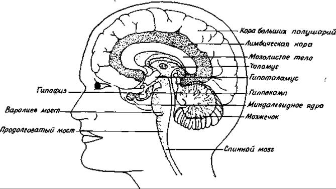
FIG. 16-1. Median sagittal section of the human brain (Pines Maya, Saturday Review Aug., 9, p. 14, 1975).
The pituitary gland consists of several lobes. As already mentioned, the anterior lobe of the pituitary (adenohypophysis) secretes hormones in response to at least nine neurohormones known as releasing factors or liberins [6, 7]. Liberins are secreted by the Hypothalamus in extremely small amounts. The Chemical Structure of several liberins has only recently been elucidated. As shown in Fig. 12-2, some of these compounds are modified simple peptides. Melanocyte-stimulating hormone (melanotropin) of the intermediate lobe of the pituitary is likewise released under the control of a specific hypothalamic factor, namely melanotropin-release-inhibiting factor (or melanoliberin).
The hypothalamus also synthesizes A number of factors that inhibit hormone release [8]. One of these, Somatostatin, suppresses the release of somatotropin, thereby counteracting The Effect of somatoliberin. Somatostatin is of great interest for yet another reason.
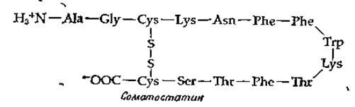
Namely, it acts not only on the pituitary gland but also on the Pancreas, where it inhibits the secretion of Insulin and Glucagon. This leads to a decrease in Blood glucose levels, opening up new approaches to the Treatment of diabetes (Supplement 11-B).
Table 16-1 Vertebrate Hormones
|
Type, name, and source of hormone |
Primary target Site of Action |
Where described |
|
A. Peptide and Protein hormones 1. Pituitary gland a. Adenohypophysis (anterior lobe) |
||
|
Somatotropin (Growth Hormone, GH) |
All Tissues |
|
|
Corticotropin (ACTH) |
Adrenal cortex, adipose tissue |
Fig. 2-2 Ch. 6, sec. E,5 |
|
Thyrotropin (thyroid-stimulating hormone, TSH) |
Ch. 14, sec. 3,5 |
|
|
Follitropin (follicle-stimulating hormone, FSH) |
Ch. 12, sec. I,3,g |
|
|
Lutropin (luteinizing hormone, interstitial Cell-stimulating hormone, ICSH or LH) |
Same |
Ch. 12, sec. I,3,g |
|
Prolactin (mammotropin) |
||
|
Lipotropin |
Ch. 16, sec. A, 1 |
|
|
b. Neurohypophysis (posterior lobe) |
||
|
Oxytocin |
Uterus, mammary glands |
Fig. 2-2 |
|
Vasopressin (antidiuretic hormone) |
Fig. 2-2 |
|
|
c. Intermediate lobe of pituitary |
||
|
Melanotropin |
Melanophores |
|
|
2. Pancreas |
||
|
Insulin |
All Cells |
Ch. 4, sec. D,7; ch. 5, |
|
sec. B,5; ch. 6, sec. E,5; ch. 12, sec. E,1 |
||
|
Glucagon |
Liver, adipose tissue |
Ch. 6, sec. E,5; ch. 11, sec. E,5; ch. 12, sec. E, 1 |
|
3. Ovary (corpus luteum) |
||
|
Relaxin |
Pelvic ligaments |
|
|
4. Thyroid gland |
||
|
Calcitonin (thyrocalcitonin) |
Bones, kidneys |
Supplement 5-D |
|
5. Parathyroid gland |
||
|
Parathyrin (parathyroid hormone) |
Same |
Supplement 5-D; ch. |
|
12, sec. 3, and supplement 12-G; ch. 16, |
||
|
6. Kidneys |
sec. A,1 |
|
|
Renin |
Renal cortex |
|
|
7. Digestive tract |
||
|
Gastrin |
||
|
Enterogastrin |
» |
|
|
Cholecystokinin |
||
|
Secretin |
Pancreas |
|
|
Pancreozymin |
Same |
|
|
B. Amino acid derivative hormones |
||
|
1. Thyroid gland |
||
|
Thyroxine and triiodothyronine |
Most cells |
Ch. 14, sec. 3,5,6 |
|
2. Adrenal medulla |
||
|
Epinephrine, norepinephrine (adrenaline, noradrenaline) |
Same |
Ch. 6, sec. E,6; ch. 12, sec. E, 1 |
|
3. Pineal Gland |
||
|
Melatonin |
Melanophores |
Ch. 14, sec. I; fig. 14-27 |
|
4. Nerve and other cells Serotonin (5-hydroxytryptamine) C. Steroids and prostaglandins 1. Testes Testosterone |
Arterioles, Central Nervous system Most cells |
Ch. 12, sec. I,3,c |
|
2. Ovaries Estrogen (17β-estradiol) |
Same |
Ch. 12, sec. I,3,g |
|
3. Corpus luteum Progesterone |
Uterus, mammary glands |
Ch. 12, sec. I,3,a |
|
4. Adrenal cortex Corticosterone, cortisol |
Most cells |
Ch. 11, sec. E,2 and E,7; ch. 12, sec. I,3,b |
|
Aldosterone |
Kidneys |
Ch. 12, sec. I,3,b |
|
5. Various tissues Prostaglandins |
Smooth Muscles |
Ch. 12, sec. D,3 |
Pituitary hormones vary significantly in the length of their peptide chains. Some of them belong to medium-molecular-weight proteins. For example, human growth hormone has a Molecular Weight of 21,500 and is characterized by high Specificity: growth hormones from other sources cannot replace it. Thyrotropin (TSH), the thyroid-stimulating hormone, is a glycoprotein with a molecular weight of 28,000. On the other hand, the neurohypophyseal (posterior pituitary) hormones Vasopressin and Oxytocin are simple peptides composed of only 9 amino acid residues (effectively eight if cystine is counted as a single amino acid; Fig. 2-2). As the name implies, the neurohypophysis consists of neural tissue whose secretory function is under the direct control of the central nervous system. Vasopressin is the primary factor regulating blood volume and ARTERIAL BLOOD PRESSURE; its secretion level is influenced by stress. Oxytocin acts on the smooth Muscle of the uterus during childbirth and also triggers Lactation. Milk ejection from the mammary glands depends to a certain extent on the infant's suckling movements, which reflexively stimulate the release of oxytocin into the bloodstream.
It is interesting to trace the relationship among the various pituitary hormones. Some of them contain an identical heptapeptide sequence with the following structure:

Corticotropin (Fig. 2-2) contains not only this heptapeptide but also the entire Amino Acid Sequence of a-melanotropin, with an additional 29 Amino Acids attached to its C-terminus. The same heptapeptide is found in lipotropins, which are likewise secreted by the pituitary gland. Lipotropin molecules contain 46 amino acids attached to one end of the heptapeptide and 37 to 5 amino acids attached to the other end (the C-terminus). The presence of a common heptapeptide indicates an evolutionary relationship among the hormones of this group. Furthermore, corticotropin can serve as a precursor for a-melanotropin and other biologically active peptides [9]. It appears that the Processing (modification) of protein and peptide hormones via post-transcriptional Cleavage by proteases or other influences is a very widespread, general biological phenomenon [10, 11] (Ch. 11, sec. D, 2).
Another example is parathyrin (parathyroid hormone), which consists of 84 amino acid residues. In secretory granules, this peptide is present as a prohormone containing 90 amino acid residues, with 6 additional amino acids attached to the N-terminus. The primary Biosynthesis product, preparathyrin, presumably contains 25 additional amino acids at the N-terminus of the molecule [11a, 11b]. Consequently, conversion into the active hormone involves at least two preliminary stages of sequential "trimming" of the biosynthetic product: pre-processing and pro-processing. A similar situation occurs in the case of insulin, where preproinsulin is first cleaved to form proinsulin (Fig. 11-9), after which proinsulin is acted upon by a protease with Trypsin-like properties.
It has been suggested that The conversion of certain prohormones into active hormones occurs via aminolysis rather than Hydrolysis of the peptide chain. Cleavage via substitution by NH3 leads to the appearance of an amide group at the C-terminus of the peptide, which is very frequently found in small-molecular-weight peptide hormones. Vasopressin, oxytocin, a-melanotropin, liberins, and certain Other Hormones are apparently formed in this manner.
It is also hypothesized that covalent modifications (e.g., via phosphorylation) participate in The regulatory mechanisms that control prohormone storage or the duration of active hormone action [11].
Among the Gastrointestinal Hormones listed in Table 16-1 are gastrin and secretin, which are relatively small Polypeptides containing 17 and 27 amino acids, respectively [12]. Particularly noteworthy is the renal hormone renin, which acts as a specific protease to cleave the decapeptide proangiotensin from serum a2-globulin [1]. Proangiotensin is then acted upon by another enzyme [13] that removes two additional amino acids from the C-terminus, yielding angiotensin, the most potent hypertensive compound known.

Angiotensin causes contraction of vascular smooth muscles. It reduces renal blood flow and decreases the Excretion of Water and salts from the body. In addition, this hormone stimulates the adrenal cortex to increase aldosterone secretion, leading to enhanced reabsorption of sodium ions.
Last update: 06/08/2026
Editorial and Educational Adaptation: This material has been compiled based on the primary/original source text. The project team performed an editorial review, corrected technical inaccuracies, structured sections, and adapted the content for an educational format.
What was processed:
- elimination of formatting defects (OCR errors, structural breaks, corrupted characters);
- editorial organization of content;
- standardization of terminology in accordance with academic sources;
- verification of factual statements against the original source text.
All mentions of the author, publication year, and origin of the primary text have been preserved in accordance with the source.