BIOLOGY Volume 2 - A Guide to General Biology - 2004
14. TRANSPORT IN ANIMALS
14.8. Functions of blood in mammals
14.8.5. Protective functions of blood
Humans possess a sophisticated system of defense mechanisms that enable the body to withstand attacks by pathogenic agents and eliminate foreign Materials. Three major protective mechanisms are associated with the Blood:

The first two mechanisms are discussed in this section, while the third is covered in section 14.9.
Blood clotting (coagulation)
When any tissue is damaged, blood escapes from it and soon clots (coagulates), forming a blood clot. The clot prevents further blood loss and hinders The entry of pathogenic microorganisms into the body—which, quite obviously, is of great survival value. Equally important is the fact that blood does not clot within undamaged vessels. The extreme complexity of The sequence of reactions involved in clotting serves precisely to ensure that this process does not occur where it is unnecessary. At least 12 different factors acting in concert are required for this process to take place. Only the most important of these are described below, and Fig. 14.34 schematically illustrates the major stages of Blood Coagulation.
Class="center">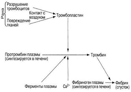
Fig. 14.34. MAIN STAGES OF the blood clotting process.
Blood escaping from a wound comes into contact with the air and mixes with substances released from damaged Cells and ruptured platelets. These substances include, in particular: 1) thromboplastin, a lipoprotein released from damaged Tissues; 2) clotting factors VII and X, which are plasma Enzymes; and 3) Calcium Ions. Together, these substances catalyze The conversion of the plasma protein prothrombin into Thrombin, an active protease (i.e., a protein-digesting enzyme). Thrombin hydrolyzes fibrinogen (another plasma protein) to form fibrin. Fibrin is insoluble and has a fibrous Structure. The fibers form a densely intertwined three-dimensional network that traps Blood Cells (Fig. 14.35). This is how a clot is formed. Upon drying, it turns into a scab covering the wound, which prevents further blood loss and creates a mechanical barrier against pathogen penetration.

Fig. 14.35. Structure of a blood clot. Scanning electron micrograph showing red blood cells entangled in fibrous strands, alongside a leukocyte (large spiky Cell) and several smaller platelets.
Given The complexity of the blood clotting process, the absence or deficiency of even a single one of its factors can lead to massive Hemorrhage. For example, if the factor initiating thromboplastin activity is entirely absent or present in minuscule amounts, even a small wound becomes life-threatening. This condition is known as hemophilia. There are two hereditary forms of hemophilia, depending on which clotting protein factor is missing and, accordingly, which Gene is defective (though they are clinically identical). Hemophilia A (85% of all cases) is caused by the loss of factor VIII, and hemophilia B (the remaining 15%) by factor IX. Both genes are located on the sex X chromosome. Since males possess only one X chromosome (their second sex chromosome, the Y, carries no genes encoding clotting factors), they are primarily the ones affected by hemophilia. For a woman to have two defective X Chromosomes for the same gene is an exceptionally rare situation. If one X chromosome is abnormal, the other usually carries a normal, dominant gene (see section 24.6.1) that ensures the Synthesis of the required clotting factor. However, such a woman remains healthy, acting as a carrier of the hemophilia gene and potentially passing it on to her offspring.
Clotting does not occur in undamaged Blood Vessels because their endothelial lining has a very smooth surface that does not trigger the destruction of platelets or blood cells. In addition, the blood contains substances that actively counteract clotting. One of these is heparin, dissolved in the plasma and produced by so-called mast cells found in Connective Tissue and the Liver. It prevents the conversion of prothrombin into thrombin and fibrinogen into fibrin, which is why it is widely used in medicine as an anticoagulant.
If a clot nonetheless forms inside a vessel, it is called a thrombus; The formation of such a clot leads to an acute circulatory disorder known as thrombosis. This can happen, for example, when the endothelium is damaged, as the roughness of the injured area promotes platelet destruction, thereby triggering the blood-clotting process. Coronary thrombosis—thrombosis in the coronary artery of The Heart (section 15.5)—is particularly dangerous and can lead to sudden death.
Phagocytosis
The primary function of certain leukocytes is to engulf microorganisms, as well as dead cell debris and other particles such as dust in the Lungs. This process is called phagocytosis, and the cells that perform it are called phagocytes. Phagocytes form the body's first line of active defense when pathogens invade.
Phagocytes are capable of a specialized form of locomotion known as ameboid movement: they effectively crawl along a substrate through the coordinated streaming of their Cytoplasm. They are directed toward the target sites by a concentration gradient of specific substances released by damaged blood cells, tissues, the blood clot, or Bacteria themselves. This movement toward a chemical stimulus is called chemotaxis. Upon arrival, phagocytes recognize harmful bacteria, aided by approximately 20 Proteins collectively known as the Complement system. These proteins are activated by invading bacteria and perform the following Functions:
1) some have The ability to attract phagocytes to the infected area (directing chemotaxis);
2) some participate in opsonization (see below);
3) some "punch holes" in the surface membrane of bacteria, causing the bacterial cells to swell and burst;
4) some stimulate the inflammatory response.
Opsonization is the process by which specific proteins called opsonins attach to The surface of a bacterium. Typically, opsonins belong either to The Complement System or to Antibodies. The Role of opsonization is illustrated in Fig. 14.36. Receptors are present On the surface of phagocytes that structurally match the opsonins attached to bacteria and are capable of binding with them. Consequently, when an opsonized target comes into contact with a phagocyte, the phagocyte recognizes it, binds to it, and then engulfs it. During this process, the phagocyte's cytoplasm flows around the stationary target from all sides until the target is enclosed within a phagocytic vacuole (Fig. 14.37). Small Lysosomes fuse with this vacuole to form a phagolysosome. Lysosomal and other hydrolytic enzymes, along with acid, are released into it to digest the bacterium. The soluble products of Digestion are absorbed into the surrounding phagocyte cytoplasm.
There are Two Types of leukocytes capable of phagocytosis: neutrophils and monocytes (Table 14.2). Neutrophils can squeeze through capillary walls and migrate through the intercellular spaces of tissues. Outside the bloodstream, monocytes transform into large amoeboid macrophages (from the Greek macros meaning large). Some macrophages move actively, patrolling the tissues—particularly in the liver, Spleen, and Lymph Nodes—while others remain stationary, surrounding the blood-filled spaces of various Organs. They phagocytose both non-living particles and microorganisms, and their targets can be much larger than those of neutrophils. For instance, macrophages engulf old erythrocytes and malaria parasites, which are Eukaryotic cells and therefore significantly larger than prokaryotic bacteria. If macrophages are unable to digest phagocytosed particles, these particles remain inside them for a long time, sometimes until The Cell dies. Together with neutrophils, macrophages form the reticulo-endothelial system of the body.
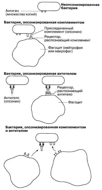
Fig. 14.36. The role of the complement system and antibodies (opsonins) in "tagging" (opsonization) a bacterium. The opsonized bacterium is recognized and engulfed by a phagocyte. (The term "opsonization" comes from the Greek word opson, meaning seasoning.)

Fig. 14.37. Phagocytosis: engulfment and digestion of a bacterium by a neutrophil.
Inflammation
When a part of the body is injured or infected, a local reaction of the surrounding tissues occurs, manifested by Swelling and pain. This condition is called inflammation; it is caused by the release of specific substances from damaged tissues, including histamine and 5-hydroxytryptamine (serotonin). These substances induce local capillary dilation, which increases blood flow to the affected area and raises its Temperature. Capillary permeability also increases, allowing plasma and leukocytes to pass into the surrounding tissues. This condition is called edema. Plasma contains substances that inhibit or kill bacterial growth, as well as antibodies and phagocytes; together, these agents prevent the spread of infection. One of the chemical substances delivered to the inflamed area is a special protein called interferon, secreted primarily by macrophages and other leukocytes in response to foreign Antigens. It non-specifically enhances the resistance of body cells to viral infection. In addition to pathogens, phagocytes engulf dead tissue debris, thereby clearing the affected area of any unwanted material. Furthermore, plasma contains fibrinogen, which promotes blood clotting if necessary, while excess tissue fluid reduces the concentration and toxicity of any potentially harmful agents.
Wound Healing
Toward the end of the inflammatory process, the affected area is colonized by cells called fibroblasts, which secrete the intercellular elements of connective tissue, mainly Collagen. Collagen is a fibrous protein; by combining with other fibroblast products—mucopolysaccharides—it forms a network of randomly interlaced scar tissue fibers. Vitamin C is essential for collagen formation, as hydroxylation cannot occur without it, leaving the collagen molecules incomplete (lacking Amino Acids such as hydroxyproline and hydroxylysine). After about two weeks, this disordered mass of fibers reorients along the tension lines of the damaged area. Numerous capillaries sprout into the region, supplying the cells involved in the wound healing process with oxygen and nutrients.
Simultaneously, the surrounding epidermis becomes involved in these processes occurring deep within the wound. Some epidermal cells migrate into the wound and digest a significant portion of the accumulated tissue debris and blood clot fibrin. Upon meeting one another, the epidermal cells firmly connect, gradually forming a continuous layer beneath the scab. When the formation of this layer is complete, the scab falls off, and the new epidermis comes into contact with the atmosphere.
CHRONOLOGICAL SEQUENCE OF WOUND HEALING STAGES
1. Bleeding occurs in the damaged area.
2. Blood clotting takes place.
3. The inflammatory process begins.
4. Leukocytes migrate into the wound to engulf foreign material, bacteria, and cellular debris.
5. Fibroblasts colonize the wound, synthesizing collagen and other elements of scar tissue.
6. Epidermal cells finally clear the wound of dead debris and begin separating the scab from the living tissues beneath it.
7. A continuous layer of epidermis forms at the wound site.
8. The scab detaches and sloughs off.
If the wound is small, phagocytes usually cope with the pathogens that have entered it. However, in cases of extensive damage, the body's immune system comes into play.
Last update: 06/08/2026
Editorial and Educational Adaptation: This material has been compiled based on the primary/original source text. The project team performed an editorial review, corrected technical inaccuracies, structured sections, and adapted the content for an educational format.
What was processed:
- elimination of formatting defects (OCR errors, structural breaks, corrupted characters);
- editorial organization of content;
- standardization of terminology in accordance with academic sources;
- verification of factual statements against the original source text.
All mentions of the author, publication year, and origin of the primary text have been preserved in accordance with the source.