Cytology, General Histology and Embryology - V. K. Napkhanyuk 2002
Tissues
Connective Tissues
Dense Fibrous Connective Tissue
Dense Fibrous Connective Tissue (textus connectivus collagenosus compactus) is a type of tissue characterized by an Abundance of densely packed fibers (predominantly Collagen) and a minimal presence of cellular elements and amorphous ground substance between them.
This structural feature gives the tissue high Shock-absorbing and mechanical properties. Depending on the arrangement of its fibrous structures, the tissue is subdivided into dense irregular and dense regular connective tissue.
Dense irregular connective tissue (textus connectivus collagenosus compactus irregularis) features a robust, non-uniform orientation of fibers.
This type of tissue forms the reticular layer of the dermis. Within it, bundles of collagen fibers run in various directions, forming a meshwork. This architecture provides the Skin with resistance to mechanical forces acting from multiple angles. Fibroblasts, macrophages, neurovascular bundles, and the extracellular ground substance reside between the collagen fiber bundles.
Dense regular connective tissue (textus connectivus collagenosus compactus regularis) is a key component of tendons, ligaments, and fibrous membranes. It is distinguished by a parallel and strictly ordered arrangement of its fibers.
Tendons (tendines) are formed by thick, densely packed collagen fibers, interspersed with fibrocytes, a small number of fibroblasts, and ground amorphous substance.
A bundle of collagen fibers enveloped by a layer of fibrocytes is referred to as a primary tendon bundle.
Several primary bundles, enclosed within thin layers of loose fibrous connective tissue, form secondary bundles. The connective tissue partition separating the secondary bundles is called the endotenon.
Secondary bundles group together to form tertiary bundles, which are divided by thicker partitions of loose fibrous connective tissue known as the peritenon.
Large tendons may also feature quaternary bundles.
Running through the endotenon and peritenon are Blood Vessels that nourish the tendon, along with nerves and proprioceptive nerve endings that Relay signals regarding tissue tension to the Central Nervous system.
The nuchal ligament also belongs to dense regular connective tissue, though its bundles are formed by elastic fibers and are less distinctly segregated.
At sites of bone attachment, certain tendons are enclosed within sheaths (vaginae) composed of two fibrous connective tissue layers containing a fluid rich in hyaluronic acid.
Fibrous membranes. This category of dense fibrous connective tissue includes fascias, aponeuroses, the central tendon of the Diaphragm, organ capsules, the dura mater, sclera, perichondrium, periosteum, and the tunica albuginea of the Testis and Ovary.
Fibrous membranes are exceptionally resistant to stretching because their bundles of collagen fibers, along with interspersed fibroblasts and fibrocytes, are arranged methodically in multiple superimpose layers. Each layer contains wavy, parallel bundles of collagen fibers, with the orientation of fibers varying across layers. Individual fiber bundles may bridge adjacent layers, tying them together. In addition to collagen fibers, fibrous membranes also contain elastic fibers.
Slides for Examination
Slide 19. Connective tissue fibers (Fig. 71).
Low magnification. Locate the clearest area of the slide.
High magnification. Against the backdrop of a transparent ground substance, observe thick, slightly wavy, pink-colored collagen fibers alongside thin, branching, violet-colored elastic fibers.
Draw the slide. Label the diagram: 1 — collagen fiber; 2 — elastic fiber.
Slide 20. Dense regular connective tissue. Cytology/practical/54.html">Longitudinal section of a tendon (Fig. 72).
Low magnification. At this magnification, tendon bundles are clearly visible, separated by partitions of loose irregular connective tissue—the endotenon—which is densely populated by the nuclei of connective tissue Cells. Cytoplasm is poorly visualized.
High magnification. Collagen fibers stained pink with eosin run parallel to one another (primary bundles). Fibrocyte nuclei lie alongside them. The endotenon groups several primary bundles into secondary bundles. The peritenon—a layer of loose fibrous connective tissue surrounding a group of collagen fibers (tertiary bundles)—contains longitudinally or transversely cut blood vessels.
Draw the slide. Label the diagram: 1) primary collagen fiber bundles; 2) fibrocytes; 3) secondary collagen fiber bundles; 4) endotenon.
Class="center">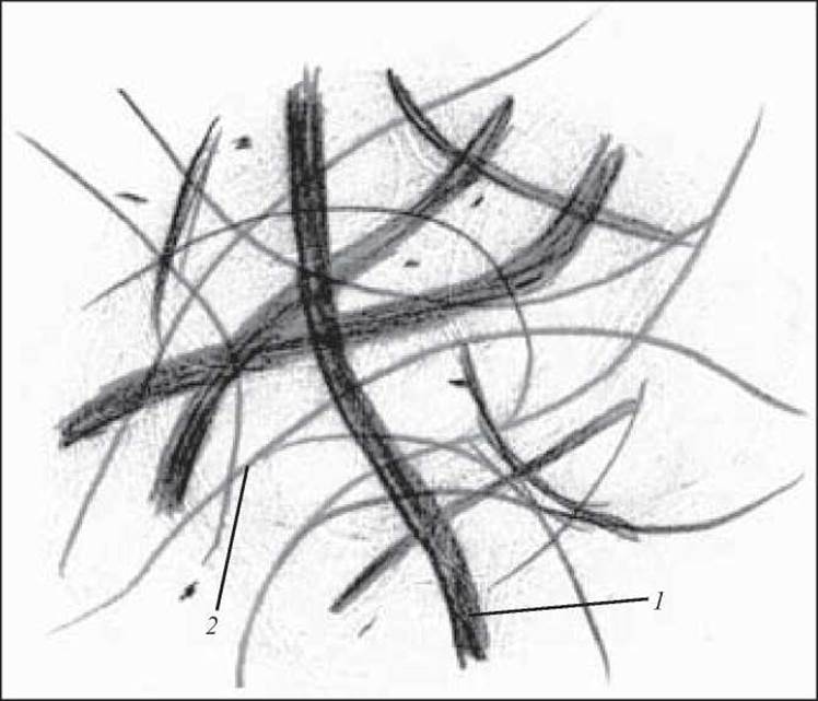
Fig. 71. Collagen and elastic fibers of loose connective tissue. Resorcin-fuchsin staining. x 400:
1 — collagen fiber; 2 — elastic fiber

Fig. 72. Dense regular connective tissue. Longitudinal section of a tendon. Hematoxylin and eosin staining. x 80:
1 — primary bundles of collagen fibers; 2 — fibrocytes; 3 — secondary bundles of collagen fibers; 4 — endotendon
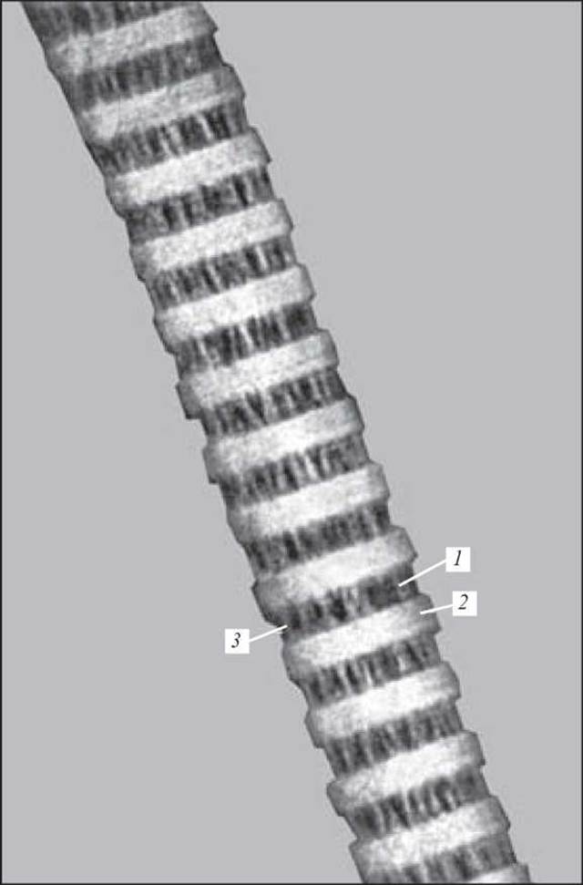
Fig. 73. Collagen fibril from a rat tendon. Electron micrograph. x 160,000. Negative staining with phosphotungstic acid at pH 7.4:
1 — dark band; 2 — light band; 3 — collagen protofibrils (after V. P. Hilyov)
1. Extracellular matrix and characteristics of its constituent components.
2. Morphofunctional characteristics of collagen fibers.
3. Levels of Organization of the collagen fiber. Types of collagen.
4. Elastic fibers. Structure, chemical composition, and functional significance.
5. Reticular fibers. Structure and Functional significance.
6. Chemical composition, functional significance, and Water/144.html">Origin of the ground amorphous substance.
7. Morphofunctional characteristics of various types of Dense connective tissue.
8. Tendon. Structure and function.
9. Fibrous membranes. Structure and function.
10. Dense irregular connective tissue, structure, and Functions.
Situational Problems
An inflammation involving blood and connective tissue cells occurs at the site of a foreign body penetration into the Organism. Which blood and connective tissue cells will be detected in the inflammation focus?
Sample Examination Questions
1. Morphofunctional characteristics of collagen fibers. Levels of collagen organization.
2. Elastic fibers. Structure and Chemical composition. Functional significance.
3. Reticular fibers. Structure. Functional significance.
4. Chemical composition, functional significance, and origin of the main amorphous component.
5. Morphofunctional characteristics of various types of dense fibrous connective tissue.
Last update: 10/08/2026
Editorial and Educational Adaptation: This material has been compiled based on the primary/original source text. The project team performed an editorial review, corrected technical inaccuracies, structured sections, and adapted the content for an educational format.
What was processed:
- elimination of formatting defects (OCR errors, structural breaks, corrupted characters);
- editorial organization of content;
- standardization of terminology in accordance with academic sources;
- verification of factual statements against the original source text.
All mentions of the author, publication year, and origin of the primary text have been preserved in accordance with the source.