Cytology, General Histology and Embryology - V. K. Napkhanyuk 2002
Tissues
Connective Tissues
Connective Tissues with Special Properties
Connective Tissues with special properties are tissues characterized by the predominance of uniform Cells, which determine the name of the tissue itself.
Accordingly, connective tissues with special properties are classified as follows: reticular Connective Tissue, adipose tissue, mucous tissue, and pigment tissue.
Reticular connective tissue (textus connectivus reticularis) is a type of connective tissue that has a net-like Structure and consists of reticular cells and reticular (argyrophilic) fibers.
Reticular cells (reticulocytus) are characterized by a large, pale Nucleus and a process-bearing Cytoplasm. Connecting via their processes, they form a network.
There are several types of reticular cells:
— reticular fibroblast-like cells;
— phagocytic cells of monocytic origin;
— unspecialized (less differentiated) cells.
Reticular fibers (fibra reticularis) are derivatives of reticular cells. Reticular fibers are revealed by impregnation with silver salts, hence they are called argyrophilic. In their chemical composition, reticular fibers are close to Collagen fibers, but differ from them in smaller thickness, branching, and anastomoses.
A distinction is made between proper reticular fibers and precollagenous fibers.
Proper reticular fibers are definitive, final formations containing type III collagen.
Precollagenous fibers represent the initial stage of collagen fiber formation during Embryogenesis and regeneration.
Reticular fibers differ from collagen fibers by a high content of sulfur, Lipids, and CARBOHYDRATES. Under an Electron microscope, the fibrils of reticular fibers do not always show a clearly defined striation with a period of 64-67 nm.
Reticular fibers are resistant to weak acids and alkalis and are not digested by Trypsin.
Most reticular cells are associated with reticular fibers; connecting with each other through their processes, they form a loose network (reticulum).
Reticular tissue forms the stroma of Hematopoietic Organs and the microenvironment of the Blood Cells developing within them.
Adipose tissue (textus adiposus) is an accumulation of fat cells found in many organs; its specific function is the storage and METABOLISM of lipids.
There are two varieties of adipose tissue: white and brown.
Cytology/practical/49.html">White adipose tissue (textus adiposus albus) consists of unilocular fat cells (adipocytes) and is located subcutaneously, in the omentum of the mesentery, retroperitoneally, and in other fat depots.
Adipocytes (adipocytus uniguttularis) contain a condensed nucleus located at the periphery. The cytoplasm forms a thin rim and accounts for approximately 1/40 of the total Cell volume, with the rest occupied by a single large fat droplet.
The Golgi complex is located near The Nucleus, while the cytoplasmic rim contains occasional Mitochondria, a small amount of granular Endoplasmic reticulum, and sparse Ribosomes.
White adipose tissue forms lobules separated by layers of Fibrous connective tissue. Each fat cell within a lobule is surrounded by a network of reticular fibers, as well as a network of Blood and Lymphatic capillaries. Fibroblasts and mast cells are located between the fat cells.
Brown adipose tissue (textus adiposus fuscus) is formed by multilocular adipocytes densely intertwined with blood capillaries; it is found in newborns on the neck, near the scapulae, behind the Sternum, along the spine, subcutaneously and between Muscles, as well as in some animals.
The multilocular adipocyte (adipocytus multiguttularis) is a cell significantly smaller than the white adipocyte. It has a polygonal shape and contains a rounded nucleus located eccentrically, but not pressed against the Plasmalemma.
The cytoplasm is filled with numerous small fat droplets and contains a small Golgi complex and numerous spherical mitochondria with well-developed cristae.
The primary function of brown Adipose tissue is thermogenesis; consequently, its oxidative capacity is approximately 20 times higher than that of white adipose tissue.
Mucous connective tissue (textus connectivus mucosus), also known as gelatinous connective tissue, is an embryonic form of loose connective tissue.
During prenatal development, it is found in many organs, particularly beneath the Skin.
A classic subject for its study is the umbilical cord of the human fetus, specifically its Wharton's jelly.
Mucous tissue consists of large, pale-staining stellate cells whose processes frequently contact one another, embedded in a gelatinous intercellular substance.
The intercellular substance is composed of an amorphous ground substance rich in hyaluronic acid and sparse, thin collagen fibrils.
As the number of collagen fibrils increases in the gelatinous tissue of the fetal body, it is gradually replaced by fibrous connective tissue characteristic of the adult Organism.
Pigmented connective tissue (textus connectivus pigmentosus) is a loose fibrous connective tissue whose cells contain large amounts of the pigment melanin. Consequently, these cells are termed melanocytes (cellula pigmentosa).
Examples of this tissue include the stroma of the iris and the choroid of the eye.
The photomicrographs (see Figs. 76 – 78) will further illustrate the cellular diversity of connective tissues.
Slides for Study
Slide 21. Reticular tissue of a Lymph node (Fig. 74).
Low magnification. Locate the clearest area in the center of the slide, where stellate cells with large, faintly stained nuclei and pale pink cytoplasm are clearly visible. Interspersed among them are cells with round, dense violet nuclei and a narrow rim of basophilic cytoplasm (lymphocytes).
High magnification. Examine the structural details of individual components and make a drawing.
Label the following in the drawing: 1) reticular cells: a — nuclei; b — cytoplasm; 2) macrophage; 3) lymphocytes; 4) lymph node trabecula.
Slide 22. Adipose tissue from human subcutaneous tissue (Fig. 75).
Low magnification. Locate clusters of fat cells arranged in cord-like rows along Blood Vessels.
High magnification. Examine The structure of unilocular fat cells. Almost the entire cell is unstained because it is filled with a lipid droplet, the visualization of which requires a special stain. The pale pink cytoplasm forms a thin peripheral rim containing a condensed, pale blue nucleus. Draw the slide.
Label the following in the drawing: 1) fat cells: a — cytoplasm; b — nucleus; c — site of the former lipid droplet dissolved by alcohol; 2) blood vessels; 3) fibrous connective tissue.
Class="center">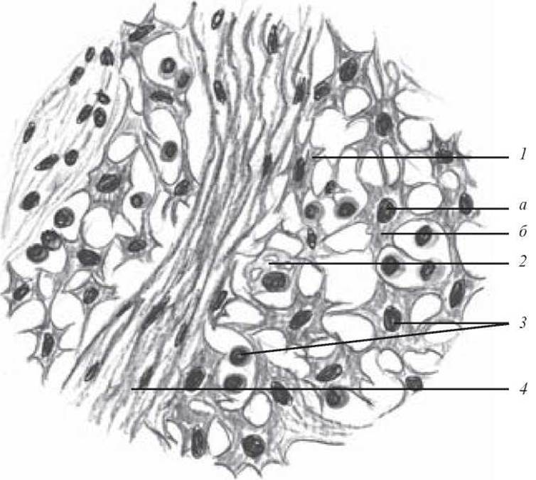
Fig. 74. Reticular tissue of a lymph node. Hematoxylin-eosin stain. × 900:
1 — reticular cells (a — nuclei; b — cytoplasm); 2 — macrophage; 3 — lymphocyte; 4 — lymph node trabecula
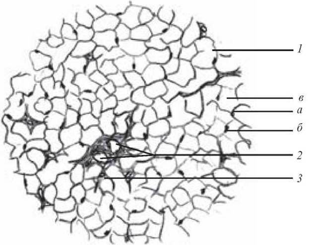
Fig. 75. Adipose tissue of the subcutaneous layer. Hematoxylin-eosin stain. × 160:
1 — fat cells (a — cytoplasm; b — nucleus; c — site of the former lipid droplet dissolved by alcohol); 2 — blood vessels; 3 — fibrous connective tissue
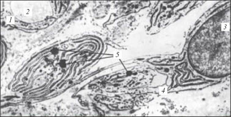
Fig. 76. Transformation of an adventitial cell into a fat cell. Initial stage. Electron micrograph. x 13,000:
1 — endothelial cell of a blood capillary; 2 — lumen of the blood capillary; 3 — Nucleus of the adventitial cell; 4 — endoplasmic reticulum; 5 — accumulation of lipid granules (after Neapolitan)
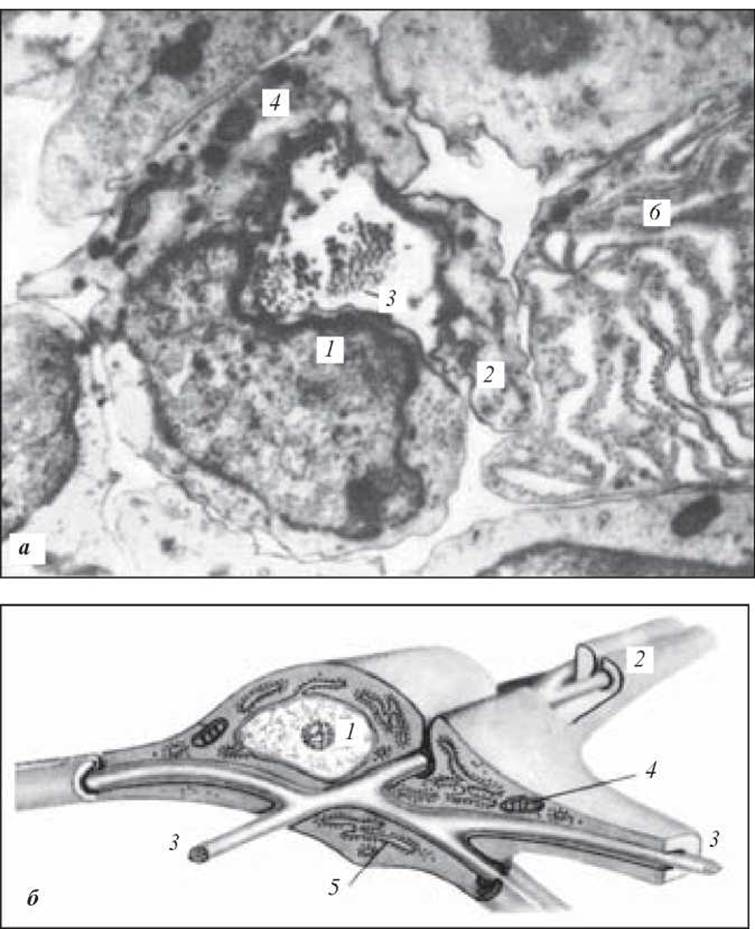
Fig. 77. Reticular tissue from a lymph node (a — electron micrograph; b — relationship between a reticular cell and reticular fibers). x 13,000:
1 — nucleus of the reticular cell; 2 — cytoplasmic processes of the reticular cell; 3 — reticular fibers; 4 — mitochondria; 5 — endoplasmic reticulum; 6 — portion of a plasma cell (after Thum and Clark)
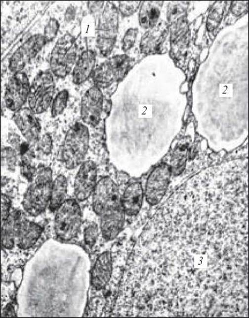
Fig. 78. Brown adipose tissue cell of a newborn rat. Electron micrograph. x 23,000:
1 — mitochondria; 2 — lipid inclusions; 3 — nucleus (after Yu. I. Afanasyev)
1. Reticular tissue. Structural features, body localization, and Functions.
2. Comparative structural and Functional Characteristics of white and brown adipose tissue.
3. Mucous tissue: localization, structural features, Chemical composition of the Extracellular matrix, and functions.
4. Pigment tissue. Structural and functional characteristics, and localization.
Case Study Problems
1. Two adipose tissue slides are stained with Sudan to detect fat. One slide contains adipocytes whose cytoplasm features numerous small lipid droplets, with a spherical nucleus located in the center of The Cell. The second slide contains adipocytes with a single large lipid droplet in the cytoplasm and a condensed nucleus shifted to the periphery. What types of adipose tissue are represented in these slides?
2. Examine slides of mucous connective tissue from two human fetuses. What feature will allow you to determine which fetus was older?
Sample Examination Questions
1. Structural and functional characteristics of various types of Connective tissue with special properties.
Last update: 10/08/2026
Editorial and Educational Adaptation: This material has been compiled based on the primary/original source text. The project team performed an editorial review, corrected technical inaccuracies, structured sections, and adapted the content for an educational format.
What was processed:
- elimination of formatting defects (OCR errors, structural breaks, corrupted characters);
- editorial organization of content;
- standardization of terminology in accordance with academic sources;
- verification of factual statements against the original source text.
All mentions of the author, publication year, and origin of the primary text have been preserved in accordance with the source.