Cytology, General Histology and Embryology - V. K. Napkhaniuk 2002
Tissues
Skeletal Tissues
Cartilage Tissues
Skeletal Tissues (textus sceletales) are tissues that primarily perform supporting, protective, and mechanical Functions, and also participate in Water-Salt METABOLISM.
Skeletal tissues include cartilaginous and Bone Tissues.
Cartilaginous tissues (textus cartilaginei) consist of Cells (chondrocytes and chondroblasts) and a large amount of Extracellular matrix, which is characterized by elasticity; they are part of certain Organs of the Respiratory system, joints, and intervertebral discs. This is a type of tissue devoid of Blood Vessels.
Cartilage tissue contains about 75% water, 10–15% organic matter, and 4–7% inorganic salts. The dry matter contains from 50 to 70% Collagen.
Depending on their Structure and Functional features, several types of cartilaginous tissues are distinguished (Fig. 79).
Histogenesis of Cartilaginous Tissues
The Development of cartilage tissue (chondrohistogenesis) takes place during the embryonic and postembryonic periods, as well as during regeneration.
Embryonic chondrohistogenesis. The source of cartilage tissue development in the Embryogenesis period is mesenchyme. The process of development of this tissue is divided into the following stages.
The First stage is The formation of the chondrogenic primordium, or chondrogenic islet. In those areas of the embryo's body where cartilage forms, the mesenchyme condenses, the cells lose their processes, proliferate intensively, and closely adhere to one another, creating a certain tension—turgor. These areas are called the chondrogenic primordium (chondrogenic islet). Mesenchymal cells that make up the chondrogenic islet differentiate into chondroblasts—cells that will give rise to the formation of cartilage tissue.
The Second Stage is the Formation of primary cartilage tissue. During this period, the cells in the central area round up and increase in size. A granular Endoplasmic reticulum develops in the Cytoplasm, through which the Synthesis and Secretion of Fibrillar Proteins (type III collagen) take place. Chondroblasts transform into first-order chondrocytes.
Class="center">
Fig. 79. Classification of cartilaginous tissues
The Third Stage is the differentiation of cartilage tissue. At this stage, primary chondrocytes transform into secondary ones and acquire The ability to synthesize, In addition to the aforementioned substances, sulfated glycosaminoglycans.
At the periphery of the cartilage rudiment, at the border with the mesenchyme, the perichondrium is formed—a sheath covering the outside of the developing cartilage. It consists of an outer fibrous and an inner chondrogenic (cambial) layer.
The growth of cartilage tissue can occur in two ways:
— appositional (external) growth, whereby cells divide intensively in the chondrogenic zone, and chondroblasts differentiate into chondrocytes that produce the extracellular matrix. In the process of secretion of synthesis products and deposition onto the existing cartilage along its periphery, the cells become embedded in the products of their own vital activity;
— interstitial (internal) growth.
Cartilage cells located in the center of the young developing cartilage retain the ability to divide mitotically for some time, remaining within a single lacuna (isogenic groups). Due to the increase in the number of these cells, the mass of the cartilage increases from within. Such growth is observed in embryogenesis and during the regeneration of cartilage tissue.
Regeneration. Physiological regeneration of cartilage tissue is carried out by poorly specialized Cells of the perichondrium and cartilage through the proliferation and differentiation of prechondroblasts and chondroblasts.
Cells of Cartilage Tissue
In the process of cartilage tissue development, a differon is formed—stem cells, semi-stem cells (prechondroblasts), chondroblasts (chondroblastocytes), and chondrocytes (see Fig. 83, 84).
Chondroblasts (chondroblastocytus) are young, poorly differentiated cells of irregular, elongated, or flattened shape, capable of proliferation and Synthesis of the extracellular matrix.
The cytoplasm stains basophilically, containing a well-developed granular and agranular endoplasmic reticulum, elements of the Golgi complex, and abundant RNA.
In the process of cartilage development, chondroblasts transform into chondrocytes. Chondroblasts mediate the peripheral (appositional) growth of cartilage.
Chondrocytes (chondrocyti) are the main type of cartilage tissue cells, which have an oval, rounded, or polygonal shape and are located in special cavities—lacunae (lacuna cartilagine) of the extracellular matrix, singly or in groups.
Cell groups originating from a single cell are referred to as isogenous groups.
An isogenous chondrocyte group contains three cell types:
— the first cell type predominates in developing young cartilage, where Cell Division is frequently observed, suggesting they serve as a source of cell renewal. These cells exhibit a high nucleocytoplasmic ratio;
— the second cell type is characterized by a reduced nucleocytoplasmic ratio, a decline in DNA Synthesis, an increased rate of RNA Synthesis, and well-developed granular Endoplasmic reticulum and Golgi apparatus;
— the third cell type features the lowest nucleocytoplasmic ratio, along with extensive development and orderly arrangement of the granular endoplasmic reticulum. This cell type maintains the ability to synthesize and secrete proteins while glycosaminoglycan synthesis decreases.
Intercellular Substance
The intercellular substance of cartilaginous tissues (substantia intercellularis) consists primarily of organic components (proteins, Lipids, glycosaminoglycans, Proteoglycans). The concentration of the latter is highest in this type of Connective Tissue. There is an Abundance of fibrillar proteins, predominantly type II collagen. The orientation of the fibers is determined by the lines of force arising from cartilage deformation during organ function. The layer of intercellular substance adjacent to the cellular lacuna, forming its wall, exhibits strong birefringence and contains a network of fibrils. This layer is known as the cartilage cell capsule.
Types of Cartilage Tissue
Hyaline cartilage tissue (textus cartilagineus hialinus) is characterized by an intercellular substance that appears homogeneous in hematoxylin-eosin stained preparations and contains no visible fibers. In reality, the intercellular substance of hyaline cartilage contains extremely thin collagen fibrils that share the same refractive index as the surrounding matrix, making them invisible in conventional histological preparations.
Hyaline cartilage tissue is externally covered by the perichondrium.
The perichondrium consists of two layers:
— an outer layer composed of Fibrous connective tissue containing blood vessels;
— an inner layer consisting mainly of pre-chondroblasts and chondroblasts.
Beneath the perichondrium, in the Superficial layer of the cartilage proper, lie young spindle-shaped chondrocytes whose long axes are oriented parallel to the cartilage surface.
In the deeper layers of the cartilage proper, chondrocytes become oval or round and aggregate into cell clusters known as isogenous groups. Young chondrocytes and isogenous groups are surrounded by chondromucoid and collagen fibers (type II collagen).
Hyaline cartilage tissue is found in the walls of the Trachea and Bronchi, at the junctions of the Ribs AND Sternum, on articular surfaces, and in epiphyseal growth plates.
The structure of hyaline cartilage across various organs shares many common features while also displaying organ-specific characteristics.
The articular hyaline cartilage lacks a perichondrium On the surface facing the joint cavity and consists of three zones:
— an outer zone containing small, flattened, poorly differentiated cells;
— a middle zone containing larger oval cells arranged in columns perpendicular to the surface;
— a deep zone consisting of calcified cartilage; blood vessels are found exclusively in this zone.
Elastic cartilage tissue (textus cartilagineus elasticus) is found in the auricle, epiglottis, and the corniculate and cuneiform cartilages of the Larynx. In its general structure, elastic cartilage resembles hyaline cartilage, with the key distinction that hyaline tissue lacks elastic networks.
Elastic fibers permeate the intercellular matrix of the cartilage in all directions. In the layers adjacent to the perichondrium, these elastic fibers merge continuously with the elastic fibers of the perichondrium. Lipids, Glycogen, and chondroitin sulfates are less abundant in elastic cartilage than in hyaline cartilage; furthermore, elastic cartilage never undergoes calcification.
Fibrocartilage tissue (textus cartilagineus fibrosa) occupies an intermediate structural position between Dense Fibrous Connective tissue and cartilage tissues. It is located in areas where fibrous connective tissue transitions into hyaline cartilage (such as tendons and ligaments) where limited motion is accompanied by strong tensile stress.
The intercellular matrix contains parallel bundles of collagen fibers that gradually fan out and merge into the hyaline cartilage. The lacunae within the matrix contain cartilage cells, either isolated or arranged in isogenous groups. Moving from the hyaline cartilage toward the tendon, fibrocartilage increasingly resembles tendinous tissue.
Slides for study
Slide 23. Hyaline cartilage (Fig. 80).
Low magnification. Locate the perichondrium. It surrounds the cartilage plate on all sides. In the perichondrium, one can distinguish the fibrous layer containing blood vessels, beneath which lies the chondrogenic layer containing chondroblasts. Beneath the perichondrium is the hyaline cartilage tissue, which consists of isolated cells, isogenous groups, and an extracellular matrix stained in pinkish-purple.
High magnification. Locate the young flattened chondrocytes situated beneath the perichondrium. Mature, oval-shaped cartilage cells are located deeper. Isogenous groups of cartilage cells (2–4 cells) lie within a single capsule in the extracellular matrix. Surrounding the isogenous cells, the extracellular matrix is basophilically stained purple—this is the territorial matrix of the cells, whereas the interterritorial matrix is weakly basophilic (pale purple). Make a drawing of the slide.
Label the following on the drawing: 1) perichondrium: a) young cartilage cells; b) extracellular matrix; c) cartilage cells; d) cartilage cell capsule; e) cell territories; f) isogenous group of cartilage cells.
Slide 24. Elastic cartilage of the auricle (Fig. 81).
Low magnification. It can be seen that the General structural layout is similar to that of hyaline cartilage.
High magnification. The boundaries of isogenous cell groups are clearly visible. Elastic fibers, which stain selectively blue, run in various directions.
Make a drawing of the slide. Label the following on the drawing: 1) perichondrium: a) ground substance; b) elastic fibers; c) cartilage cell; d) cartilage capsule; e) isogenous group of cartilage cells.

Fig. 80. Hyaline cartilage. Hematoxylin-eosin staining. x 400:
1 — perichondrium; 2 — young cartilage cells; 3 — extracellular matrix; 4 — cartilage cells; 5 — cartilage cell capsule; 6 — cell territories; 7 — isogenous group of cartilage cells
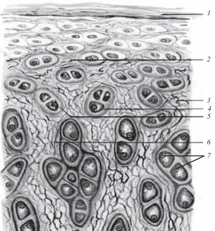
Fig. 81. Elastic cartilage of the auricle. Resorcin-fuchsin staining. x 400:
1 — perichondrium; 2 — ground substance; 3 — elastic fibers; 4 — cartilage cell; 5 — cartilage capsule; 6 — isogenous group of cartilage cells; 7 — Nucleus of the cartilage cell
Slide 25. Fibrocartilage (Fig. 82).
Low magnification. The slide shows regions of both hyaline and fibrocartilage.
High magnification. Bundles of collagen fibers run in a somewhat oblique direction and stain pink. Chondrocytes are elongated and arranged in rows (chains) between the fibers. Make a drawing of the slide.
Label the following on the drawing: 1) hyaline cartilage tissue; 2) fibrocartilage tissue; 3) chondrocytes; 4) extracellular matrix.

Fig. 82. Fibrocartilage. Hematoxylin-eosin staining. x 80:
1 — chondrocytes; 2 — extracellular matrix; 3 — collagen fibers
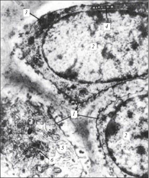
Fig. 83. Cartilage cells. Electron micrograph. x 9500:
1 — cartilage cells; 2 — large nuclei of cartilage cells; 3 — lipid granules; 4 — Mitochondria; 5 — endoplasmic reticulum; 6 — glycogen granules (from Rodin's atlas)

Fig. 84. Cartilage CELL AND EXTRACELLULAR matrix. Electron micrograph of a portion of a cartilage cell (chondrocyte) and extracellular matrix from rat cartilage regenerate. х 30,000:
1 — endoplasmic reticulum; 2 — Ribosomes; 3 — glycogen granules; 4 — lipid droplets; 5 — cell membrane (Plasmalemma); 6 — collagen fibrils in the extracellular matrix (after L. N. Mikhailova)
1. General morphofunctional characteristics of cartilage tissue. Classification.
2. Cellular elements and their characteristics.
3. Extracellular matrix. Structure. FEATURES OF CHEMICAL composition.
4. Structural Organization of hyaline cartilage. Localization.
5. Elastic cartilage. Structural features. Localization.
6. Fibrocartilage. Structural features. Localization.
7. MAIN STAGES OF chondrohistogenesis.
8. STRUCTURE AND FUNCTIONS of the perichondrium.
9. Growth of cartilage. Its regeneration and age-related changes.
Situational Problems
1. There are two slides, one containing hyaline cartilage and the other elastic cartilage. What features can be used to distinguish between them?
2. A histological section of cartilage tissue shows numerous thick bundles of collagen fibers. Which type of cartilage tissue does this represent?
Sample Exam Questions
1. Histogenesis of cartilage tissue.
2. Structure of hyaline cartilage.
3. Structure of elastic cartilage.
4. Structure of fibrocartilage.
Last update: 10/08/2026
Editorial and Educational Adaptation: This material has been compiled based on the primary/original source text. The project team performed an editorial review, corrected technical inaccuracies, structured sections, and adapted the content for an educational format.
What was processed:
- elimination of formatting defects (OCR errors, structural breaks, corrupted characters);
- editorial organization of content;
- standardization of terminology in accordance with academic sources;
- verification of factual statements against the original source text.
All mentions of the author, publication year, and origin of the primary text have been preserved in accordance with the source.