Human Histology - O.D. Lutsyk 2003
General Histology
Tissues of the Internal Environment. Morphology and Function of Blood
Tissues of the Internal Environment comprise a large group of tissues that, along with epithelium, belong to the so-called general tissues. The tissues of the internal environment include Blood, Lymph, and Connective Tissue with all its varieties. Although individual varieties of Internal Environment Tissues differ significantly in appearance (for example, blood and Bone tissue), there are ample grounds for grouping them into a single tissue type based on their common origin, Structure, and Functions.
The common origin of these tissues is their most defining feature, as they all develop from mesenchyme (Fig. 3.8). Mesenchyme is the most primitive connective tissue, existing only during Cytology/cytology/16.html">Early stages of embryonic development. Structurally, mesenchyme resembles a net because its stellate or spindle-shaped Cells are interconnected by cytoplasmic processes. The meshes of this reticular framework contain a gelatinous ground substance—the intercellular matrix—whose density fluctuates with changes in METABOLISM. Blood, lymph, and all types of connective tissue develop from mesenchyme through differentiation.
The structural commonality of these tissues lies in the presence of an intercellular matrix, which quantitatively predominates over the cells. Based on The structure of this intercellular matrix, the MAIN TYPES OF internal environment tissues can be distinguished:
Class="center">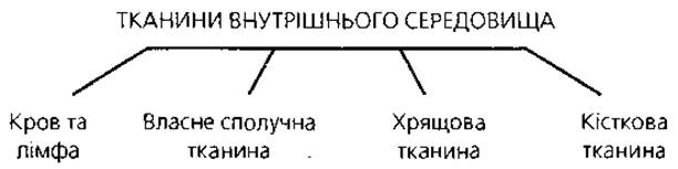
The intercellular matrix of BLOOD AND LYMPH is liquid (plasma), and fibrous structures are absent; therefore, these types of internal environment tissues have a fluid consistency, although blood can lose its fluidity when fibrinogen converts into fibrin during coagulation.
In Proper Connective Tissue, The amount of fibers can be moderate (loose Fibrous connective tissue) or substantially greater (Dense Fibrous Connective tissue). The consistency depends on The ratio of the ground intercellular substance to fibers (predominantly collagenous).
Cartilage tissue contains a well-developed ground intercellular substance and fibers, making this type of internal environment tissue exceptionally strong and resilient; thus, it belongs to the so-called Skeletal Tissues.
Bone tissue contains a well-developed ground intercellular substance. A high degree of mineralization (about 70% of bone tissue consists of calcium phosphate salts in the form of hydroxyapatite crystals) and thick parallel bundles of Collagen fibers (so-called bone lamellae) ensure the mechanical strength of the bone.
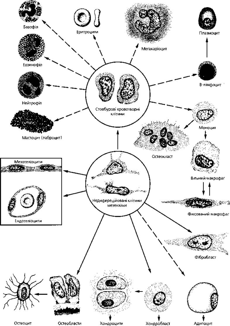
Fig. 3.8. Schematic representation of the interrelationship among mesenchyme derivatives—cellular elements of blood, connective, cartilage, and Bone Tissues, as well as endothelium and mesothelium. Dashed lines indicate the presence of intermediate Cell forms. Scale is not preserved, as the size of adipocytes, megakaryocytes, and osteoclasts significantly exceeds that of all other cell types.
The functions of internal environment tissues are diverse, but they are commonly grouped under the general term "supportive-trophic tissues." They perform the following functions: trophic, protective, and supportive (mechanical). The Functional Characteristics of various types of internal environment tissues largely depend on the PHYSICOCHEMICAL PROPERTIES OF their intercellular matrix.
Morphology and FUNCTIONS OF BLOOD
Blood (sanguis) is a fluid body tissue circulating within a system of closed vascular tubes. Blood accounts for 1/13, or 5–9%, of body mass, which in an adult equals approximately 5.0–5.5 L. Blood consists of a liquid portion—plasma, which makes up 55–60% of the volume—and formed elements, which account for 40–45% of the volume (Fig. 3.9). Plasma is the intercellular matrix of blood. The formed elements of blood include erythrocytes, leukocytes, and thrombocytes (Blood Platelets).
Blood performs A number of vital functions. Its protective function involves providing humoral and cellular Immunity. The respiratory function is carried out by transporting oxygen and carbon dioxide. The Essence of the trophic function is The transport of nutrients. The excretory function consists of removing Metabolic waste products. The humoral function is ensured through the transport of Hormones and other BIOLOGICALLY ACTIVE SUBSTANCES. The homeostatic function maintains the constancy of the body's internal environment, including immune Homeostasis.
Blood Plasma is a colloidal solution whose viscosity is 5 times higher than that of Water. Plasma contains 90-93% water and 7–10% dry residue. Of the dry residue, about 7% consists of Proteins and 3% of other organic and Mineral Substances. The total concentration of mineral substances in blood plasma is 0.9%; the plasma pH is 7.36.
Plasma Proteins include:
1) albumins, which account for about 4%; they bind and transport a wide range of substances via the bloodstream;
2) globulins, accounting for 1.1–3.1%, subdivided into alpha, beta, and gamma globulins (IMMUNOGLOBULINS); the latter fraction contains Antibodies;
3) fibrinogen, accounting for 0.2–0.4%, which is crucial because its ability to convert into an insoluble form—fibrin—drives the blood clotting process.
Plasma from which fibrin has been removed is called blood serum. It is a yellowish, transparent fluid used in the manufacture of numerous pharmaceutical preparations.
Formed Elements of Blood
Erythrocytes (Fig. 3.10), or red Blood Cells, in mammals and humans are non-motile, highly differentiated cells that, during their development, have lost their Nucleus and all cytoplasmic Organelles, becoming adapted to perform virtually a single function—Respiration—which is mediated by the presence of the respiratory pigment Hemoglobin.
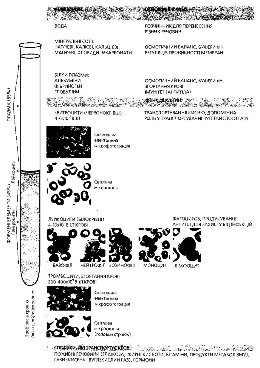
Fig. 3.9. General Overview of the composition and functions of blood
The total number of erythrocytes in an individual's blood is approximately 25x1012. The total volume of erythrocytes in humans is about 2 L. In blood tests, the content of all formed elements is expressed per unit volume—1 L. Accordingly, the erythrocyte count is 3.9x10¹² to 6.0x10¹² per 1 L in men, and 3.7x10¹² to 5.5x10¹² per 1 L in women (Table 7). A higher concentration of erythrocytes is observed in newborns—ranging from 6.0x10¹² to 9.0x10¹² per 1 L—as well as in the elderly (up to 6.0x10¹² per 1 L). The erythrocyte count in healthy individuals can fluctuate depending on physical exertion, exposure to rarefied atmospheres, Hormonal Influences, etc. Specifically, Female Sex Hormones inhibit erythrocyte development, resulting in a lower red blood cell count in women compared to men. An elevation in the number of erythrocytes per unit volume of blood is termed erythrocytosis or polycythemia, whereas a reduction is termed erythrocytopenia.
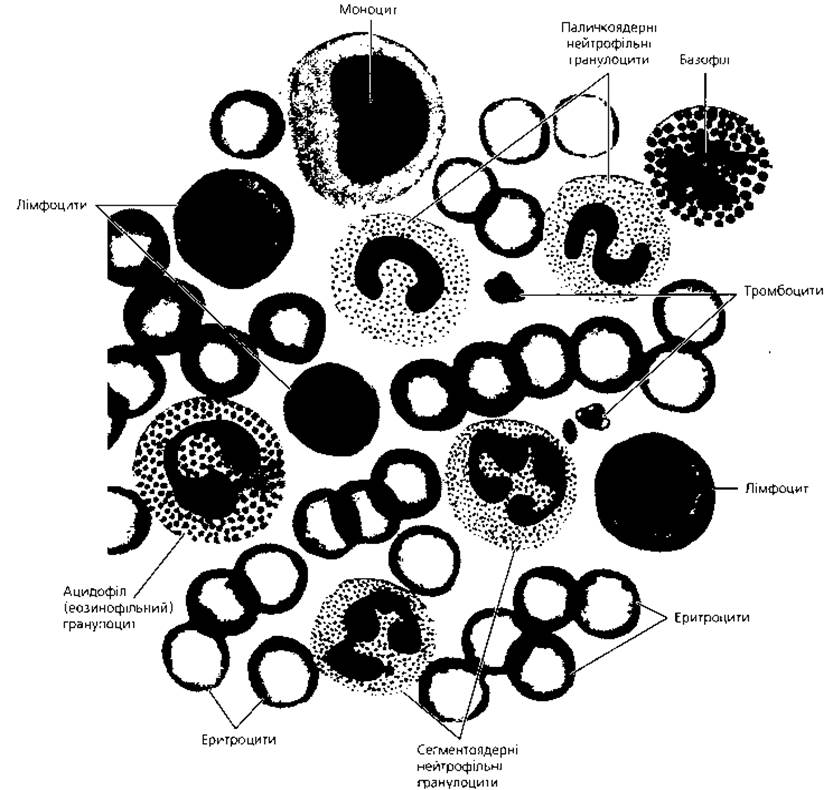
Fig. 3.10. Semi-schematic representation of a human blood smear stained with Romanowsky's method, demonstrating all major morphologically recognizable types of formed elements, which can typically be observed only by examining several fields of view, x 900
Table 7. Sizes and counts of human blood formed elements
|
Type |
||
|
of formed elements |
Size in blood smear |
Count per 1 L of blood |
|
Erythrocyte |
7.1–7.9 µm (average 7.5 µm) |
In males: 3.9–6 × 10¹² In females: 3.7–5.5 × 10¹² |
|
Leukocyte |
4–10 × 109 |
|
|
Neutrophil |
10–12 µm |
57–72 % |
|
Eosinophil |
12–14 µm |
0.5–5 % |
|
Basophil |
11–12 µm |
0–1 % |
|
Lymphocyte |
4.5–18 µm |
19–38 % |
|
Monocyte |
18–20 µm |
3–11 % |
|
Platelet |
2–3 µm |
200–400 × 109 |
In humans and mammals, erythrocytes are mostly biconcave discs known as discocytes. Normally, discocytes account for 80% of the total erythrocyte population. Other forms of erythrocytes also occur, such as planocytes (having a flat surface), spherocytes (spherical), echinocytes (spiked), etc. This normal morphological variety is termed physiological poikilocytosis (from the Greek "poikilos" meaning varied, and "kytos" meaning cell). When the proportion of modified erythrocyte shapes exceeds 20%, the phenomenon is referred to as pathological poikilocytosis. Erythrocyte shape is maintained by beta-sialoglycoprotein in The erythrocyte membrane and a specialized cytoskeletal framework composed of spectrin, which underlies the Plasmalemma from the inside and is linked to it by another protein, ankyrin.
The diameter of a human erythrocyte is 7.1–7.9 µm, with a cell thickness at the edges of 2–2.5 µm and up to 1 µm in the center. The depression in the thin central part of the erythrocyte is called the physiological excavation. This cell shape increases its surface area and accelerates the Oxygenation of hemoglobin. Under normal conditions, 75% of all erythrocytes have the aforementioned dimensions; these are the so-called normocytes. Some cells have a diameter exceeding 8 µm, known as macrocytes, accounting for 12.5%. The remaining erythrocytes may have a diameter of 6 µm or less and are termed microcytes. If the proportion of macro- and microcytes exceeds 25%, this phenomenon is designated as anisocytosis.
Under light Microscopy in blood smears, erythrocytes appear as structureless rounded discs that stain oxyphilically. This oxyphilia is due to the presence of hemoglobin. The central (thin) part of the erythrocyte stains less intensely. Electron microscopy reveals that the erythrocyte is covered by a plasmalemma approximately 20 nm thick. Its outer surface bears antigenic Oligosaccharides that determine Blood Groups, along with Phospholipids and sialic acid. Inside the erythrocyte lies an electron-dense content consisting of numerous hemoglobin granules measuring 4–5 nm.
In terms of chemical composition, erythrocytes consist of 60% water and 40% dry residue. Hemoglobin makes up 95% of the dry residue, while other substances comprise only 5%. Thus, hemoglobin accounts for one-third of the total mass of an erythrocyte. The blood of an adult human contains about 600 g of hemoglobin, which is 15 g per 100 g of blood. Hemoglobin is a complex protein composed of a protein moiety, globin, and an iron-containing non-protein group, heme. Hemoglobin is a pigment that imparts the red color to blood. It readily binds oxygen in the Lungs to form an unstable compound, oxyhemoglobin, which easily dissociates and releases oxygen to the tissues. Part of the hemoglobin binds with carbon dioxide to form carbaminohemoglobin, though the bulk of Carbon dioxide is transported by blood plasma. Hemoglobin also readily forms a compound with carbon monoxide (CO) known as carboxyhemoglobin. The affinity of hemoglobin for carbon monoxide is 300 times higher than for oxygen; therefore, in an atmosphere with a significant carbon monoxide concentration, hemoglobin becomes blocked and unavailable for oxygen, causing the Organism to die from asphyxia (oxygen deprivation).
Humans have Two Types of hemoglobin: HbA, characteristic of adults, and HbF, characteristic of the fetus. In an adult, HbA accounts for 98% and HbF for only 2%. The blood of a newborn infant contains 80% HbF and only 20% HbA. A number of blood disorders (so-called hemoglobinoses or hemoglobinopathies) are accompanied by the appearance of other hemoglobin types in the blood. Erythrocytes exhibit high elasticity and resilience, allowing them to pass through Blood Vessels of a smaller diameter than themselves, during which they can stretch in length up to 20 times and bend.
The average lifespan of human erythrocytes is 120 days. Considering the total number of erythrocytes in the body and their average lifespan, it can be calculated that 200 million erythrocytes are destroyed daily and an equal number are produced to replace them. Consequently, erythrocytes of various ages can be found in the blood: young, functionally mature, and Aging. Young erythrocyte forms are called reticulocytes. They are not fully saturated with hemoglobin and exhibit polychromatophilia. In their Cytoplasm, reticulocytes contain a reticular structure (hence the name of these cells), which can be detected by supravital staining of a blood smear with brilliant cresyl blue. Electron microscopy has proven that the reticular structure in the reticulocyte cytoplasm represents remnants of the granular Endoplasmic reticulum and free Ribosomes, where hemoglobin synthesis continues, as well as Mitochondria. Normally, the proportion of reticulocytes ranges from 1% to 5% of the total erythrocyte count. An increase in their number serves as a diagnostic sign of enhanced erythropoiesis.
Leukocytes, or white blood cells, are blood cells that, unlike erythrocytes, possess a nucleus and all cytoplasmic organelles, lack pigment, are capable of extravasation (leaving blood vessels) and active locomotion via pseudopodia formation, perform protective functions, and spend the majority of their lifespan outside The Vascular System. In an adult human, 1 L of blood contains from 4.0 × 10⁹ to 10.0 × 10⁹ leukocytes. An increase in leukocyte count is termed leukocytosis, while a decrease is termed leukopenia.
Depending on the presence or absence of specific granulation in their cytoplasm, all leukocytes are divided into granulocytes (containing granules) and agranulocytes (lacking them). Based on the staining properties of their granules with histological Dyes, granulocytes are divided into three groups: neutrophilic, acidophilic, and basophilic. Among neutrophilic granulocytes (depending on nuclear shape), band, metamyelocyte ( juvenile), and segmented forms are distinguished. Agranulocytes are subdivided into lymphocytes and monocytes.
Neutrophilic granulocytes (Figs. 3.10, 3.11) account for 65–70% of the total leukocyte count. The Cell diameter in a fresh blood drop is 7–9 µm, and 10–12 µm in a smear. The cytoplasm stains weakly oxyphilically. The granulation is fine and poorly visible in both fresh and fixed stained preparations. When stained using Romanowsky's method, the granules acquire a pinkish-purple color. Granule dimensions are 0.2–0.5 µm. Neutrophil granules are subdivided into primary (azurophilic) and secondary (neutrophilic, specific). Primary granules are Lysosomes; they contain various Hydrolases, myeloperoxidase, and proteins with bactericidal properties, notably Lysozyme (Table 8).
Secondary granules constitute the so-called specific granulation, accounting for 80–90% of all granules in mature neutrophils. The chemical composition of secondary neutrophil granules is characterized by the presence of alkaline phosphatase, major basic proteins, phagocytins, and lysozyme, whereas lysosomal Enzymes and peroxidase are absent.
Organelles are poorly developed in the neutrophil cytoplasm: there are few mitochondria, a small Golgi complex, and occasionally elements of The endoplasmic reticulum. Inclusions such as Glycogen and Lipids are typically present. Thus, neutrophils contain a full Complement of substances used to destroy phagocytized microorganisms. Neutrophilic granulocytes possess The ability to move actively, migrate through tissues toward the focus of inflammation, and phagocytize microorganisms and other small particles. I.I. Mechnikov named them microphages.
As mentioned above, Three types of neutrophils are distinguished by their nuclear shape (reflecting cell age). Juvenile (metamyelocyte) neutrophils are the youngest forms, with a bean-shaped nucleus; their proportion is small, ranging from 0–1%. Band neutrophils have a nucleus resembling the letter S or C, accounting for 1–6%. Segmented neutrophils are mature forms; their nucleus consists of several segments connected by thin strands. The number of segments ranges from 2 to 5, most commonly 3–4, and the nuclear Chromatin stains dark purple with Romanowsky's method. In female neutrophils, perinuclear satellites—small clusters of sex chromatin—are present, mostly appearing as drumsticks.
The ratio of the three neutrophil types has definite diagnostic significance and is clinically useful. For instance, an elevated count of juvenile and band forms combined with leukocytosis indicates the presence of an inflammatory focus in the body.
Eosinophilic granulocytes (Figs. 3.10, 3.11) constitute 0.5–5% of the total leukocyte count. The cell diameter in a fresh blood drop is 9–10 µm, and 12–14 µm in a smear, meaning they are larger than neutrophils. The cytoplasm stains weakly basophilically. The specific granules are large (0.7–1.5 µm) and clearly visible. In fresh preparations, they are shiny due to strong light refraction. In preparations stained with Romanowsky's method, the specific granules of acidophils appear bright pink.
Table 8. Composition of cytoplasmic granules in human leukocytes
|
Cell type |
Composition of secondary specific granules |
Composition of primary azurophilic granules |
|
Neutrophil |
Alkaline phosphatase Collagenase Lactoferrin Lysozyme Phagocytins |
Acid phosphatase α-mannosidase Arylsulfatase β-galactosidase β-glucuronidase Cathepsin 5'-nucleotidase Collagenase Myeloperoxidase Lysozyme Acid mucosubstances Cationic antibacterial proteins |
|
Eosinophil |
Acid phosphatase Arylsulfatase β-glucuronidase Cathepsin Phospholipase Histaminase RNAase Peroxidase Major basic protein |
|
|
Basophil |
Heparin Histamine Serotonin Peroxidase Histidine decarboxylase Eosinophil chemotactic factor |
The oxyphilia of the granules is due to the presence of a major basic protein rich in Arginine. Electron microscopy reveals characteristic lamellar crystalloid structures immersed in a finely granular amorphous matrix within the specific eosinophil granules. Hydrolases and peroxidases predominate among the constituents of acidophil specific granules, which is why these granules are considered a type of lysosome or peroxisome. Additionally, eosinophil granules contain the enzyme histaminase, whereas lysozyme and phagocytin are absent. Cytoplasmic organelles are poorly developed.
In the Red Bone Marrow, eosinophils undergo the same developmental stages as neutrophils; thus, juvenile, band, and segmented eosinophils exist. However, since the blood content of these cells is low, juvenile and band eosinophil forms are very rare and are not accounted for during differential counts. The Nucleus in segmented acidophils most commonly consists of two, and less frequently three, segments. These segments are larger than those in neutrophils. The nuclear structure is more delicate, and the segments have a more regular shape. Eosinophilic leukocytes are motile and capable of phagocytosis, although their phagocytic activity is lower than that of neutrophils. They participate in the body's immune responses against foreign proteins, as well as in allergic and anaphylactic reactions. Due to the presence of histaminase, eosinophils can inactivate histamine. Furthermore, they can accumulate this substance by phagocytizing histamine-containing granules and by adsorbing it onto the cytolemma, which bears histamine receptors.
The number of eosinophils increases in allergic diseases, certain infections, and helminthiases. Eosinophils circulate in the blood for 3–8 hours, after which they migrate into the connective tissue of Organs to exert their physiological activity.
Basophilic granulocytes, or basophils (Figs. 3.10, 3.11), account for 0–1% of the total leukocyte count. Their diameter is 9 µm in a blood drop and 11–12 µm in smears. The cytoplasm stains weakly oxyphilically. The specific granulation stains intensely basophilically and metachromatically (purple-violet) with Romanowsky's stain and is readily water-soluble. Granule dimensions are 0.5–1.2 µm. The metachromasia of the granules is caused by the presence of the acidic glycosaminoglycan heparin. In addition, the granules contain histamine, serotonin, peroxidase, acid phosphatase, and histidine decarboxylase, the enzyme responsible for histamine synthesis. The nucleus of basophils lacks a definite shape (segmented, bean-shaped, less frequently spherical, etc.), is centrally located, and is relatively poor in heterochromatin. The nucleus stains less intensely than the granules, causing the latter to overlie and mask it.
Basophils are sluggish cells with virtually no phagocytic capability. Their function is related to the metabolism of histamine and heparin. Heparin is a native anticoagulant; therefore, basophils participate in regulating blood clotting. Histamine causes marked vasodilation, edema, and related phenomena. Such reactions occur upon basophil degranulation, thereby involving them in allergic processes.
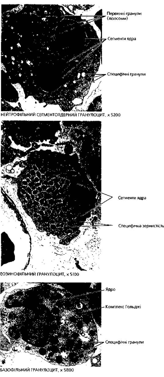
Fig. 3.11. Fine structure details of blood formed elements
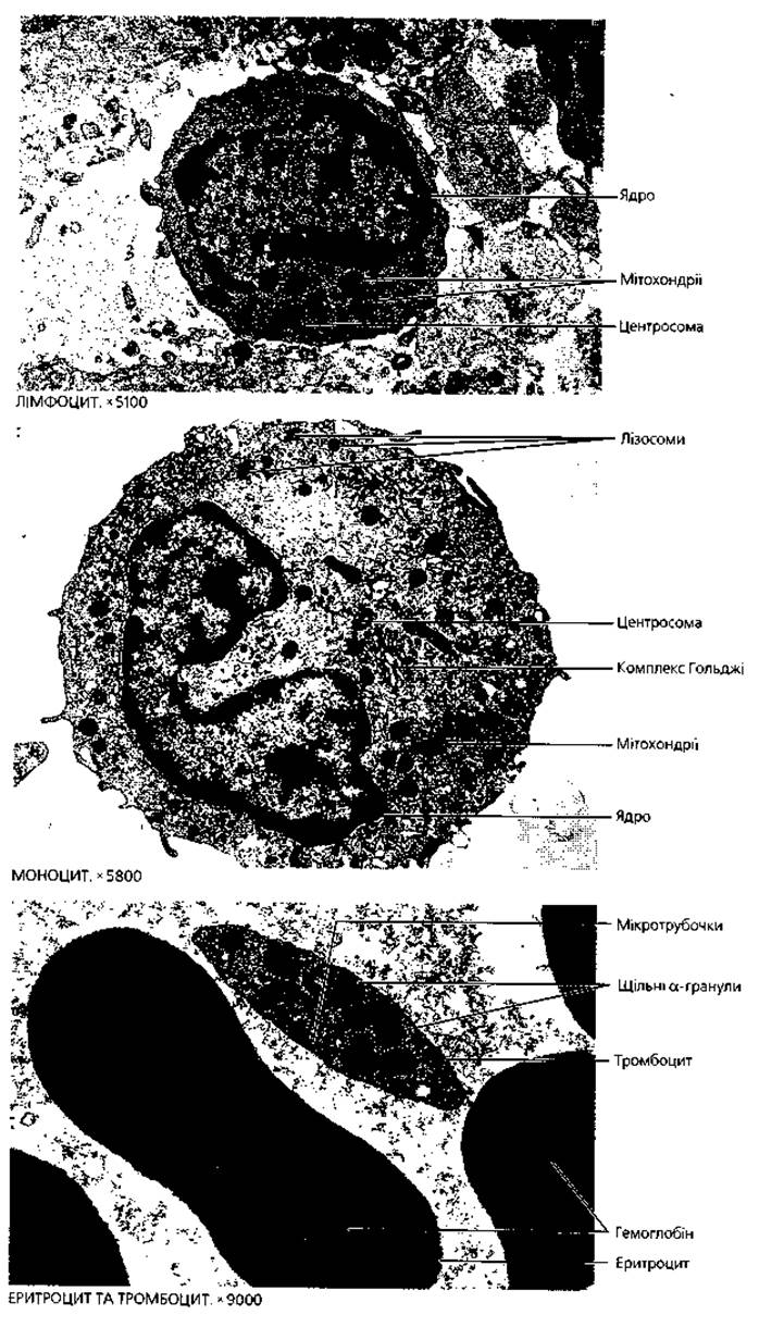
Fig. 3.11 (continued). Fine structure details of blood formed elements
Lymphocytes (Figs. 3.10, 3.11) account for 19–38% of the total leukocyte count in adult blood. Based on their size under light microscopy, lymphocytes are divided into three types: small lymphocytes have a diameter of 4.5–7 µm and make up about 2/3 of all blood lymphocytes; medium lymphocytes measure 7–10 µm in diameter and comprise 1/3 of the total; large lymphocytes, with a diameter exceeding 10 µm, are not normally observed in adult peripheral blood and can only be found in the Thoracic duct lymph. Small lymphocytes feature a large, spherical nucleus that occupies nearly the entire cell, positioned either centrally or eccentrically. The nucleus is rich in heterochromatin, clumped into compact masses. The cytoplasm stains basophilically (blue with Romanowsky stain) and surrounds The Nucleus as a narrow rim or crescent, typically showing a clear perinuclear zone. Medium and large lymphocytes possess a larger cytoplasmic volume, and their nuclei exhibit a finer Chromatin Structure.
According to electron microscopy, lymphocytes are categorized into four cell types:
1. Small light lymphocytes. These are the most abundant, making up 70–75%. They feature a pale cytoplasm with a sparse number of free ribosomes, along with all other standard organelles.
2. Small dark lymphocytes. Comprising 12–13%, these cells feature a dark, electron-dense cytoplasm packed with free ribosomes, a minimal number of mitochondria, and very rarely other organelles.
3. Medium lymphocytes. Accounting for 10–12%, they possess loosely packed chromatin and a clearly visible nucleolus. Virtually all types of organelles are present in their cytoplasm.
4. Plasmacytes or lymphoplasmacytoid cells. Making up 1–2% of lymphocytes, their characteristic feature is the presence of endoplasmic reticulum cisternae arranged concentrically around the nucleus.
Based on their origin and immune functions, lymphocytes are divided into two main varieties: T AND B lymphocytes (Table 9). T lymphocytes, or Thymus-dependent lymphocytes, originate in the thymus and mediate cell-mediated immunity as well as The regulation of humoral immunity. These are long-lived cells capable of surviving for years, or even decades, and constitute 80% of all peripheral blood lymphocytes. T lymphocytes comprise several subpopulations: T killers, or killer cells, whose specific cytotoxic effect provides antitumor and transplantation immunity; T helpers, which specifically recognize Antigens and enhance antibody production by B lymphocytes; T suppressors, which inhibit the capacity of B lymphocytes to produce antibodies; and T memory cells, which retain information about antigens over extended periods. The Effect of T lymphocytes on B lymphocytes is mediated through specialized soluble substances called lymphokines, which they produce in response to antigenic stimulation.
B (or bursa-equivalent) lymphocytes develop in the bursa of Fabricius in birds, and in the red bone marrow (and potentially the lymphoid follicles of the digestive tract) in humans. B lymphocytes are responsible for humoral immunity. They are short-lived (surviving for weeks or months) and account for about 20% of blood lymphocytes. B lymphocytes are capable of differentiating into effector cells—plasmacytes—which produce protective proteins known as immunoglobulins (antibodies).
There are no distinct morphological differences between T and B lymphocytes under light microscopy. Electron microscopic data indicate that B lymphocytes possess a better-developed granular endoplasmic reticulum, whereas T lymphocytes contain numerous lysosomes. T Lymphocytes and their nuclei are smaller in size and contain more heterochromatin. Additionally, T lymphocytes stain positive for acid phosphatase, whereas B lymphocytes are rich in alkaline phosphatase. Identification of T and B lymphocytes and their subpopulations is performed using immunological Methods, most of which rely on the specific molecular architecture of their cell membranes. B lymphocytes carry surface immunoglobulins on their membranes that act as antigen receptors, alongside Fc receptors and various other specific receptors and antigens. The Specificity of the T lymphocyte membrane is determined by the presence of specific surface antigens, receptors for certain cells (such as E-receptors for sheep erythrocytes, which facilitate rosette formation), Fc receptors for binding immune complexes, and other markers.
Monocytes (Figs. 3.10, 3.11) account for 3–11% of the total leukocyte count. They are the largest blood cells in diameter, particularly in smears due to their strong flattening on Glass (reaching 18–20 µm). In a drop of fresh blood, their dimensions are considerably smaller (10–12 µm). The cytoplasm stains basophilically, though less intensely than in lymphocytes, acquiring a smoky-gray tint. The cytoplasm contains all standard organelles and numerous lysosomes. The nucleus is most commonly Kidney-shaped (bean-shaped), though it may assume other forms (such as a figure-eight). Small clumps of heterochromatin are scattered throughout the nucleus. Monocytes are motile and capable of phagocytosis and pinocytosis; their strong adhesion property is directly related to this phagocytic activity.
Table 9. Types of lymphocytes and their principal functions
|
Lymphocyte type |
Principal functions |
|
B lymphocyte |
Carries Membrane Receptors (IgM); becomes activated upon exposure to specific antigens, proliferates via mitosis, and differentiates into antibody-secreting plasma cells (immunoglobulins) |
|
Memory B lymphocyte |
An activated B lymphocyte that mounts a faster and more robust response upon secondary exposure to the same antigen |
|
Cytotoxic T lymphocyte (T killer) |
Expresses T-cell receptors (TCR+) that are non-immunoglobulin in nature; recognizes antigens associated with Major Histocompatibility Complex class I (MHC-I); produces perforin and other proteins that destroy foreign cells, virus-infected cells, and certain tumor cells |
|
T helper cell |
Expresses T-cell receptors (TCR+); enhances The activity of other T and B lymphocytes |
|
T suppressor cell |
Expresses T-cell receptors (TCR+); suppresses the activity of other T and B lymphocytes |
|
Memory T lymphocyte |
Expresses T-cell receptors (TCR+); provides a faster and more intense response to repeated exposure to the same antigen |
|
Natural killer cell (NK cell) |
Lacks T-cell receptors (TCR-); attacks virus-infected cells and tumor cells without prior activation |
Monocytes reside in the bloodstream for a relatively short period—from 36 to 104 hours—after which they migrate out of the vessels into tissues and transform into macrophages (histiocytes), representing the terminal stage of differentiation for these blood cells. Thus, monocytes belong to the mononuclear phagocyte System of the body.
Platelets (Figs. 3.10, 3.11), or thrombocytes, are cytoplasmic fragments derived from giant bone marrow cells known as megakaryocytes. While megakaryocytes can reach sizes up to several tens of micrometers, platelets measure only 2–3 µm, prompting the biological adage that bone marrow giants give birth to blood dwarfs. The platelet count ranges from 200 to 400 × 109 per liter of blood. Counting these formed elements is challenging due to their tendency to clump together into aggregates. An elevated platelet count in peripheral blood is termed thrombocytosis and is observed in cases of major trauma and leukemias, whereas a decreased platelet count—thrombocytopenia—frequently accompanies various pathological conditions.
Each blood platelet consists of a hyalomere, which forms its structural basis and stains weakly oxyphilic, and a granulomere (or chromomere), which appears as basophilic (azurophilic) granules located in the center of the platelet. The granulomere does not contain DNA. Platelets are externally bounded by a plasmalemma. The hyalomere contains a marginal bundle of microtubules that helps maintain the platelet's discoid shape, along with Actin and Myosin microfilaments.
Electron microscopy reveals two types of granules within the granulomere: dense, dark alpha-granules (whose chemical composition is not yet fully understood) and serotonin granules. Glycogen granules and mitochondria are also present in the granulomere. Platelets possess cytoplasmic extensions of varying size and thickness (known as pseudopodia or processes), which allow platelets to interlock with one another during Blood Coagulation. The absence of these processes in platelets is associated with impaired blood clotting.
The primary function of platelets is participation in blood coagulation. Platelets contain the enzyme thromboplastin, which takes part in The conversion of fibrinogen into fibrin. In addition, platelets rapidly disintegrate and aggregate into clusters surrounded by strands of fibrin, facilitating The formation of a thrombus that seals damaged blood vessels. The clot retraction factor located in the hyalomere promotes clot compaction. Platelets also release substances that induce vasoconstriction upon vessel injury and decrease the permeability of the vascular wall.
The Main Functions of blood formed elements are summarized in Table 10.
Hemogram. Leukocyte differential count. In the blood of a healthy individual, formed elements maintain specific quantitative ratios known as a hemogram. The percentage distribution of various leukocyte types in a peripheral blood smear constitutes the leukocyte differential count (Table 11).
Table 10. Characteristics of blood formed elements based on their primary functions and synthesis products
|
Blood formed elements |
Main functions |
Synthesis products |
|
Erythrocytes |
Hemoglobin |
|
|
Neutrophils |
Phagocytosis of Bacteria |
Specific granules and lysosomes (azurophilic granules) |
|
Eosinophils |
Defense against helminths; modulation of inflammatory and allergic processes |
Specific granules, biologically active substances |
|
Basophils |
Release of histamine and other inflammatory mediators |
Specific granules containing histamine and heparin |
|
Monocytes |
Transformation into Cells of the tissue macrophage system; phagocytosis of Protozoa, Viruses, and aging body cells |
Granules containing lysosomal enzymes |
|
B lymphocytes |
Differentiation into antibody-producing cells (plasmacytes) |
Immunoglobulins |
|
T lymphocytes |
Mediation of cell-mediated immunity; participation in humoral immune responses |
Substances controlling the activity of other leukocytes (interleukins) |
|
Cytotoxic T lymphocytes (killers) |
Destruction of tumor cells or virus-infected cells |
Cell-lysing substances (perforins) |
|
Platelets |
Blood clotting (hemostasis) |
Clotting factors |
The hemogram and leukocyte differential count can vary in various diseases. These changes are used in medicine to diagnose the corresponding conditions.
Age-related blood changes. The erythrocyte count in newborns is higher than in adults, ranging from 6.0×10¹² to 9.0×10¹² per 1 L. The leukocyte count at birth is also higher, reaching 10–30×109 per 1 L. The pediatric leukocyte differential also differs from that of adults and changes throughout the first 14–15 years of life. These changes involve the ratio of neutrophils to lymphocytes. At birth, the percentage of these leukocytes is the same as in an adult, meaning the white blood profile is neutrophilic (neutrophils outnumber lymphocytes). Subsequently, the neutrophil count begins to decrease while the lymphocyte count increases, and by the 4th to 5th day of the postnatal period, the percentages of neutrophils and lymphocytes equalize (approximately 45% each). The process of decreasing neutrophil counts and increasing lymphocyte counts continues for 1–2 years until the so-called pediatric leukocyte differential stabilizes, exhibiting a lymphocytic profile (65% lymphocytes and 25% neutrophils). In the subsequent period, the lymphocyte count begins to decrease and the neutrophil count increases, which again leads to an equalization of their percentages by the 4th to 5th year of the child's life. The process of decreasing lymphocyte counts and increasing neutrophil counts continues until ages 14–15, at which point the leukocyte differential becomes identical to that of an adult.
Table 11. Samples of a healthy human hemogram and leukocyte differential count
HEMOGRAM
|
Parameter |
Value |
|
Hematocrit (formed elements-to-plasma ratio) |
45%:55% |
|
Erythrocyte count |
4-5х10,2 в 1 л |
|
Reticulocyte count |
2-10 per 1,000 erythrocytes |
|
Leukocyte count |
4-9 x 10⁹ per 1 L |
|
Platelet count |
180-320 x 109 per 1 L |
|
6-12 mm/h |
|
|
Hemoglobin |
130-160 g/L |
LEUKOCYTE DIFFERENTIAL COUNT
|
Granulocytes |
Agranulocytes, % |
|||||
|
basophils |
eosinophils |
neutrophils |
lymphocytes |
monocytes |
||
|
metamyelocytes |
band cells |
segmented |
||||
|
0-1 % |
0,5-5 % |
0,5-1 % |
1-6 % |
47-72 % |
20-40 % |
3-11 % |
Table 12. Features of the leukocyte differential count in children of various ages
|
Type of leukocyte |
Age |
||||
|
(content %) |
1 day |
5 days |
1 year |
5 years |
14 years |
|
Neutrophilic granulocytes |
64 |
45 |
25 |
45 |
60 |
|
Lymphocytes |
24 |
45 |
65 |
45 |
28 |
If the described Changes in the differential count are represented graphically, the two curves denoting the percentages of neutrophils and lymphocytes intersect twice: on the 4th–5th day and in the 4th–5th year of life. Therefore, these periods are referred to as the First and Second physiological crossovers (Table 12).
Lymph (lympha) is a yellowish fluid that circulates through Lymphatic vessels. It consists of lymphoplasma and formed elements. The Chemical composition of lymphoplasma is similar to blood plasma, but it contains less protein. Among the proteins in lymphoplasma, albumins predominate; it also contains neutral fats, sugars, and mineral substances. The formed elements of lymph are represented predominantly by lymphocytes (95–98%), a small number of other leukocyte types, and occasional erythrocytes. The composition of lymph in different PARTS OF THE body varies. For example, lymph draining from the intestine is rich in fats; lymph that has passed through Lymph Nodes is enriched with lymphocytes. A distinction is made between peripheral lymph (before entering lymph nodes), intermediate lymph (after passing through lymph nodes), and central lymph (lymph of the thoracic duct and Right lymphatic duct).
Lymph is formed by the filtration of tissue fluid into lymphatic capillaries. Tissue fluid, in turn, is formed by the passage of water, proteins, and other substances from blood capillaries into the intercellular space. From lymphatic capillaries, lymph enters peripheral lymphatic vessels, flows through them into lymph nodes, and via The system of the thoracic and right lymphatic ducts drains into the left and right subclavian Veins at the junction with the internal jugular veins.
Terms to memorize
1. Tissues of the internal environment. 2. Mesenchyme. 3. Blood plasma. 4. Albumins. 5. Globulins. 6. Fibrinogen. 7. Blood serum. 8. Erythrocytes. 9. Discocytes. 10. Poikilocytosis. 11. Normocytes. 12. Anisocytosis. 13. Hemoglobin. 14. Oxyhemoglobin. 15. Carbaminohemoglobin. 16. Carboxyhemoglobin. 17. Reticulocytes. 18. Leukocytes. 19. Leukocytosis. 20. Leukopenia. 21. Granulocytes. 22. Neutrophilic metamyelocytes. 23. Neutrophilic band granulocytes. 24. Segmented neutrophilic granulocytes. 25. Eosinophilic granulocytes. 26. Basophilic granulocytes. 27. Lymphocytes. 28. T lymphocytes. 29. Lymphokines. 30. B lymphocytes. 31. Plasma cells. 32. Monocytes. 33. Macrophages. 34. Platelets. 35. Megakaryocytes. 36. Hyalomere. 37. Granulomere. 38. Hemogram. 39. Leukocyte differential count. 40. Lymph. 41. Lymphoplasma. 42. Formed elements.
Last update: 09/08/2026
Editorial and Educational Adaptation: This material has been compiled based on the primary/original source text. The project team performed an editorial review, corrected technical inaccuracies, structured sections, and adapted the content for an educational format.
What was processed:
- elimination of formatting defects (OCR errors, structural breaks, corrupted characters);
- editorial organization of content;
- standardization of terminology in accordance with academic sources;
- verification of factual statements against the original source text.
All mentions of the author, publication year, and origin of the primary text have been preserved in accordance with the source.