Human Histology - O.D. Lutsyk 2003
General Histology
Blood Formation (Hematopoiesis)
Formed elements of the Blood, which are mostly highly specialized Cells, have a limited lifespan. For example, erythrocytes live for about 120 days, granulocytes remain in the blood for 10-20 hours and in Tissues for 24-48 hours, monocytes circulate in the blood for 30-60 hours, and platelets live for 2-3 days. The constancy of the Qualitative and quantitative composition of formed blood elements is achieved through their continuous formation and development, referred to as Hemocytopoiesis (from the Greek "haima" - blood, "kytos" - Cell, "poiesis" - creation), or hematopoiesis (hemopoiesis). During hematopoiesis, the natural loss of senescent formed elements is compensated, so hemopoiesis can be considered a process of physiological blood regeneration.
After birth, hematopoiesis occurs in Organs known as Hematopoietic organs. These include the Cytology/practical/86.html">Red Bone Marrow of flat bones and the epiphyses of long tubular bones, where erythrocytes, granulocytes, monocytes, platelets, and lymphocyte precursors are formed; and the Spleen, Lymph Nodes, and Thymus, where the differentiation and proliferation of T- and B-lymphocytes and plasma cells take place. The hematopoietic tissue of the red bone marrow is called myeloid tissue, and The process of forming erythrocytes, granulocytes, monocytes, and platelets is referred to as myelopoiesis. The hematopoietic tissue located in the spleen, lymph nodes, and thymus (as well as The system of these organs) is called lymphoid tissue, and the process of lymphocyte and plasma Cell Formation within them is called lymphopoiesis.
Various theories of hematopoiesis that existed until recently were based on identifying one or several progenitor cells from which all types of mature formed elements develop. Polyphyletic theories, according to which There are two, three, or more initial cell forms (known as dualistic, trialistic, etc.), are now of purely historical interest. Today, the unitary theory of hematopoiesis is universally accepted, according to which all mature formed elements of the blood originate from a single common progenitor cell. The foundations of this theory were first formulated in the early 20th century by the Russian histologist A.A. Maximow, who believed that such a cell exists and has the Morphology of a small lymphocyte. Currently, these concepts have been confirmed by numerous experiments based on new Research Methods that allow the generation of cell clones (a group of cells derived from a single cell), or hematopoietic colonies, in the spleen of lethally irradiated mice (the colony-forming assay; Till and McCulloch, 1961). The data obtained from these experiments formed The basis of the modern unitary theory of hematopoiesis, according to which all mature formed elements of the blood originate from a single progenitor cell (Fig. 3.12), known as the hematopoietic stem cell (HSC).
The HSC population has the following characteristics:
1) pluripotency, i.e., The ability to differentiate into all types of formed blood elements;
2) self-renewal capacity over a lifespan close to that of the human Organism: the number of mitoses a single cell undergoes can exceed 100;
3) despite its high proliferative capacity, the stem cell normally divides very rarely, remaining in the G0 phase of the Cell Cycle; however, under METABOLISM/18.html">The Influence of, for example, radiation, it can rapidly begin to proliferate;
4) HSCs are in a state of constant and intensive repopulation, meaning they migrate from some hematopoietic organs to others via the blood; this is evidenced by the fact that HSCs can always be found in the blood as cells capable of restoring hematopoiesis in irradiated animals.
In adult mammals, HSCs are concentrated mainly in the red bone marrow (there are 50 HSCs per 105 nucleated bone marrow cells). The total number of hematopoietic stem cells in humans is approximately 5х1010 (one-third of which are in the mitotic cycle). Hematopoietic stem cells originate during the Embryonic period in the yolk sac and then colonize the entire hematopoietic system: adult HSCs are their descendants.
Class="center">
Fig. 3.12. Diagram of the sequential phases of hematopoiesis (physiological blood regeneration) in an adult human (the class of maturing cells is not shown to simplify the illustration)
Morphologically, HSCs are not identified, which is due to their low concentration in the bone marrow (103—104). The Morphology of the so-called candidate hematopoietic stem cell, obtained by Special Methods, has been described. This cell is similar to bone marrow lymphocytes. Its shape is round or oval, with a diameter of 8 µm, and the nucleocytoplasmic ratio is greater than three (i.e., it is a nuclear-type cell). The Nucleus diameter is 5 µm; it is oval or round with wavy edges, featuring dense Chromatin clumps near the nuclear membrane. The cytoplasmic rim is thin, with many single Ribosomes, a small number of Mitochondria, and tubules of the rough Endoplasmic reticulum; the Golgi complex is very rarely observed.
According to the modern scheme of hematopoiesis, the following classes of cells are distinguished in all histogenetic lineages that culminate in The formation of mature formed blood elements:
Class I - pluripotent progenitor cells (HSCs);
Class II - partially determined progenitor cells (the potency of these cells is partially restricted regarding further differentiation, meaning they can no longer give rise to all types of formed elements);
Class III - unipotent progenitor cells (these cells are capable of developing in only one direction under the influence of hormone-like substances called hemopoietins; different hemopoietins exist for different histogenetic lineages);
Class IV - morphologically recognizable proliferative progenitor cells (unlike the Cells of the first three classes, which are not morphologically identified and whose existence has been proven only experimentally, Class IV cells can be recognized on bone marrow smears and are capable of mitotic division);
Class V - maturing cells (they lose the capacity for mitotic division and undergo changes associated with their transformation into mature formed elements);
Class VI - mature cells capable of entering the bloodstream.
Changes in The properties of hematopoietic cells during differentiation are summarized in Table 13.
Individual histogenetic lineages of Blood Cells are named as follows: erythropoiesis, granulocytopoiesis, monocytopoiesis, thrombocytopoiesis, and lymphopoiesis.
Erythropoiesis (erythrocyte development) occurs in the postnatal period within the red bone marrow (Figs. 3.13, 3.15). The source of erythrocyte development is the hematopoietic stem cell (Class I). Under the Influence of the specific microenvironment of the bone marrow stroma, this cell divides and differentiates into a myelopoiesis progenitor cell (Class II, partially determined; only myeloid elements can form from it). This cell is also referred to as the colony-forming unit-granulocyte, erythrocyte, monocyte, megakaryocyte (CFU-GEMM), or semi-stem cell (SSC). From this cell, more determined progenitors of two types are formed: CFU-GranE (colony-forming unit-granulocyte and erythrocyte) and CFU-MegE (colony-forming unit-megakaryocyte and erythrocyte). Thus, the next stage of erythrocyte development—their unipotent progenitor CFU-E (Class III cells, which develop only into erythrocytes)—can be formed in two ways: from CFU-GranE or CFU-MegE. The unipotent cell of erythropoiesis is also called the Erythropoietin-sensitive cell (ESC), because its further differentiation is induced by the hormone erythropoietin (Table 14). The latter is produced in the Kidneys and enhances the proliferation of ESCs and their transformation into proerythroblasts. This hormone also stimulates the development and proliferation of erythroid cells in subsequent stages.
Table 13. Changes in the properties of hematopoietic cells during differentiation
|
Stem cells |
Semi-stem and unipotent cells |
Properties |
Maturing cells |
|
Pluripotency |
Mitotic activity |
||
|
Typical morphological characteristics |
|||
|
Self-renewal capacity |
|||
|
Sensitivity to growth factors |
|||
|
Defined functional activity |
|||
Proerythroblasts (Class IV) are the first morphologically recognizable cells of the erythroid Lineage. They are round and large (cell diameter 15-25 µm). The nucleus is large, round, centrally located, has a fine reticular-granular Structure, and contains 1-3 nucleoli; the Cytoplasm stains basophilically, with a light perinuclear zone around the nucleus, numerous ribosomes, a small centrosome with two centrioles, and the characteristic presence of ferritin granules (a protein-iron complex). Proerythroblasts divide and differentiate into basophilic erythroblasts.
Basophilic erythroblasts are slightly smaller than proerythroblasts (10-18 μm). The nuclear chromatin begins to clump in a radial pattern, resembling the spokes of a wheel. The cytoplasm is intensely basophilic due to the large amount of RNA. Hemoglobin synthesis begins in these cells. They divide by mitosis and, after accumulating a certain amount of hemoglobin, differentiate into polychromatophilic erythroblasts.
Polychromatophilic erythroblasts are smaller (10-14 μm), with a smaller, denser nucleus showing a distinct wheel-spoke chromatin pattern. Nucleoli are not discernible. The cytoplasm stains polychromatically, reacting with both acidic and basic Dyes. Acidophilia is due to the presence of hemoglobin, while basophilia is due to RNA. Hemoglobin in these cells can be distributed diffusely, coloring the entire cytoplasm grayish, or in patches that take up acidic dyes, or as a rim around the nucleus. The number of ribosomes in these cells decreases, and ferritin is arranged in aggregates. Polychromatophilic erythroblasts divide by mitosis. Their late generations are called polychromatophilic normoblasts.
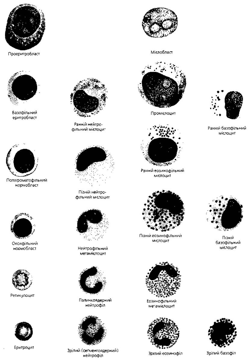
Fig. 3.13. Morphologically recognizable cell stages in The pathway of blast transformation into erythrocytes and granulocytes
Table 14. Key CHARACTERISTICS OF THE most well-studied colony-stimulating factors
|
Factor name and standard English abbreviation |
Human Gene localization and producer cells |
Primary function |
|
Erythropoietin (EPO) |
Chromosome 7 Juxtaglomerular cells of the Kidney |
Stimulates erythrocyte production |
|
Granulocyte colony-stimulating factor (G-CSF) |
Chromosome 17 Macrophages, endothelial cells, fibroblasts |
Stimulates granulocyte production, increases metabolic rate in granulocytes, stimulates malignant (leukemic) cells |
|
Granulocyte-macrophage colony-stimulating factor (GM-CSF) |
Chromosome 5 T-lymphocytes, endothelial cells, fibroblasts |
Stimulates granulocyte and macrophage production |
|
Macrophage colony-stimulating factor (M-CSF) |
Chromosome 5 Macrophages, endothelial cells, fibroblasts |
Stimulates macrophage production, enhances their antitumor activity |
|
Interleukin 3 (IL-3) |
Chromosome 5, T-lymphocytes |
Stimulates myelopoiesis |
Polychromatophilic normoblasts measure up to 10 μm, making them smaller than their predecessors; the nucleus loses its wheel-spoke chromatin arrangement and condenses, becoming pyknotic and virtually structureless. These cells lose their ability to divide (class V). At this stage, 80% of the cells lose their nuclei and transform into bone marrow reticulocytes, which continue to accumulate hemoglobin. Their maturation in the bone marrow lasts for 36-44 hours, after which they enter the bloodstream as mature erythrocytes (class VI). Some bone marrow reticulocytes leave the bone marrow before being fully saturated with hemoglobin; these are referred to as blood reticulocytes. The remaining 20% of polychromatophilic normoblasts retain their nuclei, continue to accumulate hemoglobin, and differentiate into orthochromatic (oxyphilic) normoblasts. Their cytoplasm is acidophilic (oxyphilic). They lose their nucleus either by extrusion from The Cell or by the pinching off of a nuclear fragment (the same mechanism by which polychromatophilic normoblasts lose their nuclei) and transform into erythrocytes.
The entire process of erythrocyte formation in adults, from proerythroblast to erythrocyte, takes 6-8 days (Fig. 3.14). The differentiation of nucleated erythropoietic elements lasts 100-140 hours. Morphologically recognizable cells of the erythroid lineage undergo 5-6 mitoses, while cells in the preceding classes undergo 10-15 mitoses. Each proerythroblast gives rise to 30-60 erythrocytes. The total number of erythroid cells in human bone marrow is 3x10¹¹.
Thus, during The Development of erythrocytes from a proerythroblast to a mature cell, the following key changes occur:
1) cytoplasmic basophilia, caused by a significant amount of ribosomal RNA, is replaced by polychromatophilia and then acidophilia (oxyphilia) due to increasing hemoglobin and decreasing RNA levels; all Organelles are eventually lost;
2) the nucleus condenses, becomes pyknotic, and is extruded from the cell;
3) cell size decreases during differentiation from 15-25 to 7-8 μm.
Under normal conditions, the demand for erythrocytes is met by the active proliferation of polychromatophilic erythroblasts. If the body's demand for erythrocytes increases (for example, in the case of blood loss), erythroblasts begin to develop from progenitors, and the latter from stem cells.
Granulocytopoiesis (granulocyte development). The first cell of granulocytopoiesis is the hematopoietic stem cell (Table 15) of the red bone marrow and the myeloid progenitor cell, which are analogous to those described above for erythrocyte development (classes I and II). The next stage is the formation of a more committed progenitor cell of granulocytes and monocytes-macrophages, or CFU-GM. During granulocyte development, it gives rise to unipotent progenitors (class III): basophil progenitors (CFU-B), eosinophil progenitors (CFU-Eo), and neutrophil progenitors (CFU-G). In addition, the unipotent neutrophil progenitor can also arise from CFU-NE (colony-forming unit of neutrophils and erythrocytes). The hormone that stimulates the differentiation and proliferation of this lineage is called granulopoietin (granulocyte colony-stimulating factor).
The first morphologically recognizable cell of this lineage (Fig. 3.13) is the myeloblast (class IV). It is large (up to 20 μm), with a centrally located round nucleus that occupies most of the cell, and its cytoplasm is basophilic. The nucleus has a fine reticular structure (unlike the erythroblast, it contains no chromatin clumps) and contains two to five blue nucleoli. The cytoplasm is rich in ribosomes and mitochondria, and nonspecific azurophilic granules can be detected. It has been experimentally proven that myeloblasts are committed to only a single pathway of differentiation, and basophilic, eosinophilic, and neutrophilic subtypes exist among them. They divide once by mitosis and differentiate into promyelocytes.
Promyelocytes range in size from 12 to 20 μm; unlike myeloblasts, they have a coarser nuclear structure and fewer nonspecific granules. The formation of promyelocytes is accompanied by the appearance of specific granules; depending on their nature, Three types of these cells are distinguished: basophilic, eosinophilic, and neutrophilic. Promyelocytes undergo a single mitosis and differentiate into myelocytes.
Table 15. Functional populations of neutrophils

Myelocytes range in size from 8 to 12 μm. The nucleus contains dense chromatin strands alternating with lighter areas; nucleoli are absent. The cytoplasm is weakly basophilic or weakly acidophilic (meaning cytoplasmic basophilia decreases), and the number of specific granules increases. Three distinct types of myelocytes are clearly identified: basophilic, eosinophilic, and neutrophilic. These cells divide twice by mitosis, with The ratio of early to late myelocytes being 1:2. They differentiate into metamyelocytes.
Metamyelocytes are round cells about 8 μm in diameter, with the volume of the cytoplasm exceeding that of the nucleus. The nucleus is kidney-shaped or horseshoe-shaped. These cells no longer divide and therefore belong to the class of maturing cells (class V). There are three types of these cells: basophilic, eosinophilic, and neutrophilic. Metamyelocytes can enter the peripheral blood, where they are referred to as juvenile granulocytes.
Band granulocytes develop from metamyelocytes through A change in nuclear shape (the nucleus elongates and bends). In mature segmented granulocytes, the nucleus is divided into segments (class VI). The nucleus is most highly segmented in neutrophilic granulocytes and least segmented in basophils.
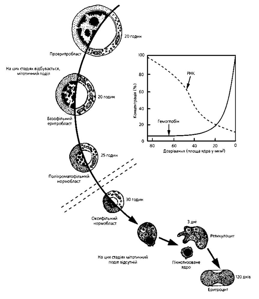
Fig. 3.14. Summary of successive stages of erythrocyte maturation: the Transformation of a proerythroblast into an erythrocyte is accompanied by hemoglobin accumulation (increasing cytoplasmic acidophilia) combined with a decrease in nuclear volume, chromatin Condensation, and subsequent extrusion of the pyknotic nucleus. The hours to the right of the cells correspond to their average lifespan
Thus, during granulocyte formation, the following morphological changes occur in the cells: cell size decreases; cytoplasmic basophilia decreases; the nucleocytoplasmic ratio shifts in favor of an increased volume of cytoplasm; the nucleus condenses and changes shape; and specific granules accumulate.
Monocytopoiesis (monocyte development). The monocyte progenitor cells of the first two classes were described above. The unipotent monocyte progenitor, or CFU-M (class III), is derived from the granulocyte-macrophage progenitor cell. The first morphologically recognizable cell is the monocytoblast (class IV). This is a large cell (up to 22 μm) with a round nucleus and a narrow rim of basophilic cytoplasm. Upon division, it differentiates into a promonocyte, which then matures into a monocyte. During this transition, the following changes occur: the volume of cytoplasm increases, its basophilia slightly decreases, and the nucleus becomes kidney-shaped. The nucleocytoplasmic ratio in a monocyte is 1:1. Monocytes, however, are not The final stage of differentiation in this lineage; they further differentiate into macrophages (histiocytes-macrophages) of Connective Tissue. There are 7-8 mitoses along the pathway from monocytoblast to macrophage.
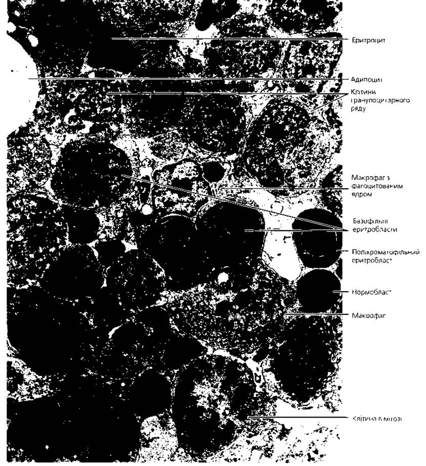
Fig. 3.15. Electron micrograph of erythropoietic islands within the red bone marrow
Thrombocytopoiesis (development of Blood Platelets, Figs. 3.16, 3.17). The first two classes of progenitor cells of thrombocytopoiesis were described above in the section on erythropoiesis (HSC→CFU-GEMM→CFU-MegE). The unipotent progenitor is the colony-forming unit-megakaryocyte (CFU-Meg), or a thrombocytopoietin-sensitive cell, named after the hormone-like factor that acts in this histogenetic lineage.
The megakaryoblast is the earliest morphologically recognizable cell of thrombocytopoiesis. It is round, with a size of 25-40 μm. The nucleus, with an even distribution of chromatin of a deep purple color, contains 1-3 nucleoli. The cytoplasm is basophilic, dark blue, and has two zones: a perinuclear zone containing organelles, and a peripheral zone penetrated by invaginations of the Plasmalemma that form complex demarcation tubules. The megakaryoblast gives rise to the promegakaryocyte. Its size is 40-80 μm. The nucleus of this cell is often indented, and its segmentation and chromatin coarsening begin. The cytoplasm becomes less basophilic, azurophilic granules appear, and the tubules of the demarcation system develop not only at the periphery but also in the middle zone of the cytoplasm. The promegakaryocyte further differentiates into a megakaryocyte.
The megakaryocyte is the largest cell of the red bone marrow, ranging in size from 50-70 to 100 μm. The nucleus is polymorphic, lobulated, with indentations and clefts, having a highly bizarre shape; its structure is coarsely reticular, and nucleoli are absent. The cytoplasm is basophilic, staining purple or pinkish-purple, and contains azurophilic granules. The cell boundary with the surrounding environment is poorly defined. Along the demarcation tubules, the cytoplasm is segmented into small fragments that detach from the cell, becoming blood platelets. A single megakaryocyte produces 3-4 thousand platelets. A unique feature of megakaryocytes is that they are polyploid, with the chromosome number reaching 32-64. The polyploidization of these cells is due to the fact that during their development from megakaryoblasts, Cell Division does not occur; instead, 4-5 endomitoses take place, resulting in an increase in both nuclear and cytoplasmic volume.
Megakaryocytes are located extravascularly (Fig. 3.16), adjacent to endothelial cells, and their cytoplasmic processes penetrate into the vascular lumen. These processes are of two types. Some do not contain organelles and anchor the megakaryocyte to the endothelium. Others measure 2.5x120 μm and, penetrating into the lumen of the sinusoids, give rise to approximately one thousand platelets each. It is also possible that megakaryocytes regulate the migration of other hematopoietic cells through the vessel wall (Figs. 3.17, 3.18).

Fig. 3.16. Megakaryocytes: A - light micrograph of two megakaryocytes near sinusoidal hemocapillaries, x 800; B - electron micrograph of a megakaryocyte with adjacent erythropoietic islands, x 2500
Lymphopoiesis (development of lymphocytes). According to the unitary theory of hemopoiesis, the source of lymphocyte development is the hematopoietic stem cell (class I), which gives rise to the lymphoid progenitor cell (class II). Further development of this cell proceeds in two directions corresponding to the Two Types of lymphocytes - T AND B. In both lineages, unipotent progenitors arise, which differentiate through lymphoblasts (T and B) into lymphocytes (T and B) (Fig. 3.12, Table 16). The development of T lymphocytes occurs in the thymus under the influence of the specific microenvironment of its stroma and the hormone of this organ. In humans, the development of B lymphocytes takes place in the red bone marrow and, possibly, in the lymphatic nodules of the digestive tract. Progenitors of T and B lymphocytes are also formed in the red bone marrow.
A characteristic feature of these lineages is that mature cells are not terminal elements, and their further histogenesis depends on the presence of Antigens. Upon antigen exposure, they revert to blast forms and begin to divide. Upon repeated antigenic stimulation, B lymphocytes, for example, can produce clones with an astronomical number of cells, undergoing up to 90 mitoses. This so-called antigen-dependent process of lymphocyte differentiation occurs in the peripheral hematopoietic organs - the spleen and lymph nodes. Here, antigen-stimulated T lymphocytes differentiate through T lymphoblasts, large, and medium lymphocytes into T killers, T suppressors, and T memory cells. Stimulated B lymphocytes transform through plasmablasts and proplasmacytes into plasma cells and B memory cells.

Fig. 3.17. Diagram of the release of formed blood elements from the red bone marrow into the bloodstream. The migration of erythrocytes occurs along a pressure gradient across the wall of the sinusoidal capillary; leukocytes enter the capillary lumen due to their active motility and under the influence of specific permeability factors; platelets pinch off from the processes of megakaryocytes located within the capillary lumen

Fig. 3.18. Release of formed elements into the bloodstream. Electron micrograph of a bone marrow sinusoidal capillary, x 4000
Table 16. Approximate content of lymphocytes in hematopoietic organs
|
Hematopoietic organ |
T-lymphocytes, % |
B-lymphocytes, % |
|
Thymus |
100 |
0 |
|
Red bone marrow |
10 |
90 |
|
Spleen |
45 |
55 |
|
Lymph nodes |
60 |
40 |
|
Blood |
80 |
20 |
Embryonic hemopoiesis. In the course of embryonic hemopoiesis, blood develops as a tissue. In humans, foci of hemopoiesis are first observed during the second to third week of embryonic development in the wall of the yolk sac. Initially, condensed areas of mesenchyme arise here, known as blood islands. Cells at the periphery of the island flatten, connect with each other, and form the vascular wall. Central cells lose their processes, round up, and transform into HSCs. Some HSCs differentiate into primary blood cells (blasts) - large cells with basophilic cytoplasm and large, prominent nucleoli in the nucleus. These cells divide mitotically and transform into primary erythroblasts (megaloblasts) - large nucleated cells with basophilic cytoplasm. They rapidly accumulate hemoglobin, transforming into oxyphilic erythroblasts, which then lose their nuclei to become primary erythrocytes - megalocytes. However, nuclear loss does not occur in all cells: some primary erythrocytes function as nucleated cells.
Thus, at this stage of Embryogenesis, erythrocytes are formed via an abbreviated pathway, are large in size, and arise within Blood Vessels (intravascularly). This type of hemopoiesis is called megaloblastic and is normal for embryogenesis. The appearance of such hemopoiesis in the postnatal period indicates pathology (pernicious anemia). Simultaneously with megaloblastic hemopoiesis, the process of normoblastic hemopoiesis begins in the wall of the yolk sac, leading to the appearance of normocytic erythrocytes. In addition, a small number of granulocytes - neutrophils and eosinophils - are formed here extravascularly from some of the primary blast cells. This described type of embryonic hemopoiesis is called mesoblastic (extraembryonic).
Some HSCs remain in an undifferentiated state and are carried by the blood to various Organs of the embryo, where the process of hemopoiesis begins. For example, during the fifth week of embryogenesis, hemopoiesis starts in the Liver. Here, erythrocytes and granulocytes, predominantly neutrophilic and eosinophilic, are formed. Development occurs extravascularly. In addition to granulocytes, giant cells - megakaryocytes - are formed in the liver. The process of hepatic hemopoiesis ceases by the end of the prenatal period.
At the beginning of the 2nd month of embryogenesis, the thymus emerges, and during the 7th-8th week, it is colonized by stem cells, from which the first lymphocytes develop. In the 3rd month, hemopoiesis begins in the spleen. Here, all formed elements of the blood are formed from stem cells. Thus, during embryogenesis, the spleen acts as a universal hematopoietic organ. After the 5th month, lymphopoiesis begins to predominate in it. This period of embryonic hemopoiesis is called the hepato-thymosplenic period.
From the 4th month of embryogenesis, the bone marrow begins to function, and from the 6th month, it becomes the primary universal hematopoietic organ. During this period, hemopoiesis also occurs in the thymus, lymph nodes, and spleen, which is why it is called the medullo-thymo-lymphoid period.
Key terms
1. Hemocytopoiesis. 2. Myeloid tissue. 3. Myelopoiesis. 4. Lymphoid tissue. 5. Lymphopoiesis. 6. Unitary theory of hemopoiesis. 7. Hematopoietic stem cell. 8. Hemopoietins. 9. Erythropoiesis. 10. Progenitor cell (myeloid progenitor cell). 11. Erythropoietin-sensitive cell. 12. Proerythroblast. 13. Basophilic erythroblast. 14. Polychromatophilic erythroblast. 15. Polychromatophilic normoblast. 16. Reticulocyte. 17. Oxyphilic normoblast. 18. Erythrocyte. 19. Granulocytopoiesis. 20. Myeloblast. 21. Promyelocyte. 22. Myelocyte. 23. Metamyelocyte. 24. Monocytopoiesis. 25. Monocytoblast. 26. Promonocyte. 27. Monocyte. 28. Histiocyte-macrophage. 29. Thrombocytopoiesis. 30. Thrombocytopoietin-sensitive cell. 31. Megakaryoblast. 32. Promegakaryocyte. 33. Megakaryocyte. 34. Lymphopoiesis. 35. Lymphoblast. 36. Lymphocyte. 37. Primary erythroblast (megaloblast). 38. Megalocyte. 39. Megaloblastic hemopoiesis. 40. Normoblastic hemopoiesis. 41. Mesoblastic hemopoiesis.
Last update: 09/08/2026
Editorial and Educational Adaptation: This material has been compiled based on the primary/original source text. The project team performed an editorial review, corrected technical inaccuracies, structured sections, and adapted the content for an educational format.
What was processed:
- elimination of formatting defects (OCR errors, structural breaks, corrupted characters);
- editorial organization of content;
- standardization of terminology in accordance with academic sources;
- verification of factual statements against the original source text.
All mentions of the author, publication year, and origin of the primary text have been preserved in accordance with the source.