Human Histology - O.D. Lutsyk 2003
General Histology
Connective tissues. Connective tissue proper. Connective tissues with special properties
Connective Tissue (textus connectivus) is widely distributed throughout the body, accounting for about 50% of total body mass. It forms the Skeleton, Skin, Cartilage, tendons, ligaments, and the stroma of Organs. Connective tissue is classified into connective tissue proper, cartilage, and bone. Connective tissue proper, in turn, is divided into Fibrous connective tissue and Connective Tissues with special properties. The latter include reticular, adipose, pigment, and mucous tissues. Depending on the density of fibrous structures, fibrous connective tissue is classified as loose or dense. Loose connective tissue contains relatively more Cells and ground substance, whereas Cytology/practical/45.html">Dense connective tissue is richer in fibers. Depending on the arrangement of these fibers, dense connective tissue is subdivided into regular and irregular: in regular dense connective tissue, the fibers run parallel to each other, while in irregular dense connective tissue, they run in various directions, forming a network.
Among all the types of connective tissue mentioned in the Classification, loose fibrous connective tissue is the most common and contains all types of tissue elements. It is present in almost all Internal Organs, forming their sheaths, filling the spaces between organs, underlying the epithelium, and accompanying Blood Vessels and nerves. It performs all the Functions characteristic of the Tissues of the Internal Environment, namely: trophic, protective, and supportive-mechanical. In addition, loose connective tissue performs a replacement function, filling in defects in organs in case of injury.
Loose fibrous connective tissue (textus connectivus fibrosus laxus) consists of cells and Extracellular matrix (Fig. 3.19). The latter, in turn, includes fibrous structures (Collagen, elastic, and reticular fibers) and ground substance. A similar structural plan is characteristic of all Other Types of connective tissue. The cellular elements of loose connective tissue include fibroblasts, macrophages, plasma cells (plasmocytes), mast cells (tissue basophils), adipocytes, pigment cells (pigmentocytes), adventitial cells, as well as leukocytes migrating from the blood (Fig. 3.8, Table 17).
Table 17. Functions of connective tissue cells
|
Cell type |
Main substance produced or type of activity |
Main function |
|
Fibroblast, chondroblast, osteoblast, dentinoblast |
Formation of fibers and ground substance |
Structural |
|
Plasma cell |
Antibody production |
Immune |
|
Lymphocyte |
Differentiation into immunocompetent cells |
Immune |
|
Eosinophil |
Phagocytosis of antigen-antibody complexes |
Immune |
|
Macrophage, neutrophil |
Phagocytosis of foreign substances and Bacteria |
Protective |
|
Mast cell (tissue basophil), blood basophil |
Release of pharmacologically active substances (histamine, etc.) |
Protective |
|
Adipocyte (lipocyte) |
Accumulation of neutral fats, heat production |
Energy storage, thermogenic |
Class="center">
Fig. 3.19. Light Microscopy of connective tissue types: A - loose connective tissue (subcutaneous): abundant ground substance, diverse cellular elements, collagen and elastic fibers, x 400; B - dense irregular connective tissue (reticular layer of the dermis): thick, multidirectional bundles of collagen fibers separated by thin layers of ground substance and fibrocyte cell bodies, x 200; C - dense regular connective tissue (Longitudinal section of a tendon): thick bundles of collagen fibers showing parallel orientation, separated by fibrocytes, x 200
Cellular elements of connective tissue
Fibroblasts (Fig. 3.20) are the cells responsible for producing the extracellular matrix. They synthesize both the fibrous structures and the Main Components of the ground substance. In a sense, fibroblasts build connective tissue. Due to their ability to form the primary supportive structures of the body, fibroblasts are often called mechanocytes. Their name itself reflects this fiber-producing capacity ('fibra' - fiber, and 'blastos' - germ or bud). The activity of these cells drives wound healing, scar development, and The formation of capsules around foreign bodies. Fibroblasts comprise a large group of cells at various stages of differentiation, forming the so-called fibroblastic Lineage (or differon): stem cells - committed progenitor cells - minimally specialized (immature) fibroblasts - mature fibroblasts - fibrocytes. In addition, myofibroblasts also belong to this lineage.
Minimally specialized, or immature, fibroblasts are round or spindle-shaped cells with basophilic Cytoplasm containing A large number of free Ribosomes. Other Organelles (Endoplasmic reticulum, Mitochondria, Golgi apparatus) are poorly developed. These cells are capable of mitotic division and exhibit low Levels of Protein Synthesis and Secretion. Their size does not exceed 20-25 μm.
Mature fibroblasts are large, branched cells. In flat-mount preparations, they can reach 40-50 μm or more in their spread state, while being very thin. The Nucleus of these cells is large, oval, and pale, containing finely dispersed, evenly distributed Chromatin, against which 1-2 large nucleoli are clearly visible. The cytoplasm stains basophilically. In whole-mount preparations, The Cell body of the fibroblast can be seen divided into two zones: an intensely stained central endoplasm and a much more weakly stained peripheral ectoplasm, which lacks distinct boundaries and merges with the surrounding extracellular matrix.
The cytoplasm of a fibroblast contains all general organelles. The rough endoplasmic reticulum is particularly well-developed, occupying up to 35 % of the cell volume; this is where procollagen and Elastin are synthesized. The Golgi apparatus is also well-developed, occupying about 10 % of the cell volume and appearing as cisternae and vesicles scattered throughout the cell; this is where glycosaminoglycans are synthesized. The latter, along with Fibrillar Proteins, are exported into the extracellular space and incorporated into fibers and ground substance. Fibroblasts also synthesize fibronectin, a fibrillar glycoprotein of the extracellular matrix that mediates cell binding to their microenvironment and regulates migration. Mitochondria are large and moderate in number, as are Lysosomes.
Microfilaments with a thickness of 5-6 nm are located in the peripheral layer of the cytoplasm; they contain contractile Proteins of the Actin and Myosin types, enabling cell motility. It is believed that two populations of fibroblasts exist: one with a short life cycle (several weeks) and another with a long life cycle (several months).

Fig. 3.20. Fibroblasts: A - light microscopy of fibroblasts in the lamina propria of the Small Intestine mucosa, x 400; B - transmission Electron microscopy of a mature fibroblast of subcutaneous connective tissue, x 3000; C - transmission electron microscopy of corneal fibrocytes, x 2000
Fibrocytes are the definitive (terminal) forms of fibroblast development. They are spindle-shaped and may have wing-like processes. They contain few organelles, and their synthetic processes are sharply reduced.
Myofibroblasts are a cell type into which fibroblasts can differentiate. They are functionally similar to smooth Muscle cells but, unlike them, possess a well-developed endoplasmic reticulum. Such cells can be observed in the Uterus during Pregnancy, as well as in granulation tissue (during wound healing).
Macrophages (macrophagocytes). These cells (Fig. 3.21) are also referred to as histiocytes. In terms of Abundance in loose connective tissue, macrophages rank second only to fibroblasts. Compared to the latter, they have smaller cell bodies (10-15 μm) that are well-demarcated from the ground substance. Their shape varies, being round, elongated, or irregular. The nucleus is also smaller and less regularly shaped than that of a fibroblast, contains more heterochromatin, appears dense, and stains quite intensely. The cytoplasm of macrophages is basophilic, heterogeneous, and mottled, containing numerous lysosomes, phagosomes, and pinocytotic vesicles. Other organelles (mitochondria, rough endoplasmic reticulum, Golgi apparatus) are moderately developed.
The Plasmalemma of macrophages forms deep folds and long microvilli, which these cells use to engulf foreign particles. The surface of the macrophage plasmalemma contains receptors for tumor cells, erythrocytes, T- and B-lymphocytes, Antigens, and IMMUNOGLOBULINS. The presence of immunoglobulin receptors mediates their participation in immune responses.
Macrophages play an important role in both innate (natural) and adaptive (acquired) Immunity. Their role in innate immunity is demonstrated by their phagocytic capacity and their synthesis of various active substances—such as phagocytin, Lysozyme, interferon, pyrogen, and Complement system components—which are key factors of innate immunity. Their role in adaptive immunity involves antigen presentation to immunocompetent cells (lymphocytes) after Processing the antigen from a particulate to a molecular form (participating in the cooperative three-cell System of the Immune Response alongside T- and B-lymphocytes). Furthermore, macrophages produce monokine mediators that facilitate specific antigen responses, as well as cytolytic factors that selectively destroy tumor cells. Macrophages originate from promonocytes in the Red Bone Marrow, arising from hematopoietic stem cells, and represent the terminal stage of the monocytic lineage. Together with other Cells of the same origin, they form the macrophage system of the body (Table 18).
The macrophage system comprises all cells capable of engulfing foreign particles, dead cells, non-cellular structures, bacteria, and other elements from the tissue fluid. Once phagocytosed, this material undergoes enzymatic degradation within the lysosomal apparatus. This eliminates harmful agents, whether generated locally or introduced from the outside. These cells can be identified using Vital Staining Methods, such as the in vivo administration of trypan blue, colloidal silver, or India ink. All these colloidal substances are phagocytosed by macrophages because they form macromolecular aggregates, making the cells clearly visible in histological preparations.

Fig. 3.21. Macrophages: A - light microscopy of a group of free macrophages (dust cells) in the lumen of a terminal bronchiole of the lung, x 100; B - transmission electron microscopy of a free macrophage, x 5000; C - scanning electron microscopy of a murine macrophage phagocytosing opsonized erythrocytes, x 4000
Table 18. Distribution and Main Functions of cells of the macrophage system
|
Cell type |
Localization |
Main functions |
|
Monocyte |
Blood |
Differentiates into macrophage, phagocytosis |
|
Macrophage |
Connective tissue, Lymphoid organs, Lungs, serous membranes |
Production of cytokines, chemotactic factors, and other molecules involved in inflammation; antigen presentation to lymphocytes |
|
Kupffer cell |
Phagocytosis |
|
|
Microglia |
Nervous Tissue of the Central Nervous system |
Phagocytosis |
|
Epidermal dendritic cell (Langerhans cell) |
Skin |
Antigen processing for the immune response |
|
Osteoclast |
Resorption of bone and cartilage |
|
|
Foreign-body giant cell |
Connective tissue |
Digestion of foreign particles |
The cells of the macrophage system include histiocytes-macrophages of loose connective tissue, free and fixed macrophages of Hematopoietic organs (so-called dendritic cells), stellate cells of hepatic sinusoidal capillaries (Kupffer cells), alveolar macrophages of the lungs (so-called dust cells), peritoneal macrophages, glial macrophages of nervous tissue (microglia), osteoclasts of bone tissue, and foreign-body giant cells. All of them are capable of active phagocytosis, possess immunoglobulin receptors on their surface (rendering them capable of immune phagocytosis), and originate from promonocytes of the red bone marrow and blood monocytes. Unlike macrophages, which I.I. Mechnikov termed "professional phagocytes," other cell types—such as fibroblasts, reticular cells, endothelial cells, and neutrophilic leukocytes—possess the capacity for facultative phagocytosis. However, these cells do not belong to the macrophage system because they cannot perform specific immune phagocytosis and also differ in their origin.
METABOLISM/2.html">THE CONCEPT OF phagocytosis was first proposed by I.I. Mechnikov. He concluded that phagocytosis, which arose in evolution as intracellular digestion and became established in many cells, is an important defense mechanism. He justified the integration of these cells into a single system and proposed calling it the macrophage system. In the 1930s–1950s, this protective system was referred to as the reticuloendothelial system (RES), erroneously including some types of facultative phagocytes. Recently, it has been called the mononuclear phagocyte system, which, however, is not entirely accurate, as this system also includes multinucleated cells (osteoclasts and foreign-body giant cells).
The macrophage system is a powerful defense apparatus involved in both systemic and local protective Reactions of the body. In the Organism as a whole, the macrophage system is regulated by local mechanisms, as well as by the nervous and endocrine systems.
Plasma cells (plasmacytes) (Fig. 3.22) range in size from 7-10 μm, although they can be slightly larger; their shape is round or polygonal if they are adjacent to each other. The nucleus is small, round, eccentrically located, and contains predominantly condensed chromatin, the clumps of which form a pattern characteristic of a plasmacyte—resembling a spoke wheel or clock face. The cytoplasm is intensely basophilic, against which a well-defined "pale halo" or perinuclear zone with lighter staining is visible near the nucleus. The ultrastructure of these cells is characterized by the presence of a well-developed granular endoplasmic reticulum in the cytoplasm, which is arranged concentrically and occupies most of the cell. A large number of ribosomes (RNA) accounts for the basophilia of the cytoplasm. In the "pale halo" region, centrioles surrounded by cisternae of the Golgi complex are located. Immunoglobulins (Antibodies) are synthesized within the cisternae of the granular endoplasmic reticulum of plasmacytes. Part of the carbohydrate component of immunoglobulins is synthesized in the Golgi complex. This organelle, which is quite well developed in plasmacytes, is also responsible for the secretion of synthesized immunoglobulins out of the cell; they then enter the bloodstream via the Lymph.
Thus, plasmacytes provide humoral immunity, i.e., The production of specific proteins—immunoglobulins (antibodies)—in response to the penetration of an antigen into the body, which will be neutralized by the antibodies. Plasma cells originate from hematopoietic stem cells (via the B-lymphocyte stage). Plasma cells are mostly found in the loose connective tissue of the lamina propria of the intestinal and respiratory mucosa, in Lymph Nodes, the Spleen, and in the interstitial connective tissue of various glands.
Tissue basophils (Fig. 3.23) have many names, which are worth mentioning to help navigate the literature: mastocytes, labrocytes, mast cells. The latter name was given to these cells by P. Ehrlich, who in 1877 first described cells that were overfilled with granules, as if they had "gorged" on them. This name is very common in the literature. The term "tissue basophils" indicates that the cells have granulation similar to the granules of blood basophilic leukocytes. Tissue basophils are often localized along the Blood Vessels of the microcirculatory bed, forming perivascular sheaths. A large number of these cells are found in the walls of the digestive tract organs, in the uterus, mammary gland, Thymus, and Tonsils.
The shape of tissue basophils is diverse, as are their sizes. They can be round, oval, or have broad processes. Their sizes range from 10-20 to 35 and even up to 100 μm. The nuclei are relatively small, round, and of typical Structure. The cytoplasm contains a large number of mitochondria, a few elements of granular and agranular endoplasmic reticulum, and a well-developed Golgi complex. The main feature of these cells is the presence of a large number of characteristic granules measuring 0.2-0.8 μm, each surrounded by a membrane. By electron microscopic structure, the granules of human tissue basophils are crystalloid or lamellar (Species Differences in granule structure are observed). The granulation stains basophilically and metachromatically. The granules contain several substances of great physiological importance. The first of these substances is heparin, which accounts for 30% of the granule content and is mainly responsible for their basophilia and metachromasia. The second substance is histamine, which makes up 10% of their content. The granule matrix consists of protein (tissue basophil chymase) and heparin, which form a stable meshwork; histamine is attached to it by ionic bonds. The granules also contain chondroitin sulfate and hyaluronic acid; in some animals (but not in humans), serotonin has also been found.

Fig. 3.22. Electron micrograph of a bone marrow plasmacyte, x 6000
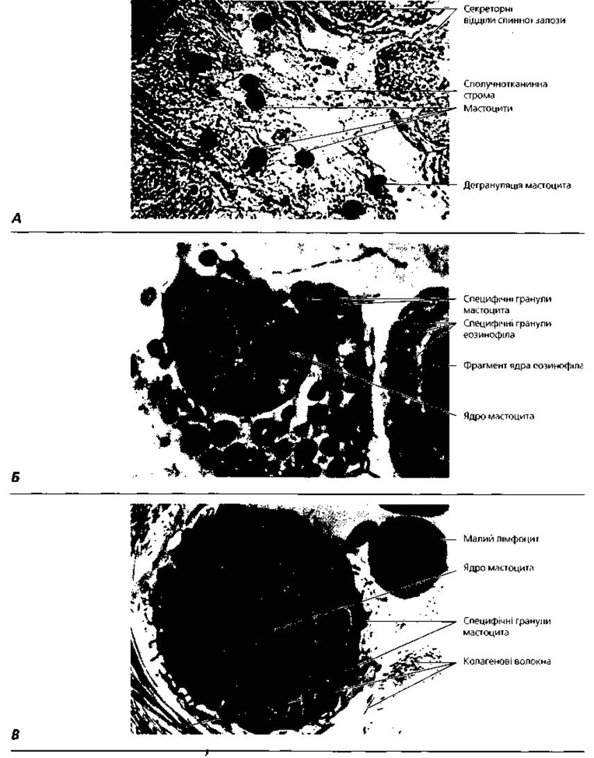
Fig. 3.23. Tissue basophils: A - light microscopy of tissue basophils of the connective tissue stroma of the Parotid salivary gland. Histochemical reaction with peanut lectin, x 280; B - electron microscopy of the interaction between a tissue basophil and an eosinophil, x 5000; C - electron microscopy of the interaction between a tissue basophil and a small lymphocyte, x 5000
Heparin is a sulfated glycosaminoglycan that was first isolated from the liver (hence its name) and prevents blood clotting. It has been found that tissue basophils synthesize heparin in the Golgi complex. They can lose their granules (The process of degranulation), releasing heparin into the intercellular substance. Heparin reduces its permeability, has an anti-inflammatory effect, and acts as an anticoagulant. In addition, heparin stimulates the activity of the enzyme lipoprotein lipase, thereby promoting The breakdown of plasma chylomicrons.
Histamine is synthesized in tissue basophils with the participation of Histidine decarboxylase (a marker enzyme of these cells), which converts histidine to histamine. Histamine acts on smooth Muscles, causing them to contract, and also promotes the leakage of plasma from venules and capillaries by dilating and increasing the permeability of their walls. As a result of plasma leakage, blisters form in the loose connective tissue beneath the epidermis. This symptom is known as urticaria (hives). The described action of histamine can be observed during anaphylactic Shock or allergy. The Development of these processes and the involvement of tissue basophils in them is explained as follows. In response to the penetration of certain antigens, called allergens, into the body, specific antibodies belonging to the class of immunoglobulins E (IgE) are produced. Tissue basophils, like basophilic leukocytes, have receptors for antibodies of this type and bind them so that the variable Regions of the immunoglobulin molecules remain free. Upon re-exposure to the antigen, the latter binds to the antibodies On the surface of the tissue basophils. Following the Formation of the antigen-antibody complex, histamine is released from the granules of these cells. Symptoms of allergy or anaphylaxis can be alleviated by administering antihistamines. Under normal conditions, such hypersensitivity reactions involving tissue basophils tend to be self-limiting due to the release of eosinophil chemotactic factor by these cells. The eosinophil Enzymes histaminase and arylsulfatase degrade the substances released by tissue basophils during immune reactions.
It is known that tissue basophils originate from hematopoietic stem cells. Undifferentiated progenitors of tissue basophils migrate through the blood into the connective tissue, where they proliferate and differentiate into mature cells. T-lymphocytes participate in these processes. Some authors believe that tissue basophils are formed from blood basophils when they migrate into the connective tissue. Mitotic division of tissue basophils is observed quite rarely. Since there is Evidence of the ability of tissue basophils to synthesize DNA, mitoses may occur in them more frequently, but they are difficult to visualize due to the large number of granules contained in the cytoplasm of these cells.
Adipocytes (fat cells, Fig. 3.24). Previously, these cells were called lipocytes. Adipocytes are capable of accumulating reserve fat in their cytoplasm, which is important for Nutrition, energy production, and Water Metabolism. In loose connective tissue, they are arranged in groups, less frequently individually, and mostly near blood vessels. When they accumulate in large numbers, they form adipose tissue.
The shape of an individual adipocyte is spherical, but when There are many of them, they press against each other and assume a polygonal shape. A mature fat cell contains a single large lipid droplet that distends the entire cell so that the cytoplasm surrounds the fat in only a thin layer. The nucleus changes its shape, becoming flattened. The diameter of a fat cell can reach 120 μm. In cross-section, such a cell resembles a signet ring: the nucleus is the signet, and the ring is the thin layer of cytoplasm surrounding the fat. Lipids stain well with Sudan orange or osmic acid black. Organelles are located primarily around the nucleus. The fat cell contains free ribosomes, both types of endoplasmic reticulum, the Golgi complex, and mitochondria. Accumulations of such fat cells, which are called unilocular, form White adipose tissue.
Lipid droplets that enter the lymph and then the blood from the epithelial cells of the small intestine, measuring about 1 μm, are called chylomicrons (from the Greek "chylos" - juice, "micron" - small). These particles contain triglycerides, as well as Phospholipids, Cholesterol esters, and a small amount of proteins that form Lipoproteins with lipids. Under the action of lipoprotein lipase enzymes produced by the vascular endothelium, chylomicron triglycerides are cleaved into Fatty acids and glycerol, which can be absorbed by the fat cell. Under the action of adipocyte glycerokinase, triglycerides are resynthesized from fatty acids and glycerol. Fat stored in adipocytes is metabolized under the action of lipolytic Hormones (epinephrine, Insulin) and the tissue enzyme lipase, which cleaves triglycerides into glycerol and fatty acids. The latter bind to blood albumin and are transported to other tissues that require nutrients.
In addition to the discussed unilocular adipocytes, multilocular adipocytes are also distinguished, which are characterized in more detail below in the "Adipose Tissue" section. By origin, fat cells apparently represent a distinct cell lineage. Fat cells are long-lived. Mitoses in adipocyte progenitor cells cease 2-3 weeks after birth. In adults, fat cells do not divide, but there is evidence that new adipocytes can be formed from adventitial cells through the accumulation of lipid within them.
Pigment cells (melanocytes) contain the pigment melanin in their cytoplasm. They are observed not only in connective tissue but also within the epithelium, particularly in the basal layer of the epidermis. Connective tissue melanocytes usually do not produce melanin (as evidenced by a negative DOPA reaction), but only phagocytose melanin produced by epithelial melanocytes. The only exception is individuals of Mongoloid descent, in whom melanin-synthesizing pigment cells occur in the dermis of the coccygeal region, forming the so-called Mongolian spot. Unlike other cell populations of connective tissue, melanocytes originate from neural crest cells rather than mesenchyme.
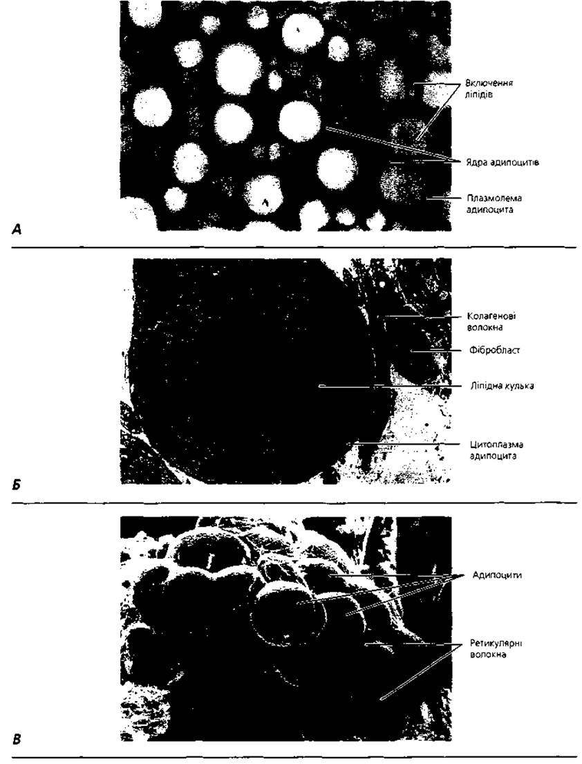
Fig. 3.24. Adipocytes: A - light microscopy of unilocular adipocytes of subcutaneous adipose tissue, x 300; B - transmission electron microscopy of a unilocular adipocyte of human fetal subcutaneous tissue, x 1600; C - scanning electron micrograph of an accumulation of adipocytes, x 400
Adventitial cells are a population of poorly specialized cells localized along blood vessels. They have a flat or spindle-like shape, weakly basophilic cytoplasm, an oval nucleus, and a small number of organelles. In the process of differentiation, these cells can transform into fibroblasts and adipocytes. Some authors believe that adventitial cells and pericytes of blood capillaries represent the same population of poorly differentiated cells of mesenchymal origin.
Fibrous structures
Collagen fibers (Fig. 3.25). In loose connective tissue, collagen fibers are arranged in various directions and appear as wavy, spirally twisted, round, or flat bundles with a thickness of 1-10 μm. They are capable of forming bundles whose thickness can reach 150 μm. In their Native State, collagen fibers are colorless; on histological preparations, they stain oxyphilically, and upon silver impregnation, they acquire a brownish-yellow color. These fibers do not branch or anastomose with each other.
A collagen fiber is composed of bundles of fibrils cemented by glycosaminoglycans and Glycoproteins. The thickness of the fibrils is 50-100 nm. Fibrils consist of microfibrils about 10 nm thick, which can be seen under an Electron microscope as slightly wavy threads. Microfibrils are built from even thinner elements—protofibrils, and the latter from tropocollagen molecules (Fig. 3.27). Tropocollagen molecules are about 280 nm long and 1.4 nm thick. They are composed of three polypeptide chains of the collagen precursor, procollagen. The synthesis of collagen, as well as glycosaminoglycans and glycoproteins, occurs in the cells of loose connective tissue—fibroblasts (Fig. 3.26), which secrete these substances into the extracellular environment. Outside the cell, fibrils are formed from collagen molecules (Fig. 3.27), which exhibit a characteristic transverse striation in the form of alternating dark and light bands with a repeating period of 64 nm. The marker Amino Acids of mature collagen are hydroxyproline and hydroxylysine.
According to molecular Organization, organ localization, and tissue Specificity, 12 types of collagen are distinguished (Table 19). Type I collagen is present in the connective tissue of the skin, bones, cornea, sclera, arterial walls, etc.; type II is found in hyaline and fibrous cartilage, and the vitreous body; type III is in the dermis of fetal skin, the walls of large blood vessels, and reticular fibers; type IV is in basement membranes and the lens capsule; type V is located around the cells that synthesize it, forming an exocytoskeleton. Types VI and VII collagens are referred to as microfibrillar; types VIII, IX, X, and XI collagens are the so-called minor varieties, found in small amounts in the endothelium, cartilage, and vitreous body. Collagen fibers contain about 65 % water. They are capable of binding water and Swelling both in vivo and in vitro. In running water, their thickness increases by 50 % due to swelling, and in an acidic medium, up to 500 times, while the length of the fibers does not increase. These properties of collagen fibers determine their function in the body as a water reservoir. This property of collagen fibers is responsible for the development of edema under pathological conditions. In the event of blood loss, they release water, restoring blood volume. Upon boiling, collagen fibers yield glue (hence their name, from the Greek "kolla" - glue, and "gen" - to produce). They have low resistance to acids, alkalis, and Proteolytic Enzymes. Collagen fibers are highly durable but have low elasticity, with an elastic modulus of 60-70 kg/mm. They are the strongest structures in the body, and their main function is supportive and mechanical. Impaired collagen synthesis leads to various pathological conditions (Table 20).

Fig. 3.25. Fibrous structures of connective tissue: A - electron micrograph of a longitudinally sectioned bundle of collagen fibrils, x 75 000; B - electron micrograph of transversely sectioned collagen fibrils of a tendon, separated by processes of a fibrocyte, x 20 000; C - network of reticular fibers of a lymph node. Silver impregnation, x 200

Fig. 3.26. Diagram of type I collagen Biosynthesis by a fibroblast
Table 19. Main characteristics of Different types of collagen
|
Collagen type |
Localization |
Light microscopy |
Electron microscopy |
Synthesizing cells |
Interaction with glycosaminoglycans |
Main function |
Molecular organization |
|
I |
Dermis, tendons, bones, fibrocartilage, cornea, sclera, Arteries |
Thick, striated, non-argyrophilic |
Densely packed thick microfibrils, variable in diameter |
Fibroblasts, odontoblasts, osteoblasts, chondroblasts |
Weak; primarily with dermatan sulfate |
Resistance to stretching |
Fibril-forming |
|
II |
Hyaline and fibrocartilage, intervertebral discs, vitreous body |
Loose bundles of fibrils, visible in polarized light |
Thin microfibrils embedded in the ground substance |
Chondroblasts |
Intense interaction with chondroitin sulfates |
Resistance to pressure |
Fibril-forming |
|
III |
Smooth muscle, reticular connective tissue, blood vessels, fetal dermis |
Thin, transversely striated, argyrophilic |
Loosely packed thin microfibrils of uniform diameter |
Smooth myocytes, reticular cells, neurolemmocytes |
Moderate with heparan sulfate |
Structural support of expandable organs |
Fibril-forming |
|
IV |
Basement membranes, lens capsule |
Thin, PAS-positive, argyrophilic |
Microfibrils absent |
Endothelial cells, epithelial cells, myocytes, neurolemmocytes |
Moderate with heparan sulfate |
Structural support, filtration |
Network-forming |
|
V |
Dermis, tendons, bones, fibrocartilage |
Similar to type I |
Thick microfibrils |
Fibroblasts |
Similar to type I |
Fibril-forming |
|
|
VII |
Dermis |
Invisible |
Visible microfibrils |
Anchoring cells to the underlying connective tissue |
Anchoring collagen |
||
|
IX |
Hyaline cartilage |
Chondroblasts |
Lateral association of fibrils |
Fibril-associated |
|||
|
XI |
Hyaline cartilage, intervertebral discs |
Similar to type II |
Thin microfibrils |
Chondroblasts |
Similar to type II |
Fibril-forming |
|
|
XII |
Tendons, ligaments |
Fibroblasts |
Lateral association of fibrils |
Fibril-associated |

Fig. 3.27. Diagram of tropocollagen molecule self-assembly into collagen filaments, microfibrils, fibers, and their bundles, illustrating The Mechanism of transverse striation (64 nm periodicity)
Table 20. Examples of clinical disorders caused by defects in collagen synthesis
|
Disorder |
Defect |
Symptoms |
|
Ehlers-Danlos syndrome type IV |
Defect in Transcription or Translation of type III collagen |
Aortic and/or intestinal rupture |
|
Ehlers-Danlos syndrome type VI |
Defect in Lysine hydroxylation |
Altered skin elasticity, Eyeball damage |
|
Ehlers-Danlos syndrome type VII |
Decreased procollagen peptidase activity |
Decreased joint mobility, frequent dislocations |
|
Scurvy |
Gum ulceration, bleeding |
|
|
Single nucleotide defect in type I collagen genes |
Spontaneous bone fractures, Heart Failure |
Elastic fibers (Fig. 3.28), unlike collagen fibers, have a yellowish color in their native state, branch and anastomose with each other, are always found individually, and do not form bundles. Their thickness ranges from 0.3 to 10-18 µm.
The main chemical component of elastic fibers is the globular protein elastin, which is synthesized by fibroblasts. Elastin contains a large amount of the amino acids proline and Glycine, while cystine is absent. In addition, it is characterized by the presence of two Amino Acid Derivatives—desmosine and isodesmosine—which account for its elasticity. Elastin molecules are globular in shape with a diameter of 2.8 nm. Extracellularly, they assemble into chains 3–3.5 nm thick, termed elastin protofibrils, which combine with glycoproteins to form microfibrils 8–10 nm thick. According to electron microscopy data, an elastic fiber consists of two components: an amorphous core in the center and a microfibrillar component on the periphery. The ratio of these two components varies in different types of elastic fibers. The most mature elastic fibers contain about 90 % elastin in the form of an amorphous component. The microfibrillar component is more developed where the demands for mechanical strength are greater than those for elasticity.

Fig. 3.28. Electron microscopy of successive stages of elastic fiber formation (A, B, C): A — oxytalan fibers formed by fine-fibrillar glycoprotein conglomerates; B — elaunin fibers: accumulation of amorphous elastin between glycoprotein fibrils; C — elastic fibers: an elastin core surrounded by peripheral glycoprotein fibrils; D — light microscopy of skin stained for elastin: specific topography and gradual increase in thickness of oxytalan, elaunin, and elastic fibers
In addition to mature elastic fibers, less mature fibers, namely oxytalan and elaunin fibers, are distinguished during elastogenesis. In elaunin fibers, the ratio of microfibrils to the amorphous component is approximately equal, whereas oxytalan fibers consist solely of microfibrils (Fig. 3.28).
Elastic fibers contain less water compared to collagen fibers (containing 47% water). They are resistant to boiling, acids, alkalis, maceration, and putrefaction, and persist longer in cadaveric material. Their tensile strength is much lower than that of collagen fibers, but they possess high elasticity. They are excellent shock absorbers that ensure the return of structures to their original position (Fig. 3.29). With age, the elasticity of these fibers decreases, and they fragment. Elastic fibers stain poorly with routine histological Dyes; they can be selectively visualized using orcein or resorcin-fuchsin.
Reticular fibers can be observed in specimens impregnated with silver salts, which is why they are also called argyrophilic fibers. Among the latter, 2 types of fibers are distinguished: reticular fibers proper, which are definitive structures composed of type III collagen; and precollagen fibers, representing the initial stage of collagen fiber formation during Embryogenesis and regeneration. Reticular fibers are very similar to collagen fibers in their composition but differ in being thinner, branched, and forming anastomoses. Reticular fibers, together with the reticular cells that produce them, form reticular tissue.
Electron microscopy reveals protofibrils 40 nm thick in reticular fibers, cemented by an amorphous ground substance. The protofibrils do not always show distinct striation, with a period of 64-67 nm (which is identical to collagen fibers). Unlike collagen fibers, reticular fibers have a high concentration of lipids, CARBOHYDRATES, and sulfur. They are resistant to weak acids, alkalis, and Trypsin. In terms of extensibility, they occupy an intermediate position between collagen and elastic fibers.
Ground substance. Cells and fibers of connective tissue are embedded in the ground (intercellular) substance. Ground substance accounts for about 20 % of body weight. It is more abundant in childhood than in adulthood or old age.
The content of ground substance varies among different types of connective tissue. Physically and chemically, it is a gel of variable viscosity and chemical composition. Connective tissue cells, primarily fibroblasts, participate in the formation of the ground substance. The chemical composition of the ground substance is characterized by the Presence of water, proteins, lipids, Polysaccharides, and minerals. The polysaccharide content is 0.5-5 %. These include glycosaminoglycans (GAGs): sulfated—heparan sulfate, chondroitin-4-sulfate, chondroitin-6-sulfate, dermatan sulfate, as well as non-sulfated, represented by hyaluronic acid (Table 21). Sulfated GAGs form proteoglycan complexes with proteins (Fig. 3.30). Glycosaminoglycans determine the consistency and Functional Properties of the ground substance, which, in turn, influences the Functional Characteristics of the connective tissue as a whole. The denser the ground substance, the more pronounced the mechanical, supportive function of the connective tissue. A more fluid consistency of the ground substance better serves the trophic function. Histamine and hyaluronidase increase the permeability of the amorphous component (many microorganisms contain hyaluronidase, which helps them make their way through connective tissue). Conversely, an increase in the concentration of GAGs (particularly hyaluronic acid) decreases the permeability of the ground substance.
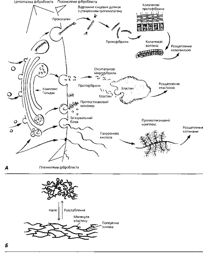
Fig. 3.29. A — diagram of the formation of collagen and elastic fibers, as well as proteoglycan complexes within the extracellular matrix of connective tissue. Under the action of exopeptidases, procollagen and proelastin synthesized by fibroblasts are converted into insoluble forms, and under the action of lysyl oxidase, cross-linking of molecules occurs to form protofibrils; B — two states of elastic fibers: relaxed and stretched
The ground substance provides a pathway for the migration of actively motile cells and serves to transport nutrients and metabolic products.
Table 21. Composition and distribution of glycosaminoglycans in connective tissue and their interaction with collagen fibers
|
Glycosaminoglycan |
Repeating Disaccharides |
Distribution |
Electrostatic interaction with collagen |
|
|
Hexuronic acid |
Hexosamine |
|||
|
Hyaluronic acid |
D-glucuronic acid |
D-glucosamine |
Umbilical cord, synovial fluid, vitreous body, cartilage |
|
|
Chondroitin 4-sulfate |
D-glucuronic acid |
D-galactosamine |
Cartilage, bone, cornea, skin, notochord, aorta |
Strong interaction, predominantly with type II collagen |
|
Chondroitin 6-sulfate |
D-glucuronic acid |
D-galactosamine |
Cartilage, umbilical cord, aorta (tunica media) |
Strong interaction, predominantly with type II collagen |
|
Dermatan sulfate |
D-glucuronic or L-iduronic acid |
D-galactosamine |
Skin, tendon, aorta (tunica adventitia) |
Weak interaction, predominantly with type I collagen |
|
Heparan sulfate |
D-glucuronic or L-iduronic acid |
D-galactosamine |
Aorta, lung, liver, basement membranes |
Moderate interaction, predominantly with type III and IV collagens |
|
Corneal keratan sulfate |
D-galactose |
D-galactosamine |
Cornea |
No interaction |
|
Skeletal keratan sulfate |
D-galactose |
D-glucosamine |
Cartilage, nucleus pulposus, annulus fibrosus of intervertebral discs |
No interaction |
Dense Fibrous Connective tissue (textus connectivus fibrosus compactus). This type of connective tissue is characterized by the predominance of fibrous structures, primarily collagen fibers. This feature provides high shock-absorbing and mechanical properties. Depending on the spatial orientation of the collagen fibers, dense fibrous connective tissue is classified into dense regular and dense irregular connective tissue.

Fig. 3.30. Proteoglycans: A - semi-schematic representation of a proteoglycan aggregate; B - structure of a proteoglycan monomer; C - diagram of the interaction between a proteoglycan aggregate and collagen fibers of the extracellular matrix of connective tissue
Dense regular fibrous connective tissue is localized within fibrous membranes, ligaments, and tendons. The latter, connecting muscles to bones, are subjected to a force vector acting primarily in one direction. This factor determines the parallel spatial orientation of collagen fiber bundles. Highly differentiated cells of the fibroblastic lineage (fibrocytes) are located between individual fiber bundles, and their synthetic activity ensures the physiological regeneration of tendon bundles. A bundle of collagen fibers surrounded by a layer of fibrocytes is called a primary tendon bundle. Fibrocytes separate adjacent primary tendon bundles and appear as small dashes in longitudinal sections of the tendon. The alternating pattern of collagen fiber bundles and rows of fibrocytes is characteristic.
In a transverse section of a tendon, characteristic lamellar processes of fibrocytes can be seen, which result from the compression of the cell body by adjacent collagen fibers. Several primary tendon bundles form secondary tendon bundles, which are separated by layers of loose connective tissue called endotendineum. Within large tendons, secondary bundles merge to form tertiary and even quaternary tendon bundles. Externally, the tendon is surrounded by the peritendineum, which is composed of loose connective tissue.
An example of dense irregular fibrous connective tissue is the reticular layer of the dermis. Within it, thick bundles of collagen fibers are oriented in various directions, which ensures the strength of the skin under mechanical forces acting from diverse directions. Fibroblasts, macrophages, neurovascular bundles, and the ground substance lie between the collagen fiber bundles.
Connective tissues with special properties. This group of connective tissues is characterized by the predominant development of a particular type of cellular elements, as well as Specific features of the extracellular matrix.
Adipose tissue (textus adiposus). A characteristic feature of Adipose tissue is the predominance of fat cells, or adipocytes. There are two types of adipose tissue: white and brown. White adipose tissue is composed of the unilocular adipocytes described at the beginning of this chapter, which contain a single large lipid droplet in their cytoplasm. Adipocytes form lobules of various Sizes and Shapes. Between them are narrow layers of loose connective tissue containing fibroblasts, mast cells, lymphocytes, and thin collagen fibers. Blood and Lymphatic capillaries are also located here, wrapping their loops around the adipose lobules. White adipose tissue serves as a depot for high-energy nutrient material, which for the body is neutral fats. It also participates in water metabolism and performs cushioning functions, protecting vital organs from mechanical damage. In humans, white fat is located mainly in the anterior abdominal wall, thighs, buttocks, Peritoneum, and subcutaneous adipose tissue. During starvation, subcutaneous and perirenal adipose tissues, as well as the omentum, rapidly lose their fat stores. In contrast, the adipose tissue of the palms, soles, and Orbit of the eye loses almost no lipids even during prolonged starvation, as its primary function in these areas is mechanical rather than metabolic.
Brown adipose tissue consists of adipocytes containing a large number of small lipid droplets in their cytoplasm. The nucleus in these cells occupies a central position, and the cytoplasm contains a significant number of mitochondria, whose Cytochromes give the tissue its brown color. Multilocular adipocytes have a high oxidative capacity; their metabolism releases heat that warms the blood in the numerous capillaries between the cells. Thus, The primary function of this tissue is thermoregulation. It is believed that brown fat in humans is present only in childhood; it is most commonly localized in the interscapular region, on the neck, in the axillae, and in the perirenal fat. Its stores in newborns amount to about 30 g. However, there is evidence that brown fat has been found in the perinephric fat depots, which are the main site of this tissue in humans, in individuals up to 50 years of age.
Reticular tissue (textus reticularis) forms the connective tissue stroma of hematopoietic organs, providing a microenvironment for maturing Blood Cells. The basis of reticular tissue consists of reticular cells and reticular fibers. Reticular cells have processes through which they contact each other, forming a network. This network is supplemented by reticular fibers, which are closely associated with the cells. Among reticular cells, fibroblast-like cells, phagocytes of monocytic origin, and poorly differentiated cells are distinguished.
Pigment tissue (textus pigmentosus), compared to other types of connective tissue, is enriched with pigment cells - melanocytes, or more precisely, melanophorocytes. Pigment tissue is abundant in the iris of the eye, the skin of the mammary gland nipples, and around the anus. Due to their high melanin content, which can absorb ultraviolet rays, pigment cells play a protective role against the damaging effects of solar radiation.
Mucous connective tissue (textus mucosus), or Wharton's jelly, is located within the embryonic umbilical cord. Its characteristic feature is the absence of fibrous structures and a high content of high-molecular-weight Biopolymers in the ground substance, which provide turgor (elasticity) to the umbilical cord tissues and prevent potential compression of the blood vessels supplying the embryo.
Terms to remember
1. Connective tissue. 2. Loose connective tissue. 3. Collagen fibers. 4. Collagen. 5. Elastic fibers. 6. Elastin. 7. Elaunin fibers. 8. Oxytalan fibers. 9. Reticular fibers. 10. Ground substance. 11. Glycosaminoglycans. 12. Hyaluronic acid. 13. Chondroitin sulfate. 14. Heparan sulfate. 15. Dermatan sulfate. 16. Fibroblastic cell lineage. 17. Poorly differentiated fibroblasts. 18. Mature fibroblasts. 19. Fibrocytes. 20. Myofibroblasts. 21. Macrophages. 22. Macrophage system of the body. 23. Plasma cells (plasmocytes). 24. Humoral immunity. 25. Mast cells (tissue basophils). 26. Heparin. 27. Histamine. 28. Adipocytes (lipocytes). 29. Melanocytes. 30. Melanin. 31. Adventitial cells. 32. Dense regular connective tissue. 33. Tendon bundles. 34. Endotendineum. 35. Peritendineum. 36. Dense irregular connective tissue. 37. Adipose tissue. 38. White adipose tissue. 39. Brown adipose tissue. 40. Reticular tissue. 41. Pigment tissue. 42. Mucous tissue.
Last update: 09/08/2026
Editorial and Educational Adaptation: This material has been compiled based on the primary/original source text. The project team performed an editorial review, corrected technical inaccuracies, structured sections, and adapted the content for an educational format.
What was processed:
- elimination of formatting defects (OCR errors, structural breaks, corrupted characters);
- editorial organization of content;
- standardization of terminology in accordance with academic sources;
- verification of factual statements against the original source text.
All mentions of the author, publication year, and origin of the primary text have been preserved in accordance with the source.