Human Histology - O.D. Lutsyk 2003
General Histology
Skeletal Tissues: Cartilage and Bone
Cartilage tissue (textus cartilagineus). A characteristic feature of cartilage tissue is its high Water content (up to 75%), which, by binding to giant proteoglycan molecules, provides the resilient and Elastic properties of cartilage. Organic substances make up about 15% of cartilage tissue, and inorganic salts account for 8%. This is the only type of Connective Tissue that lacks Blood Vessels (Fig. 3.31). Nutrients enter the cartilage by diffusion from the perichondrium. The cellular elements are chondroblasts and chondrocytes. The Extracellular matrix contains chondrin fibers composed of type II Collagen or Elastin. Depending on The Structure of the extracellular matrix, Three types of cartilage tissue are distinguished: hyaline, elastic, and fibrocartilage (Fig. 3.32). The Main Functions of all types of cartilage are supportive and form-giving.
Chondroblasts (Figs. 3.31, 3.32) are poorly differentiated, irregularly shaped, elongated Cells capable of proliferation and Synthesis of the cartilage extracellular matrix. They develop from progenitor cells (prechondroblasts) derived from mesenchymal stem cells. Stem cells, progenitor cells, chondroblasts, and chondrocytes collectively form the differon (histogenetic Lineage) of cartilage tissue cells. The Cytoplasm of chondroblasts contains a well-developed rough Endoplasmic reticulum and Golgi complex elements, as well as abundant RNA, which indicates intensive synthetic processes and accounts for cytoplasmic basophilia.
Class="center">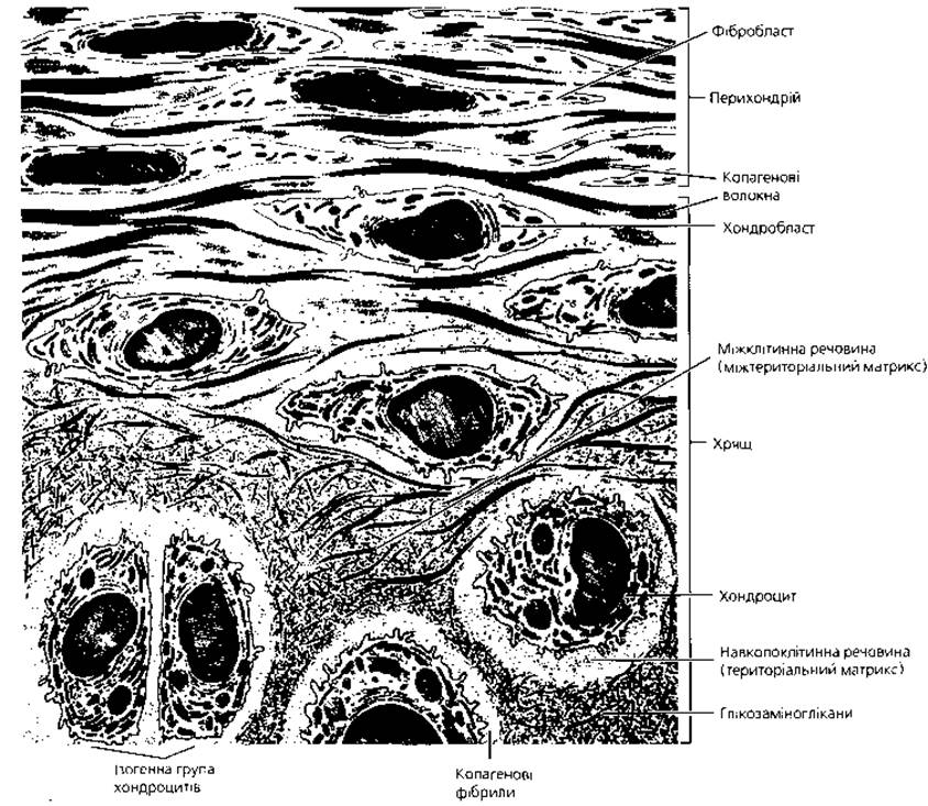
Fig. 3.31. Schematic representation of cartilage tissue Organization. The transition zone from the perichondrium to hyaline cartilage. Glycosaminoglycans predominate in the pericellular matrix, while the interterritorial matrix is enriched with thin collagen fibrils
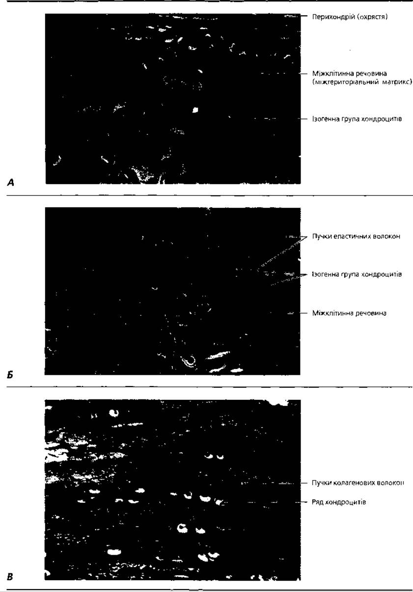
Fig. 3.32. Light Cell/15.html">Microscopy of Different types of cartilage tissue: A - hyaline cartilage, x 200; B - elastic cartilage, stained for elastin, x 300; C - fibrocartilage, x 500
Chondrocytes (Figs. 3.31, 3.32, 3.33) are irregularly rounded or polygonal cells located in cavities called lacunae within the extracellular matrix, either singly or in groups of 2-4 cells. The latter are called isogenous cell groups because they are formed by the division of a single parent cell. Three types of chondrocytes are distinguished. The first type, consisting of relatively poorly differentiated cells, is observed mainly in young, so-called primary cartilage tissue. These cells are characterized by a high nucleocytoplasmic ratio, well-developed Golgi complex elements in the cytoplasm, and numerous Mitochondria and free Ribosomes. In type II chondrocytes, the nucleocytoplasmic ratio is lower, and the cytoplasm contains increased amounts of RNA, rough endoplasmic reticulum elements, and Golgi complex elements, which ensure the Synthesis and Secretion of Proteoglycans and Glycosaminoglycans into the extracellular space. Type III chondrocytes are characterized by the lowest nucleocytoplasmic ratio and a highly developed rough endoplasmic reticulum. The rate of proteoglycan and glycosaminoglycan synthesis is reduced compared to type II chondrocytes, but the synthesis of Glycoproteins, collagen, and elastin Proteins is higher.
The organic Components of the ground substance of cartilage tissue—chondromucoid—are represented by proteins, Lipids, glycosaminoglycans, and proteoglycans. The latter are the most characteristic feature of cartilage tissue. Giant macromolecular complexes with a molecular weight in the range of tens and hundreds of millions of daltons and a molecular length of several micrometers have been found in cartilage proteoglycans (Fig. 3.30). They are composed of a long strand of hyaluronic acid, to which about a hundred polypeptide chains are attached by non-covalent bonds; A large number of polysaccharide chains of sulfated glycosaminoglycans (chondroitin sulfate, keratan sulfate, dermatan sulfate), as well as oligosaccharide molecules, are linked to the middle amino acid residues of these polypeptide chains. Overall, the proteoglycan molecule resembles a spruce twig, and the resilience (turgor) of the cartilage depends on its degree of Hydration.
Chondrin fibers are composed of type II collagen (in hyaline and fibrocartilage) or elastin (in elastic cartilage). The orientation of the fibers is determined by the lines of force that arise during the functioning of the organ. A particularly high concentration of chondrin fibers is found around the lacunae, forming the so-called capsule of the cartilage cell (or cells).
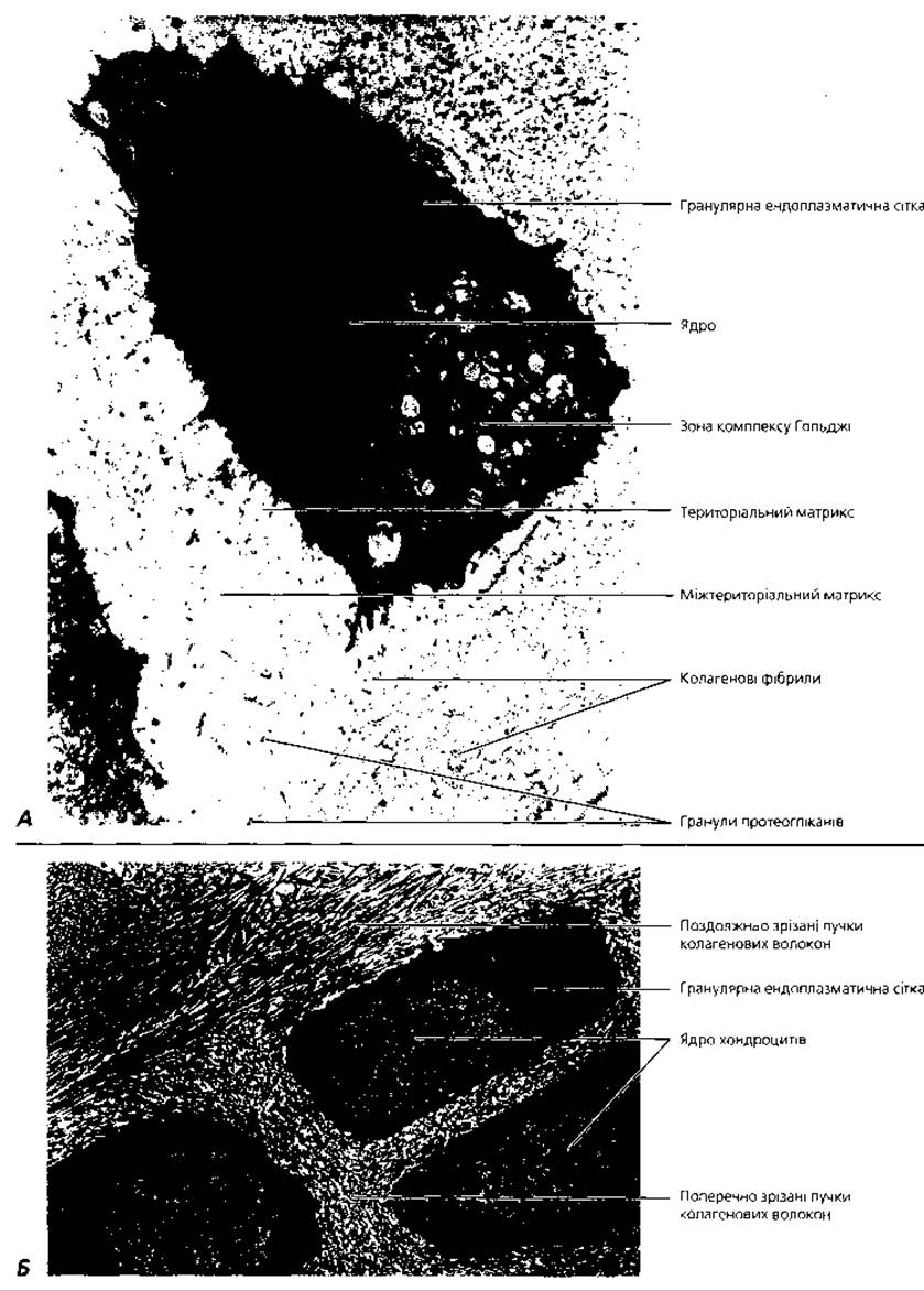
Fig. 3.33. Electron microscopy of chondrocytes: A - chondrocyte of hyaline cartilage, x 2500; B - a group of chondrocytes of fibrocartilage, x 3000
Hyaline cartilage (textus cartilagineus hyalinus) is localized in the walls of the Trachea and Bronchi, at the junctions of the Ribs with the Sternum, on articular surfaces, and in the meta-epiphyseal growth plates of bones. During the Embryonic period, the hyaline cartilage of the primordia forms The basis of the vast majority of skeletal bones. With age, it is replaced by Bone tissue.
In its Native State, hyaline cartilage is light blue and translucent. Histologically, it consists of the perichondrium and the cartilage proper. The perichondrium consists of a superficial fibrous layer (predominantly collagen fibers) and a deep cellular layer (containing chondroblasts and prechondroblasts). The Superficial layer of the perichondrium is highly vascularized, providing nourishment to the cartilage. The deep cellular layer of the perichondrium is responsible for physiological regeneration and appositional (peripheral) growth of the cartilage. The cartilage proper consists of isogenous groups of chondrocytes, as well as young solitary chondrocytes, surrounded by chondromucoid and chondrin fibers. The chondromucoid located around young chondrocytes stains acidophilically (oxyphilically), while that surrounding more differentiated isogenous cell groups acquires basophilic properties. The chondrin fibers of hyaline cartilage are composed of type II collagen.
A morphological feature of articular hyaline cartilage (Fig. 3.34) is the absence of a perichondrium on the articular surface. Chondrocytes deep within the articular cartilage are rounded and arranged in rows oriented perpendicular to the articular surface. Superficial chondrocytes are flattened and do not form isogenous groups. Collagen fibers deep in the cartilage are oriented perpendicular to the articular surface, while closer to the surface they run parallel to it. Between the articular surfaces is the synovial fluid, secreted into the joint cavity by Cells of the synovial layer of the Joint Capsule (more detailed information on joint structure can be found in anatomy textbooks). The synovial layer is a special type of connective tissue adapted to constant stretching, displacement, and pressure during joint movements. It consists of deep and superficial collagen-elastic layers and a lining layer (Fig. 3.34, C).
Elastic cartilage (textus cartilagineus elasticus) is found in the auricle (pinna), auditory tube, external acoustic meatus, and the corniculate and cuneiform cartilages of the Larynx. Its characteristic features are its yellow color and elasticity (ability to stretch). Elastic cartilage does not undergo calcification. Unlike hyaline cartilage, the chondrin fibers in elastic cartilage are composed of elastin rather than collagen. Elastic fibers form capsules around the chondrocytes and also weave into the perichondrium.
Fibrocartilage (textus cartilagineus collagenofibrosus) forms the intervertebral discs and the Pubic Symphysis, and is located at the transition zones of tendons into hyaline cartilage tissue. Chondrocytes in fibrocartilage are arranged in distinctive rows (cell columns), and collagen fibers form thick parallel bundles. In structure, fibrocartilage resembles a tendon, but its cells are typically cartilaginous.
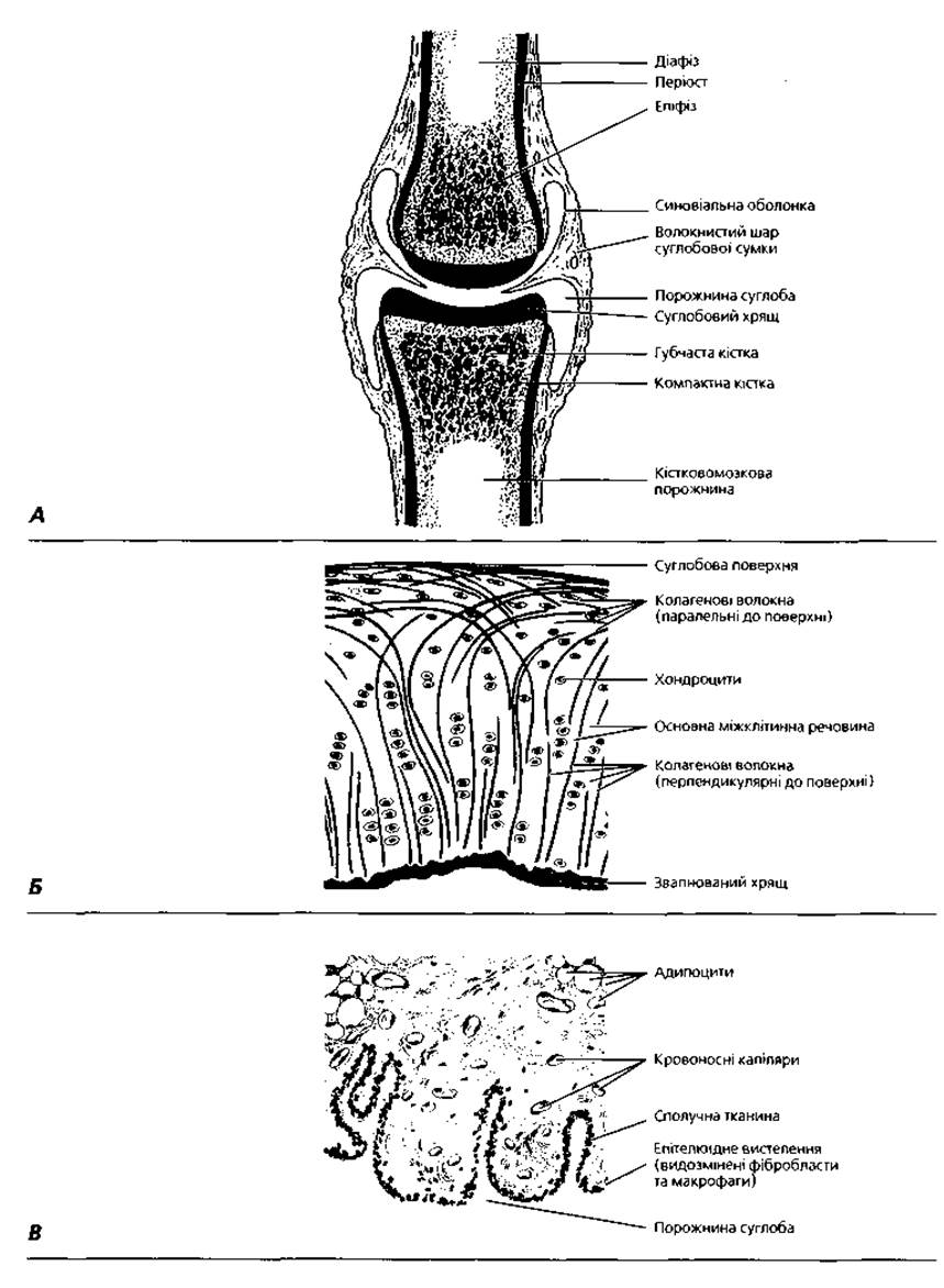
Fig. 3.34. Diagram of tissue interaction during joint formation: A - General structural plan of the joint; B - STRUCTURE OF THE articular cartilage; C - MICROSTRUCTURE OF THE synovial membrane
Histogenesis, regeneration, and age-related changes of cartilage tissue. The source of cartilage tissue formation in ontogeny is the mesenchyme—embryonic connective tissue. During chondrohistogenesis, some mesenchymal cells lose their processes, round up, and form a cartilage primordium—a chondrogenic islet. Mesenchymal cells within it differentiate into chondroblasts (Fig. 3.38). In the next stage of primary cartilage tissue formation, as chondroblasts transform into type I chondrocytes, collagen synthesis increases, and collagen fibers appear, causing the extracellular matrix to acquire acidophilic (oxyphilic) properties. The maturation of chondrocytes and their transformation from type I to type II cells leads to increased synthesis of proteoglycans and, consequently, to increased basophilia of the extracellular matrix.
There are two mechanisms of cartilage growth: internal (interstitial) and by deposition (appositional). Interstitial growth of cartilage occurs As a result of the proliferation of young chondrocytes and The formation of new isogenous cell groups. Appositional growth occurs due to the perichondrium—through the proliferation of chondroblasts in the deep layer, their transformation into chondrocytes, and their production of extracellular matrix.
Physiological regeneration of cartilage tissue occurs due to The activity of chondrocytes and chondroblasts, which produce chondromucoid, collagen, and elastin, contributing to the formation of new chondrin fibers. With age, the cellular content in cartilage tissue decreases, while The amount of extracellular matrix increases. As type I and type II chondrocytes transform into type III chondrocytes, the amount of proteoglycans in the cartilage extracellular matrix decreases, chondromucoid is replaced by albumoid, and the content of collagen fibers increases. The latter have The ability to accumulate calcium salts and undergo calcification. All these changes lead to a decrease in hydration, loss of elasticity, and increased brittleness of the cartilage tissue. Blood vessels are also observed to grow into the calcified cartilage, and the cartilage tissue is replaced by bone.
Bone tissue (textus osseus), along with cartilage, belongs to the Skeletal Tissues of the body. The primary role of bone tissue is supportive and mechanical: due to their significant strength, bones protect vital Organs from mechanical damage, provide support, and enable body movement in space. Elements of bone tissue form a scaffold and microenvironment for Blood Cells within the Cytology/practical/86.html">Red Bone Marrow. Bone tissue serves as a reservoir for Calcium and phosphorus in the body.
Bone tissue is composed of cellular elements (osteoblasts, osteocytes, and osteoclasts) and an extracellular matrix (ossein fibers and osseomucoid). Osseomucoid contains glycoproteins (including osteonectin, a specific bone tissue protein) and proteoglycans. The uncalcified extracellular matrix of bone tissue is called osteoid (prebone). A characteristic feature of bone tissue is the exceptionally high content (up to 70%) of Inorganic Compounds in its extracellular matrix, predominantly calcium salts—hydroxyapatites (Са10(РО4)6(ОН)2) and phosphates (Са3(РО4)2). The strength of bones depends on the high content of ossein fibers composed of type I collagen, which form bundles.
Osteoblasts are irregularly cuboidal or polygonal cells (Figs. 3.35, 3.36) measuring about 15-20 µm. Their cytoplasm is basophilic due to a high RNA content and features well-developed elements of the rough endoplasmic reticulum and Golgi complex. These are relatively poorly differentiated mononuclear cells in which the synthesis of glycoproteins and proteoglycans of the osseomucoid takes place. Osteoblasts are found mainly in areas of bone tissue formation; in the adult Organism, these are the deep layers of the periosteum, as well as areas of bone tissue regeneration.
Osteocytes develop from osteoblasts. These are highly differentiated, mononuclear, elongated cells measuring about 15x45 µm. Osteocytes are located in bone lacunae (cavities) within the calcified extracellular matrix of bone tissue. Branched processes extend from The Cell bodies of osteocytes, penetrating the extracellular matrix and contacting the processes of neighboring cells. The cytoplasm of osteocytes is weakly basophilic, indicating a decreased level of synthetic processes compared to osteoblasts. Osteogenic stem cells, progenitor cells, osteoblasts, and osteocytes form the differon (histogenetic lineage of cells) of bone tissue.
Osteoclasts are large, multinucleated, irregularly rounded cells (Figs. 3.35, 3.36), which can originate from poorly differentiated bone marrow cells as well as blood monocytes. The primary function of osteoclasts is the resorption (breakdown) of bone tissue. These cells have a diameter of 90 μm or more, and their cytoplasm contains from three to several dozen nuclei. The cytoplasm of osteoclasts is acidophilic or weakly basophilic, containing a significant number of Lysosomes and mitochondria. Osteoclasts reside in depressions On the surface of the bone matrix known as Howship's lacunae. On the cell surface adjacent to the site of bone destruction, two zones are distinguished: the adsorption and enzyme secretion zone covered with Plasmalemma folds (the so-called ruffled border) and the sealing zone, which isolates the contact area from the surrounding tissue. The Mechanism of the destructive action of osteoclasts on bone tissue is associated with the release of carbon dioxide by these cells, from which carbonic acid is formed under the Influence of the enzyme Carbonic anhydrase, capable of dissolving calcium salts. Osteoid prevents the interaction of osteoclasts with the Inorganic Components of bone. Successful resorption requires the secretion of collagenase by osteoblasts—an enzyme that degrades the osteoid layer and provides osteoclasts with access to the mineral matrix of the bone.
Depending on the organization of collagen fibers, bone tissue is classified into two types: lamellar and woven (coarse-fibered). Lamellar bone tissue is characterized by the parallel arrangement of collagen fiber bundles, forming the so-called bone lamellae. Depending on the spatial orientation of these lamellae, lamellar bone tissue is divided into compact bone (which lacks cavities) and spongy (cancellous) bone (where bone lamellae form trabeculae arranged at angles to each other, creating a characteristic spongy structure). Compact bone tissue makes up the diaphyses of long bones, while spongy bone tissue forms flat bones and the epiphyses of long bones. Lamellar bone tissue constitutes the vast majority of the bones in the body.
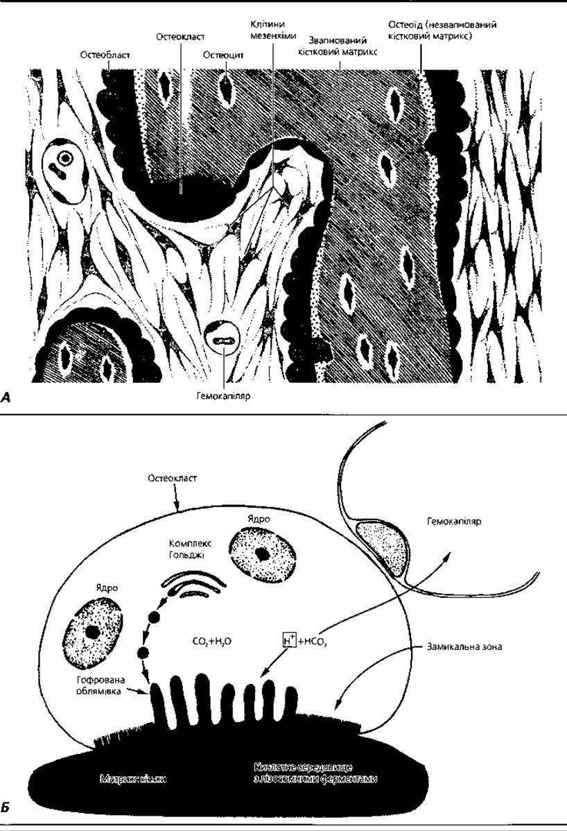
Fig. 3.35. A — comparative micromorphology of the main bone-forming cellular elements; B — ultrastructural organization and functional principle of an osteoclast
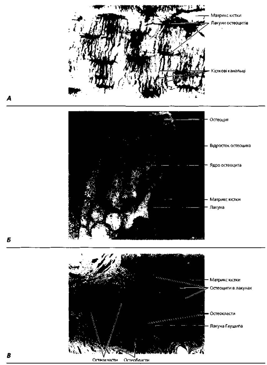
Fig. 3.36. Micromorphology of bone tissue: A — light microscopy of a bone section showing The system of osteocyte lacunae and canaliculi, x 600; B — transmission electron microscopy of an osteocyte within a lacuna, x 6,000; C — micrograph of a bone resorption site with four osteoclasts, x 600
Woven (coarse-fibered) bone tissue is characterized by a random (multidirectional) arrangement of collagen (ossein) fiber bundles surrounded by calcified osseomucoid. Osteocytes reside in lacunae within the osseomucoid between the bundles of ossein fibers. Woven bone tissue is localized primarily in the embryonic Skeleton, while in the adult organism, it is found only in the cranial sutures and at the sites of tendon attachment to bones.
Structure of long bones. The diaphysis (Fig. 3.37) is the central shaft, and the epiphysis is the peripheral end of long bones. In the region of the diaphysis, there are three layers: the periosteum, the bone proper, and the endosteum (inner layer). The periosteum consists of an outer fibrous layer, formed by bundles of collagen fibers, and an inner osteogenic layer (containing osteoblasts and osteoclasts). The periosteum, which is richly vascularized, provides nourishment to the bone tissue; the cellular elements of the deep osteogenic layer ensure bone growth in thickness, as well as its physiological and reparative regeneration.
The bone proper of the diaphysis is composed of compact bone substance, in which bone lamellae (4 to 15 μm thick) form three layers: the outer circumferential (general) lamellae, the osteonal layer, and the inner circumferential (general) lamellae.
Beneath the periosteum lies the layer of outer circumferential lamellae. These lamellae run around the diaphysis but do not form complete rings, being overlapped by other layers. The main thickness of the bone wall is the osteonal layer. Each osteon (Haversian system) is a bone cylinder with a diameter of 20 to 300 μm, in whose central canal (Haversian canal) runs a nutrient vessel and where osteoblasts and osteoclasts are located. Around the central canal, 5 to 20 bone lamellae are arranged concentrically. Collagen fibers within the bone lamellae of each layer have a parallel orientation. The bundles of collagen fibers in adjacent lamellae are oriented at an angle to each other, which strengthens the osteon as a structural element of the bone.
Between the bone lamellae, within the lacunae, lie the cell bodies of osteocytes, which anastomose via their processes located in the bone canaliculi. The osteonal layer can be envisioned as a system of parallel cylinders (osteons), the spaces between which are filled with interstitial lamellae. Perforating (Volkmann's) vessels, as well as bundles of collagen fibers, pass from the periosteum into the osteonal layer. The osteonal layer is separated from the endosteum by a layer of inner circumferential lamellae. The latter is analogous to the outer circumferential lamellae but is less developed. The endosteum is a thin Fibrous connective tissue enriched with osteoblasts and osteoclasts that lines the medullary cavity. The Haversian canals of the diaphysis communicate with each other, as well as with the medullary cavity and the bone surface, through a system of transverse canals (Volkmann's canals, Fig. 3.37, B).
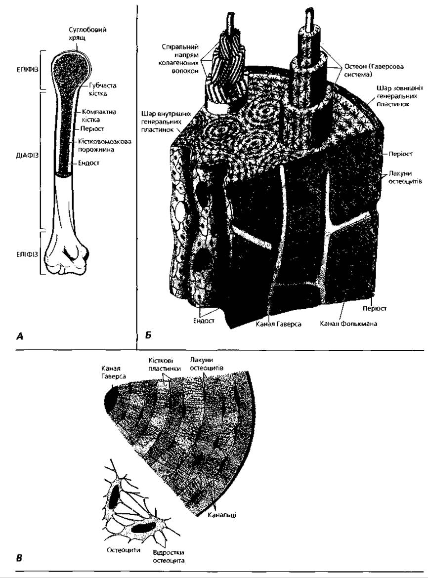
Fig. 3.37. Diagram of the Structural organization of bone tissue: A — general structural plan of a long bone; B — three-dimensional reconstruction of a diaphysis segment of a long bone; C — part of a transversely sectioned osteon showing osteocyte Morphology
The epiphysis of the bone is formed by spongy (cancellous) bone tissue. It is superficially covered by the periosteum, beneath which lies a layer of circumferential lamellae. Within the epiphysis, the bone lamellae form a system of trabeculae arranged at angles to each other, with the cavities between them filled with reticular tissue and hematopoietic cells. Flat BONES OF THE skeleton have a structure similar to that of the epiphysis.
Histogenesis of bone tissue. There are two modes of bone tissue development: directly from the mesenchyme (intramembranous osteogenesis) and on the site of a cartilage model (endochondral osteogenesis). Intramembranous osteogenesis (Fig. 3.38) is characteristic of the early weeks of embryonic development, while endochondral osteogenesis occurs during later stages of embryonic development and postnatal ontogeny. The process of bone development directly from the mesenchyme involves the following stages. The First stage is the Formation of the so-called osteogenic islet, or bone blastema, within the mesenchyme. During this stage, local proliferation of mesenchymal cells occurs, accompanied by the ingrowth of blood vessels into the skeletogenic islet. The second, osteoid stage is characterized by the secretion of collagen (forming ossein fibers) and high-molecular-weight Biopolymers (glycoproteins, proteoglycans, lipids) of the osseomucoid into the extracellular space by osteogenic cells. The Third Stage—the formation of woven bone—involves the calcification of the extracellular matrix (deposition of calcium salts). For this process to occur, the presence of alkaline phosphatase, produced by osteoblasts, and the protein osteonectin in the extracellular matrix is required. The latter, by binding collagen to hydroxyapatite, determines the site of calcium phosphate crystal growth and their attachment to the organic matrix of the bone. The Fourth Stage is associated with the resorptive activity of osteoclasts and the replacement of coarse, multidirectional bundles of ossein fibers with bone lamellae (replacement of woven bone tissue with lamellar bone tissue).
Endochondral osteogenesis (Figs. 3.39, 3.40) is characterized by a somewhat different sequence of morphofunctional changes. The first stage consists of the formation of a cartilaginous model of the future bone. It is constructed of hyaline cartilage covered by perichondrium. The next stage is perichondral ossification. As osteogenic cells exit the Vessels of the perichondrium, intensive production of ossein fibers and osseomucoid occurs on The surface of the cartilage model, followed by calcification. This forms a so-called bone collar around the cartilage. The third stage—endochondral ossification—proceeds with the formation of a diaphyseal center of ossification. This process begins with the ingrowth of vessels from the bone collar into the diaphysis of the cartilage model and the migration of osteogenic cells beyond them. Due to the activity of osteoclasts, resorption cavities arise in the cartilage, which merge to form the medullary cavity. Bone lamellae form around the vessels, and osteons are laid down. The final, fourth stage of endochondral osteogenesis is the ingrowth of blood vessels into the epiphyseal part of the cartilage model and the formation of an epiphyseal center of ossification. Consequently, a so-called metaephyseal growth plate (epiphyseal plate) is formed between the epiphyseal and diaphyseal centers of ossification. Three main zones with distinct morphofunctional characteristics are distinguished within it.
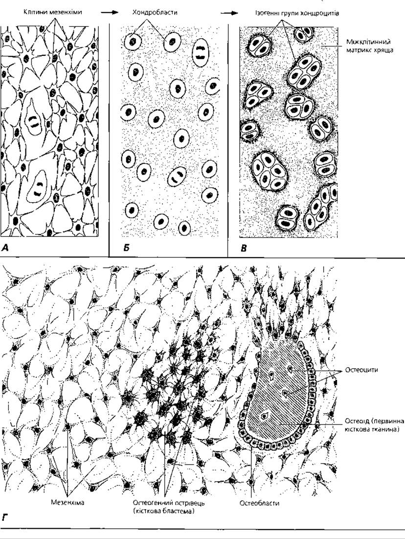
Fig. 3.38. Diagram of the transformation of mesenchyme into cartilaginous (A, B, C) and bone (A, D) tissues
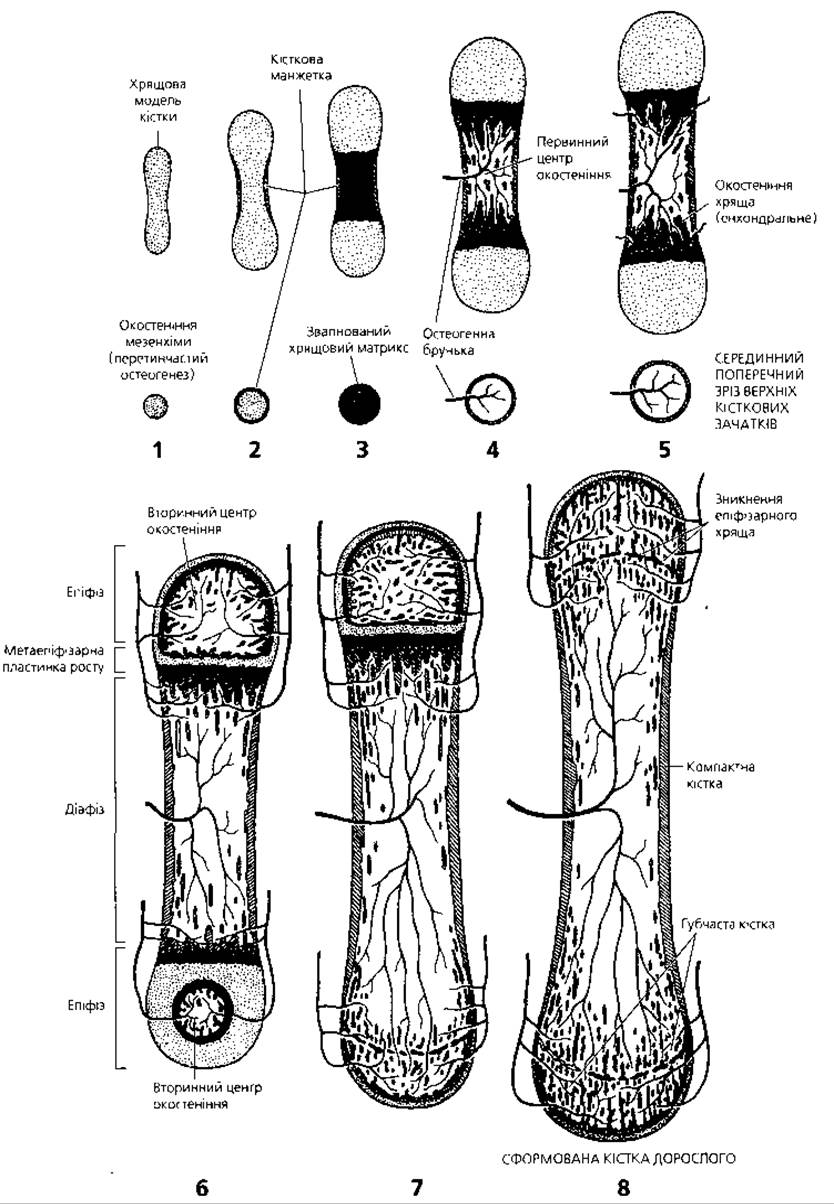
Fig. 3.39. Diagram of the successive stages of endochondral osteogenesis (Bone Formation on the site of cartilage, 1-8)
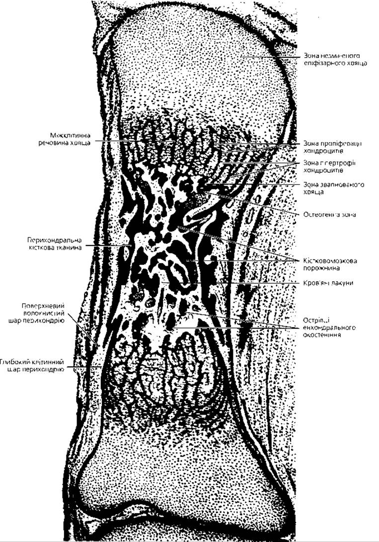
Fig. 3.40. Semi-schematic representation of bone development on the site of cartilage. Midsagittal section of a finger phalanx of a 3.5-month-old human fetus, x 35
The first zone, furthest from the diaphyseal center of ossification, is the zone of reserve (resting) cartilage. Closer to the diaphysis is the zone of proliferating (columnar) cartilage, whose Cell Division drives the longitudinal growth of the bone. The chondrocytes within it are arranged in parallel rows, the so-called cell columns. Even closer to the diaphyseal center of ossification lies the zone of hypertrophic (vacuolated) cartilage. It is characterized by chondrocyte degeneration and cartilage matrix resorption. Directly adjacent to the hypertrophic zone is the zone of cartilage resorption and immature bone tissue, where the processes of bone matrix formation and calcification occur intensively. When the epiphyseal and diaphyseal ossification centers fuse (with the disappearance of the cartilage cell proliferation zone), longitudinal bone growth ceases. In humans, this typically occurs at the age of 18-20 years.
Growth, regeneration, and age-related changes of bone tissue. Unlike cartilage, bone tissue grows only by apposition—the deposition of newly formed bone tissue onto the existing surface. Bone growth in thickness occurs via the periosteum as a result of the proliferation and synthetic activity of osteoblasts in its deep osteogenic layer. Longitudinal growth is ensured by the proliferation of cells in the columnar zone of the epiphyseal plate. It should be noted that a high level of oxygenation (oxygen saturation) is an important factor in bone tissue formation, as vascularization (vessel ingrowth) always precedes the onset of osteogenesis in cartilaginous tissue.
Physiological regeneration of bone tissue consists of the continuous (throughout the individual's life) replacement of old bone lamellae with newly formed ones, and the formation of new osteons in place of resorbed ones. These opposing processes are driven by the activity of osteoclasts and osteoblasts. The mechanisms of bone remodeling are based on the constant change in the direction of the force vector acting on the bone, which produces a so-called piezoelectric effect (generating a potential difference on the concave and convex surfaces of the bone lamellae). The concentration of osteoblasts and the processes of appositional bone formation are associated with negative charges, while the concentration of osteoclasts and resorption processes are linked to positive charges on the surface of the bone tissue.
Age-related changes in bone tissue involve a gradual loss of the inorganic bone matrix after the age of twenty. Notably, in men, the loss of bone mineral components is a steady lifelong process, with an annual loss of inorganic matrix amounting to approximately 0.4% of bone tissue mass. In women, the onset of menopause—apparently due to estrogen deficiency—accelerates demineralization, which reaches a rate of 1–1.5% annually.
Terms to remember
1. Cartilage tissue. 2. Hyaline cartilage. 3. Elastic cartilage. 4. Fibrocartilage. 5. Perichondrium. 6. Chondroblast. 7. Chondrocyte. 8. Isogenous group of chondrocytes. 9. Chondromucoid. 10. Proteoglycans. 11. Glycosaminoglycans. 12. Chondrin fibers. 13. Chondrogenic islet. 14. Primary cartilage tissue. 15. Interstitial growth of cartilage. 16. Appositional growth of cartilage. 17. Albumoid. 18. Bone tissue. 19. Osteoblast. 20. Osteocyte. 21. Osteoclast. 22. Ossein fiber. 23. Osseomucoid. 24. Calcium hydroxyapatite. 25. Bone lacuna. 26. Bone lamella. 27. Lamellar bone tissue. 28. Compact bone tissue. 29. Spongy lamellar bone tissue. 30. Woven bone tissue. 31. Diaphysis. 32. Epiphysis. 33. Periosteum. 34. Outer circumferential lamellae layer. 35. Osteon, osteonal layer. 36. Inner circumferential lamellae layer. 37. Endosteum. 38. Haversian canal. 39. Volkmann's canal. 40. Medullary cavity. 41. Intramembranous osteogenesis. 42. Endochondral osteogenesis. 43. Osteogenic primordium. 44. Osteoid stage. 45. Osteonectin. 46. Perichondral ossification. 47. Cartilage bone model. 48. Bony collar. 49. Endochondral ossification. 50. Diaphyseal ossification center. 51. Epiphyseal ossification center. 52. Epiphyseal growth plate. 53. Zone of columnar cartilage. 54. Zone of hypertrophic cells. 55. Zone of cartilage resorption.
Last update: 09/08/2026
Editorial and Educational Adaptation: This material has been compiled based on the primary/original source text. The project team performed an editorial review, corrected technical inaccuracies, structured sections, and adapted the content for an educational format.
What was processed:
- elimination of formatting defects (OCR errors, structural breaks, corrupted characters);
- editorial organization of content;
- standardization of terminology in accordance with academic sources;
- verification of factual statements against the original source text.
All mentions of the author, publication year, and origin of the primary text have been preserved in accordance with the source.