Principles of Biochemistry, Volume 1 - A. Lehninger 1985
Biomolecules
Globular Proteins: Structure and Function of Hemoglobin
Oxygenation of hemoglobin induces a change in its spatial conformation.
The answer to these questions was obtained after it was revealed that the deoxyhemoglobin molecule undergoes conformational changes upon binding oxygen. The first indication of such changes dates back to the discovery that deoxyhemoglobin crystals grown in an oxygen-free atmosphere shatter as soon as they come into contact with oxygen. This observation suggested that upon binding oxygen, Hemoglobin molecules change in size and no longer fit into the crystal lattice of deoxyhemoglobin. This assumption was fully confirmed by comparative X-Ray Diffraction Analysis, which showed that deoxyhemoglobin and oxyhemoglobin have different spatial Conformations (Figs. 8-10 and 8-18). Upon oxygenation of deoxyhemoglobin, the Tertiary Structure of the α- and β-chains remains virtually unchanged, as they remain tightly fitted together to form α1β1 and α2β2 dimers. However, as soon as Oxygen binds to the heme groups of deoxyhemoglobin, the α1β1 and α2β2 halves of the molecule, while retaining their characteristic rigid conformation, shift their positions relative to each other and draw closer together. In other words, the oxygenation of hemoglobin causes A change in its quaternary structure, i.e., the packing of its subunits. As a result, the oxyhemoglobin molecule assumes a somewhat more compact structure compared to deoxyhemoglobin, and the central cavity shrinks. The Hemes of the two β-chains move closer together, while the hemes of the two α-chains move further apart, which leads to the sigmoidal shape of the oxygen saturation curve. Due to these changes, The amino acid residues in the α- and β-chains that bind H+ ions move from a relatively hydrophilic environment to a more hydrophobic one, facilitating the release of H+ ions from protonated groups; in other words, upon oxygenation of hemoglobin, the protonated groups acquire The properties of stronger acids, which explains the Bohr effect. Thus, The change in The quaternary structure of hemoglobin resulting from its oxygenation is directly related to the reciprocal relationship between the affinity of hemoglobin for oxygen and its affinity for CO2 and H+ ions.
Class="center">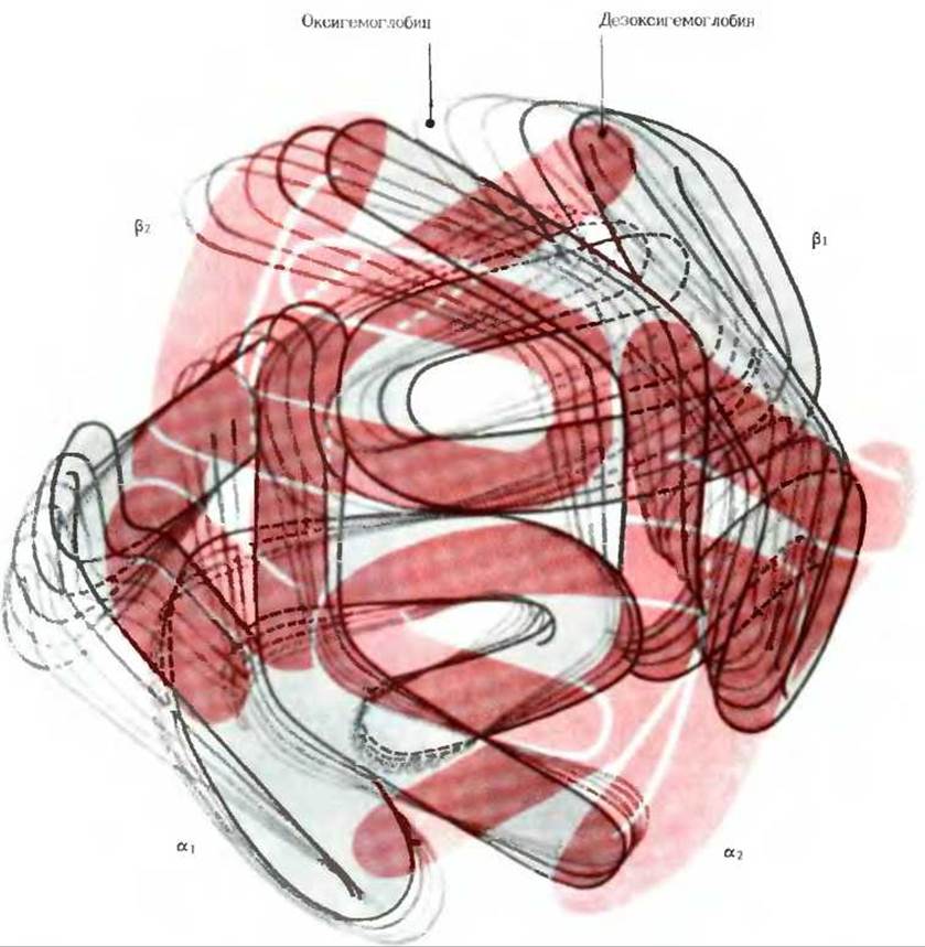
Fig. 8-18. Schematic representation (in the form of "freeze-frames") of Changes in the Quaternary Structure of hemoglobin caused by the movement of the α1β1 subunit pair relative to the stationary α2β2 pair during the release of oxygen from oxyhemoglobin as it transitions to the deoxyhemoglobin form.
Finally, another feature of the regulatory properties of hemoglobin was revealed by Reinhold Benesch and Ruth Benesch, who discovered a fourth Ligand of hemoglobin—2,3-bisphosphoglycerate. This interesting feature is described in Box 8-1.
Box 8-1. Bisphosphoglycerate and the Oxygen Affinity of Hemoglobin
It has long been known that 2,3-bisphosphoglycerate (Fig. 1) is present in erythrocytes in fairly high concentrations, but the function of this compound remained a mystery until it was discovered that it has a strong effect on the affinity of hemoglobin for oxygen. When 2,3-bisphosphoglycerate (BPG) is added to a solution of pure hemoglobin, the oxygen affinity of hemoglobin decreases significantly. This effect is due to the fact that BPG itself binds to deoxyhemoglobin. Consequently, we can write the equation for yet another (the fourth) ligand-binding reaction of hemoglobin:
Hb-BPG + O2⇄ HbO2 + BPG.
Thus, there is an inverse relationship between the two processes—the binding of oxygen and BPG (which bind to different sites on the hemoglobin molecule).

Fig. 1. A. Structure of 2,3-bisphosphoglycerate (BPG). Charged groups interacting with the two β-chains are highlighted in red. B. Location OF THE BPG molecule (highlighted in red) in the central cavity of hemoglobin. The negatively charged groups of BPG are attracted to the nearby positively charged R-groups of the β-chains (shown in bold black lines).
The regulatory effect of BPG on the oxygen affinity of hemoglobin in erythrocytes depends on the partial pressure of oxygen in the Lungs. After a healthy person ascends, say, to an altitude of 4,000 m above sea level, the concentration of BPG in their erythrocytes will increase within the first few hours; as a result, the number of BPG molecules bound to hemoglobin will increase, and the oxygen affinity of hemoglobin will decrease. At high altitudes, the partial pressure of oxygen is significantly lower than at sea level. Therefore, the partial pressure of oxygen in the Tissues also decreases. The increase in BPG content in erythrocytes during ascent to high altitudes facilitates the release of oxygen from hemoglobin in the tissues. Changes of the opposite nature are observed in people acclimated to high-altitude conditions, such as residents of the Himalayas or the Andes, when they descend into valleys. An increase in BPG concentration in erythrocytes is also observed in individuals with Hypoxia; this pathological state arises from an insufficient supply of oxygen to the tissues, which may be caused by respiratory diseases or Circulatory Disorders.
To which site on the hemoglobin molecule does BPG bind? There is an open central cavity, or channel, in the hemoglobin molecule, which is clearly visible in Fig. 8-10. This channel, lined with many positively charged R-groups, serves as the binding site for BPG, which binds to deoxyhemoglobin and forms a cross-link (salt bridge) between the two β-subunits. When hemoglobin binds oxygen, BPG is displaced from the cavity. Hemoglobin binds only one molecule of BPG (Fig. 1); recall that it can bind up to four molecules of O2 or CO2 and approximately four H+ ions.

Fig. 2. Effect of BPG on the oxygen saturation curve of hemoglobin.
When hemoglobin is normally isolated from Blood, it contains a fairly large amount of BPG, which is difficult to remove completely. Upon complete removal of BPG from hemoglobin, its oxygen-binding curve largely loses its sigmoidal shape, and hemoglobin acquires a much higher affinity for oxygen. After adding an excess of BPG to hemoglobin, the oxygen-binding capacity of the latter decreases (Fig. 2). Thus, the presence of BPG is essential for the normal release of oxygen from hemoglobin in the tissues. The erythrocytes of some birds contain not BPG, but another phosphate-containing compound—Inositol hexaphosphate, which reduces the oxygen affinity of hemoglobin even more effectively than BPG.
Hemoglobin can be viewed as an automatic molecular control system that responds to changes in the concentration of any of its four ligands, transmits the corresponding information by converting it into conformational changes of the molecule, and in this way regulates its affinity for other ligands. It is believed that the binding of oxygen to one or two subunits induces small Conformational Changes in them, which facilitate conformational rearrangements in the empty (unoxygenated) subunits, thereby altering the quaternary STRUCTURE OF THE entire molecule; as this happens, its affinity for oxygen increases, while its affinity for CO2 and H+ ions decreases. Conversely, upon the release of oxygen, the quaternary structure returns to its original state, which favors the binding of CO2 and H+ ions. Various theories have been proposed to explain in detail all the structural changes of hemoglobin observed during its oxygenation and deoxygenation, as well as the accompanying changes in its ligand-binding capacity (Fig. 8-19). But whatever the details of these changes, one thing is clear—hemoglobin can serve as a model of an automatic regulatory system when considering other Oligomeric Proteins, such as Enzymes, especially those possessing both catalytic and regulatory activity. Many proteins endowed with such regulatory properties consist of two or more tightly fitted polypeptide chains forming a characteristic quaternary structure capable of changing as the protein transitions from one active state to another during its function.

Fig. 8-19. Symmetric ("all-or-none") and sequential (induced-fit) models of Cooperative oxygen binding by hemoglobin. In both models, subunits can exist in two different states. Circles represent the state in which subunits have a low affinity for oxygen, and squares represent the high-affinity state. A. According to the symmetric ("all-or-none") model, hemoglobin can exist in only two forms: one in which all subunits have a low affinity for oxygen, and another in which they have a high affinity. In the absence of oxygen, both forms are in equilibrium, but the low-affinity form dominates. If oxygen is present, it binds preferentially to the high-affinity form of hemoglobin; this shifts the equilibrium to the right, increasing the probability of the remaining oxygen binding to hemoglobin. The symmetric model does not envision the existence of intermediate forms relative to the low- and high-affinity states. B. In the sequential model (induced-fit model), there are several intermediate conformations that sequentially approach the high-affinity form. The binding of an oxygen molecule to one of the low-affinity subunits induces the transition of this subunit to the high-affinity form. This transition, in turn, increases the probability that the sequential binding of oxygen to other subunits will induce their transition to the high-affinity form.
Last update: 06/08/2026
Editorial and Educational Adaptation: This material has been compiled based on the primary/original source text. The project team performed an editorial review, corrected technical inaccuracies, structured sections, and adapted the content for an educational format.
What was processed:
- elimination of formatting defects (OCR errors, structural breaks, corrupted characters);
- editorial organization of content;
- standardization of terminology in accordance with academic sources;
- verification of factual statements against the original source text.
All mentions of the author, publication year, and origin of the primary text have been preserved in accordance with the source.