Principles of Biochemistry, Volume 1 - A. Lehninger 1985
Biomolecules
Globular Proteins: Structure and Function of Hemoglobin
The quaternary structure of several other oligomeric proteins has also been established.
X-ray crystallographic studies have also been conducted on several other Oligomeric Proteins. One example is the Yeast enzyme hexokinase, which catalyzes the reaction
Class="center">ATP + D-glucose → ADP + D-glucose-6-phosphate.
This vital reaction occurs in almost all organisms, as it is an essential step in glucose METABOLISM. Yeast hexokinase has a Molecular Weight of about 102,000 and contains two polypeptide chains. X-ray crystallographic analysis was used to determine both the Tertiary Structure of these two polypeptide chains and the Quaternary Structure of hexokinase, which is formed by the assembly of the two chains into a single, tightly packed globule (Fig. 8-13). Of particular interest is The structure of the Active Site of this enzyme—the region of the molecule where both ATP and glucose, which participate in the catalytic process, bind. As we will see in Ch. 9, the conformation of the hexokinase molecule changes its geometry during the catalytic cycle.
Another oligomeric protein whose structure has also been determined is the enzyme Lactate dehydrogenase from Skeletal Muscle, which catalyzes the final step in the metabolic conversion of glucose to lactate. Lactate dehydrogenase has a molecular weight of 140,000 and contains four polypeptide chains. In terms of tertiary structure, these chains differ significantly from those of Hemoglobin.

Fig. 8-12. Evolution of Myoglobin and hemoglobin from an ancestral oxygen-binding hemoprotein. In all myoglobins, as well as in the α- and β-chains of all modern Hemoglobins (a total of 145 sequences investigated), there are six invariant residues and A large number of Amino Acids with similar properties occupying identical positions in these proteins. It can be assumed that the Gene encoding the ancestral single-chain hemoprotein underwent duplication. One of the resulting copies gave rise to the myoglobin gene, and the other to the ancestral hemoglobin gene. Both of these genes subsequently underwent independent Mutations. The hemoglobin gene may have duplicated once more at some point, giving rise to the modern genes for the α- and β-chains.
In addition to the genes encoding the α- and β-chains of normal adult hemoglobin, There is a gene encoding the γ-chain, which is part of fetal hemoglobin. This hemoglobin, with the composition α2γ2, has a higher affinity for oxygen than adult hemoglobin. Adult erythrocytes also contain a very small amount of hemoglobin with δ-chains, which has the composition α2δ2.
Another protein with a known quaternary structure is the enzyme Glutamine Synthetase from E. coli, which catalyzes The formation of glutamine from glutamate and ammonia at the expense of ATP energy (Ch. 19). Compared to hemoglobin or hexokinase, this is a much more complex oligomeric protein. Fig. 8-14 shows the relative arrangement of the 12 subunits of glutamine synthetase.
These three oligomeric proteins—all of them Enzymes—share one common property with hemoglobin. They are all involved in some type of biological regulation, which is an integral part of their function. Hexokinase, lactate dehydrogenase, and glutamine synthetase from E. coli are members of a special class of enzymes called regulatory enzymes. As we will see in the next chapter, they not only catalyze specific reactions but also help regulate The rate of the metabolic pathway in which they participate as catalysts. Hemoglobin also plays a regulatory role. It not only transports oxygen from the Lungs to peripheral Tissues but also regulates oxygen binding in the lungs and its release in tissues in response to specific signals, in particular, changes in pH and CO2. Such a regulatory function is apparently characteristic of many oligomeric enzymes.
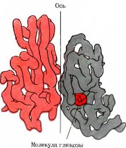
Fig. 8-13. STRUCTURE OF THE oligomeric protein hexokinase isolated from yeast. The two subunits of the protein are related by a screw axis of Symmetry. If the molecule is rotated 180° around its vertical axis and simultaneously translated upward, the lower subunit will superimpose on the upper subunit. Hexokinase is a regulatory enzyme that controls the rate of glucose entry into cellular metabolism. The right subunit contains a glucose molecule bound to the active site of the enzyme. Another view of hexokinase is shown in Fig. 12 of Box 9-4 in Ch. 9.
We will now consider hemoglobin in more detail and attempt to understand what determines its ability to transport oxygen from the lungs to the tissues, and H+ ions and CO2 molecules from the tissues to the lungs. We will see how the quaternary structure of hemoglobin helps regulate these vital transport Functions. Hemoglobin serves as a prototype or model for many other regulatory oligomeric proteins.

Fig. 8-14. Subunit structure of E. coli glutamine synthetase. This regulatory enzyme consists of 12 subunits, the relative arrangement of which is shown in the figure.
Last update: 06/08/2026
Editorial and Educational Adaptation: This material has been compiled based on the primary/original source text. The project team performed an editorial review, corrected technical inaccuracies, structured sections, and adapted the content for an educational format.
What was processed:
- elimination of formatting defects (OCR errors, structural breaks, corrupted characters);
- editorial organization of content;
- standardization of terminology in accordance with academic sources;
- verification of factual statements against the original source text.
All mentions of the author, publication year, and origin of the primary text have been preserved in accordance with the source.