Principles of Biochemistry, Volume 1 - A. Lehninger 1985
Biomolecules
Globular Proteins: Structure and Function of Hemoglobin
In their tertiary structure, the а- and ß-chains of hemoglobin are highly similar to myoglobin.
X-Ray Diffraction Analysis and chemical studies of Hemoglobin have revealed several important features. First, it was found that the α- and β-chains of hemoglobin have almost identical tertiary structures. Both consist of more than 70% α-helices, and all of these α-helical segments are nearly equal in length and form approximately the same angles at the bends.
The second pattern is that Hemoglobins from different vertebrate species share a very similar Tertiary Structure of their polypeptide chains. Furthermore, they are also highly similar in their quaternary structure.
A third important Conclusion is that the tertiary STRUCTURE OF THE α- and β-chains of hemoglobin shares many features with that of Myoglobin. This structural similarity correlates with the ability of both Proteins to bind oxygen, which is fundamental to their biological function.
The evolutionary relationship of Myoglobin and hemoglobin chains to the same protein family is further supported by a comparison of the Amino acid sequences of sperm whale myoglobin and the α- and β-chains of horse hemoglobin. As shown in Fig. 8-11, all three chains share 27 equivalent positions occupied by identical amino acid residues; moreover, in another 40 positions, the residues are chemically similar, such as aspartic and glutamic acids, or isoleucine and valine. Thus, we see once again that The amino acid sequences of homologous proteins contain A number of invariant amino acid residues, and that homologous proteins typically share a highly similar three-dimensional structure.
Another conclusion can be drawn from the structural data on myoglobin and hemoglobin. It appears highly probable that myoglobin and hemoglobin arose from a common ancestral oxygen-binding hemoprotein (Fig. 8-12), which was likely a single-chain polypeptide. At some point in subsequent evolutionary history, the Gene encoding this ancestral oxygen-binding protein underwent duplication. The resulting two gene copies then mutated independently; one gradually evolved into a gene encoding a myoglobin-type protein adapted for intracellular oxygen storage, while the other, through a different series of Mutations, came to encode the α- and β-chains of hemoglobin, which are specialized for Oxygen transport by red Blood Cells. We will see many other Examples of functionally and structurally similar proteins that have diverged from common evolutionary ancestors.
Class="center">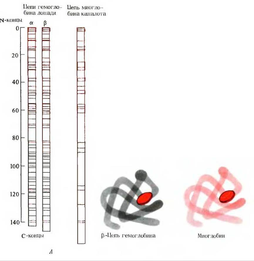
Fig. 8-11. A. Positions of invariant amino acid residues (red dashes) shared by the α- and β-chains of horse hemoglobin and sperm whale myoglobin. Black dashes indicate positions occupied by identical amino acid residues in the α- and β-chains of hemoglobin. B. Similarity between the tertiary structures of the horse hemoglobin β-chain and sperm whale myoglobin. The red disc represents the heme group.
Last update: 06/08/2026
Editorial and Educational Adaptation: This material has been compiled based on the primary/original source text. The project team performed an editorial review, corrected technical inaccuracies, structured sections, and adapted the content for an educational format.
What was processed:
- elimination of formatting defects (OCR errors, structural breaks, corrupted characters);
- editorial organization of content;
- standardization of terminology in accordance with academic sources;
- verification of factual statements against the original source text.
All mentions of the author, publication year, and origin of the primary text have been preserved in accordance with the source.