Principles of Biochemistry, Volume 1 - A. Lehninger 1985
Biomolecules
Globular Proteins: Structure and Function of Hemoglobin
X-ray crystallography has made it possible to determine both the tertiary and quaternary structures of hemoglobin.
Hemoglobin (mol. wt. 64,500) was the first oligomeric protein to be analyzed by X-ray crystallography; it contains four polypeptide chains and four prosthetic heme groups in which the iron atoms are in the ferrous state [Fe (II)]. The protein portion of the molecule, called globin, consists of two α-chains (141 residues each) and two β-chains (146 residues each). Because the hemoglobin molecule is four times larger than Myoglobin, solving its three-dimensional Structure—accomplished by Max Perutz and his colleagues in Cambridge, England—required much more time and effort. According to X-ray crystallographic data, the hemoglobin molecule is nearly spherical, with a diameter of about 5.5 nm. Each of the four chains has its own characteristic tertiary structure. Like myoglobin, the α- and β-chains of hemoglobin contain several α-helical segments separated by nonhelical regions that form bends in the chain. The four polypeptide chains are arranged relative to one another in an approximately tetrahedral configuration, resulting in the characteristic Quaternary Structure of hemoglobin (Fig. 8-10). One heme group is associated with each chain. The Hemes of the different chains are relatively far apart (about 2.5 nm) and are oriented at different angles. Each heme is partially buried in a pocket lined with hydrophobic R groups and is linked to the polypeptide chain by a coordinate bond between the iron atom and the R group of a Histidine residue, as shown in Fig. 8-2. The sixth coordination position of the iron atom in each heme is vacant and is used to bind an O2 molecule.
Class="center">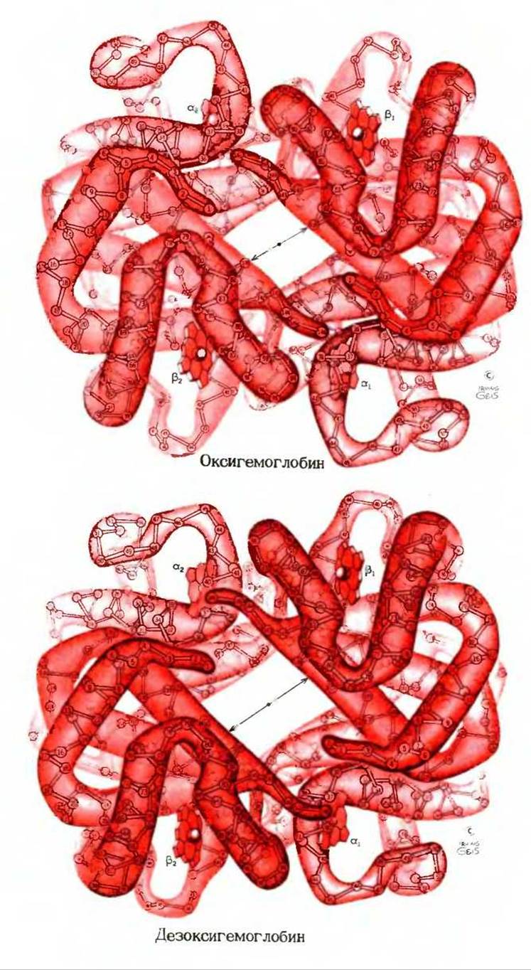
Fig. 8-10. Three-dimensional structure of oxy- and deoxyhemoglobin determined by X-ray crystallography. The quaternary structure of the molecule is shown, i.e., the spatial arrangement of the four subunits. The subunits are paired as α1β1 and α2β2.
There are few contacts between identical subunits of the two types (α and β), whereas There are many contacts between unlike subunits, stabilizing the α1β1 and α2β2 pairs. Despite its irregular shape, the molecule has a twofold axis of Symmetry: a 180° rotation about an axis passing through the center of the molecule perpendicular to the plane of the drawing brings subunit α1 into coincidence with subunit α2, and β1 with β2. The residue numbers are indicated in each chain. The central cavity plays an important role, as described in Box 8-1. Note the relatively large distances between the heme groups. The differences between oxyhemoglobin and deoxyhemoglobin are small but highly important for hemoglobin function. These differences will be discussed later in this chapter.
A close Analysis of the quaternary structure of hemoglobin using models shows that there are few direct contacts between the two α-chains or the two β-chains, whereas there are numerous contacts of the α1β1 and α2β2 type between the α- and β-chains. These contacts are formed primarily by the hydrophobic R groups of amino acid residues. Because of the irregular shape of The polypeptide chains, the two pairs of subunits, α1β1 and α2β2, do not fit tightly together, leaving a central channel (or cavity) that runs through the entire hemoglobin molecule, as is clearly visible when looking at the molecule from the top (Fig. 8-10). We will return to the function of this channel later.
Last update: 06/08/2026
Editorial and Educational Adaptation: This material has been compiled based on the primary/original source text. The project team performed an editorial review, corrected technical inaccuracies, structured sections, and adapted the content for an educational format.
What was processed:
- elimination of formatting defects (OCR errors, structural breaks, corrupted characters);
- editorial organization of content;
- standardization of terminology in accordance with academic sources;
- verification of factual statements against the original source text.
All mentions of the author, publication year, and origin of the primary text have been preserved in accordance with the source.