Human Histology - O.D. Lutsyk 2003
Embryology
Gastrulation. Histo- and Organogenesis. Extraembryonic Organs
Gastrulation is the period of Embryogenesis during which the germ layers—ectoderm, endoderm, and mesoderm—form, and the embryo acquires a trilaminar Structure. Concurrently with Changes in the embryonic body, extraembryonic Organs develop to create the conditions necessary for the embryo's viability (providing trophic support, gas exchange, etc.). In humans, gastrulation spans from the 14th to the 17th day of Prenatal ontogenesis.
It is preceded by processes occurring during the second week of development. At this stage, the embryoblast divides into two layers—the epiblast and the hypoblast—a process known as delamination. The hypoblast is a layer of small cuboidal Cells facing the blastocoele. Hypoblast cells segregate from the inner Cell mass due to weak adhesive interactions between them; the hypoblast gives rise to the extraembryonic endoderm. Proliferating hypblast cells migrate along the inner surface of the trophoblast and form a tightly adherent wall of the primary yolk sac, also known as Heuser's membrane; the primary yolk sac is alternatively called the exocoelomic cavity.
Among the embryoblast cells remaining after the segregation of the hypoblast, a small cavity appears. It expands and transforms into the amniotic cavity. The layer of high columnar cells forming the floor of the amniotic cavity and adjacent to the hypoblast is termed the epiblast. The remaining wall of the amniotic cavity, adjacent to the trophoblast, is formed by amnioblast cells.
Together, the epiblast and hypoblast form the bilaminar embryonic disc (Fig. 2.5, G; 2.6). Subsequently, between the trophoblast (now differentiated into an inner cellular layer, the cytotrophoblast, and an outer symplastic layer, the syncytiotrophoblast) and the exocoelomic membrane, a new population of cells emerges, forming a delicate loose Connective Tissue known as extraembryonic mesoderm (its exact origin is currently debated). It fills all the spaces between the trophoblast externally and the amnion and exocoelomic membrane internally. Later, cavities appear within the extraembryonic mesoderm, which coalesce to form the extraembryonic coelom, or chorionic cavity.
By the 13th–14th day, cells delaminate from the hypoblast, proliferating and gradually forming a new cavity known as the secondary or definitive yolk sac. The latter is significantly smaller than the primary exocoelomic cavity or primary yolk sac. During The formation of the definitive yolk sac, large portions of the exocoelomic cavity are compressed, forming exocoelomic vesicles that are frequently observed within the expanding chorionic cavity. The portion of the extraembryonic mesoderm lining the trophoblast and amnion is termed the extraembryonic somatopleure, while the part surrounding the yolk sac is called the extraembryonic splanchnopleure. The extraembryonic mesoderm strand, which essentially suspends the embryo within the chorionic cavity, connects it to the chorion and is designated as the amniotic or connecting stalk. It subsequently develops into the umbilical cord (Fig. 2.13).
Class="center">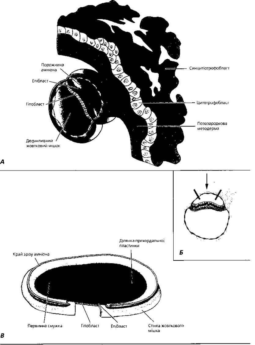
Fig. 2.6. Bilaminar embryonic disc on the 14th day of development. A - general view of the embryo; B - access scheme to The surface of the embryonic disc shown in fragment C; C - view of the embryo from the amniotic cavity side, the primitive streak forms a shallow groove in the caudal region of the epiblast
Gastrulation begins around the 14th day of development with the Formation of the primitive streak On the surface of the epiblast. Initially poorly defined, by the 15th–16th day it becomes clearly visible as a thickening with a narrow groove in the middle (Fig. 2.7). It is localized in the caudal region along the midline of the embryonic disc, extending in a craniocaudal direction. Cranially, the streak ends in a rounded thickening—the primitive node (Hensen's node)—in the center of which lies a depression, the primitive pit. Epiblast cells migrate toward the primitive streak, where they acquire a flask-like shape, detach from the epiblast, and slip beneath it. The first cells migrate inward through the primitive streak, displace the hypoblast, and form the embryonic endoderm. Later, cells migrate to occupy a position between the epiblast and the newly formed endoderm, establishing the mesoderm. Cells remaining within the epiblast form the ectoderm. Thus, during gastrulation, the epiblast gives rise to all three embryonic germ layers. Subsequently, the mesoderm expands laterally and cranially as mesodermal wings.
Cells that invaginate into the primitive pit move in a cranial direction, forming the notochord—the primordium of the embryonic Axial Skeleton. The space between the germ layers is filled with embryonic connective tissue, or mesenchyme. Structurally, it resembles a network formed by stellate cells contacting via their processes; intercellular spaces are filled with a semi-fluid Extracellular matrix and fine fibrils. The primary source of mesenchyme is the mesoderm, and to a lesser extent, the ectoderm and endoderm. Accordingly, a distinction is made between entomesenchyme (developing from the endoderm and mesoderm) and ectomesenchyme (of ectodermal origin). Morphologically, there is no difference between these types of mesenchyme, yet they give rise to distinct embryonic structures: entomesenchyme forms Internal Environment Tissues, whereas ectomesenchyme gives rise to the auditory ossicles, HEAD connective tissue, etc.
At the end of the second week of development, Blood islands and primary blood vessel primordia begin to form in the wall of the yolk sac. A finger-like pouch, the allantois, grows from the posterior part of the intestinal endoderm into the amniotic stalk. Blood Vessels of the yolk sac sprout into the wall of the allantois and chorionic villi. The latter are bathed in maternal blood. The resulting uteroplacental unit, the allantoic chorion, ensures fetal Nutrition and Respiration at an early stage of development.
Histogenesis and Organogenesis of the embryo proceed through cell proliferation, migration, differentiation, the establishment of intercellular contacts, and programmed cell death. From the 17th to the 20th day, the presomitic period continues, followed by the onset of the somitic period of development on the 20th day (Fig. 2.8). On the 20th day of embryogenesis, through the formation of body folds (cephalocaudal and lateral), the embryo proper separates from the extraembryonic organs, and its flat shape transforms into a cylindrical one (Fig. 2.12). Simultaneously, the dorsal Regions of the embryonic mesoderm divide into discrete segments located on either side of the notochord—the somites. On the 21st day, the embryo possesses 2–3 pairs of somites. Somite formation initiates with the III pair, while the I and II pairs appear somewhat later. The number of somites progressively increases: by the 23rd day of development, there are 10 pairs of somites (Fig. 2.9); on the 25th day, 14 pairs; on the 27th day, 25 pairs; and by the end of the fifth week, the somite count reaches 43–44 pairs. Counting somites allows for an approximate Determination of the developmental age (somite age) of the embryo.
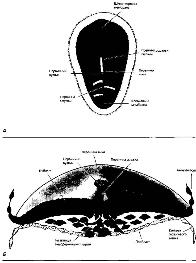
Fig. 2.7. Gastrulation: formation of the embryonic endoderm and mesoderm on the 16th day of development. A - direction of movement of surface epiblast cells through the primitive streak and primitive node (solid lines) followed by migration between the epiblast and hypoblast (dashed lines); B - invagination of epiblast cells resulting in the formation of embryonic endoderm and mesoderm
The outer part of each somite gives rise to the dermatome, the inner part to the sclerotome, and the middle part to the myotome (Fig. 2.10). The dermatome becomes the source of the dermis, the sclerotome forms cartilaginous and osseous tissues, and the myotome gives rise to the skeletal Muscles of the dorsal region of the embryo. The ventral regions of the mesoderm—the splanchnotome—do not segment, but instead split into visceral and parietal layers, which develop into the serous membranes of Internal Organs, Cytology/cytology/34.html">Cardiac Muscle tissue, and the adrenal cortex. Blood vessels, Blood Cells, connective tissue, and Smooth muscle tissue of the embryo differentiate from the mesenchyme of the splanchnotome. The mesodermal region connecting the somites to the splanchnotome subdivides into segmental stalks (nephrotomes), which serve as the source for the Kidneys, Gonads, and paramesonephric ducts. The epithelium of the Uterus and uterine tubes develops from the latter.
During the Differentiation of the embryonic ectoderm, the neural tube, neural crests, placodes, surface ectoderm, and prechordal plate are formed. The process of neural tube formation is termed neurulation. It involves the creation of a slit-like depression on the surface of the ectoderm; the thickened edges of this depression (neural folds) fuse to form the neural tube. Brain vesicles, which serve as the primordium of the brain, develop from the cranial portion of the neural tube. Groups of cells separate from both sides of the neural tube (between the latter and the surface ectoderm) to form the neural crests. Neural crest cells are capable of migration. Cells migrating toward the dermatome give rise to pigment cells (melanocytes); those migrating toward the Abdominal cavity give rise to sympathetic and parasympathetic ganglia, as well as The adrenal medulla. Non-migrating neural crest cells form ganglionic plates, which develop into spinal and peripheral autonomic ganglia. The head ganglia and the sensory Neurons of The Organ of Hearing and Equilibrium form from the placodes.
The prechordal plate serves as the source for the epithelium of the Trachea, Bronchi, Lungs, as well as the Oral Cavity and Esophagus.
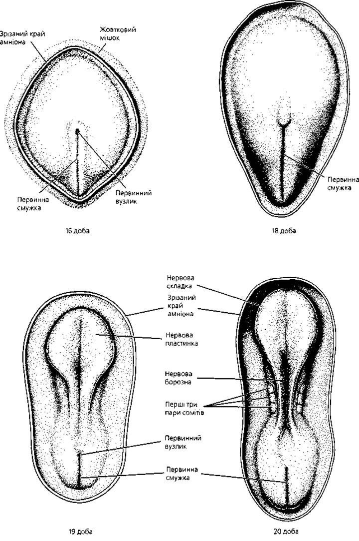
Fig. 2.8. Formation of the neural plate and somites. Dorsal surface of human embryos on the 16th–20th day of development, viewed from the amniotic cavity side (scale not maintained)
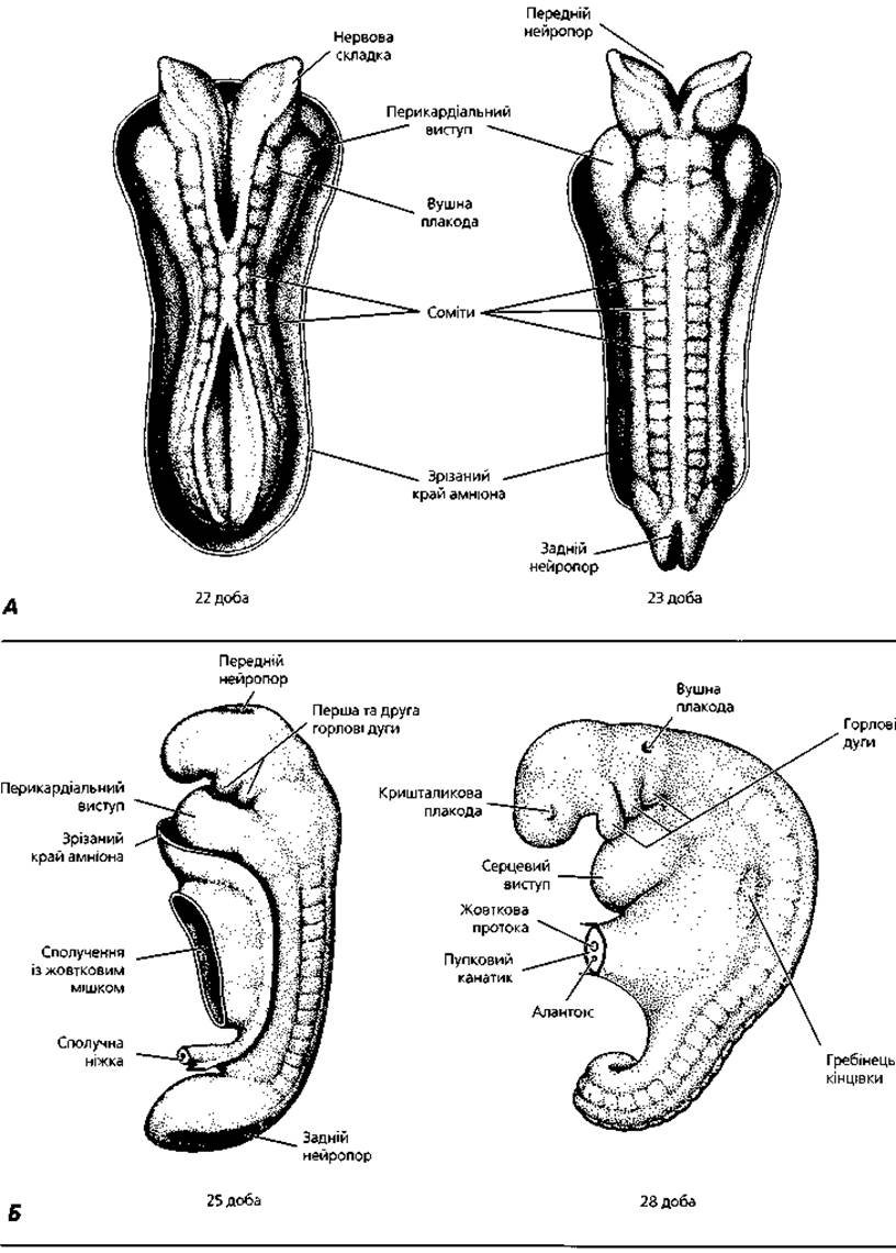
Fig. 2.9. Formation of the neural tube and continuation of somitogenesis: A - dorsal surface of human embryos on the 22nd–23rd day of development, viewed from the amniotic cavity side. B - view from the side of 14-somite (25 days of development) and 25-somite (28 days of development) human embryos
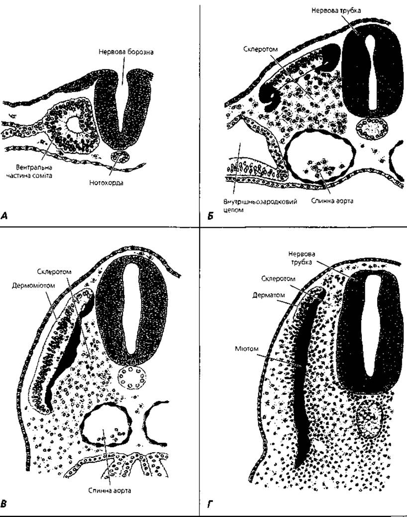
Fig. 2.10. Schematic representation of somitogenesis stages (transverse section of the dorsal region of the embryo): A - mesodermal cells aggregate around a small cavity; B - Cells of the ventral and medial PARTS OF THE somite migrate toward the notochord, forming the sclerotome. Cells of the dorsolateral part of the somite migrate to form progenitor cells for the limb and body wall muscles. Dorsomedial cells migrate beneath the remnants of the dorsal somitic portion to form the myotome; C - myotome cells continue to spread ventrally beneath the dorsal somite region; D - following the ventral expansion of the myotome, dermatome cells flatten beneath the ectoderm, forming the dermal layer of the Skin
The surface ectoderm gives rise to the epidermis and its derivatives, dental enamel and cuticle, and the epithelium of the oral vestibule, anal canal, and Vagina. The embryonic endoderm is the source of the epithelium lining the midgut, Liver, and Pancreas (Fig. 2.11).
As a result of these processes, the crown-rump length of the embryo increases in the following sequence: end of the third week of development – approximately 1.5 mm; end of the fourth week – 3.5 mm; end of the fifth week – 6.5 mm; sixth week – 10 mm; seventh week – 17 mm; end of the eighth week – 30 mm. The embryonic mass reaches 5 g by the end of the eighth week, by which time the primordia of all organs have already formed.
With the beginning of the third month of development, the Embryonic period ends, and the third, fetal, period of prenatal ontogenesis begins. A three-month fetus has a crown-heel length (measured along the crown-heel line, or so-called standing length) of approximately 9 cm; a four-month fetus, 16 cm; and a five-month fetus, 25 cm. Starting from the sixth month, fetal length is calculated by multiplying the gestational age in months by five: at six months it is approximately 30 cm; at seven months, 35 cm; at eight months, 40 cm; at nine months, 45 cm; and at birth, it reaches 50 cm. This is the standard length for a normal full-term newborn infant. The body mass increases a billion-fold from the zygote to the newborn infant—from 3x10⁻⁶ g to 3200 g. The Development of individual fetal organs and systems is discussed below in the relevant chapters of this book.
Multiple Pregnancy. Normally, a single fetus develops in the mother's uterus; however, in about 1% of pregnancies, multiple fetuses (twins) develop and are born simultaneously. The simultaneous development of up to six fetuses has been reported in humans, although multiple pregnancies involving three or more fetuses are extremely rare. There are two MAIN TYPES OF multiple pregnancy: monozygotic (identical) and dizygotic (or polyzygotic, fraternal).

Fig. 2.11. Histogenesis: transformation of the three germ layers into the Tissues of the body
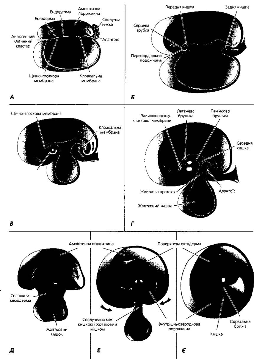
Fig. 2.12. Formation of lateral folds and Body Cavities
Monozygotic twins develop from a single fertilized egg. In humans, monozygotic twinning most likely originates at the blastocyst stage as a result of the division of the embryoblast into two symmetrical parts. Some embryologists consider a symmetrical division of the embryo possible during gastrulation—at the stage of primitive streak development. An independent Organism develops from each newly formed half. Monozygotic twins account for about 15% of all multiple pregnancies. Monozygotic twins share a common Placenta, as well as shared or separate amniotic membranes; they are remarkably similar in physical and mental characteristics and are always of the same sex.
In some cases, monozygotic multiple pregnancy results in the birth of fetuses fused at certain parts of the body. Such twins are called conjoined twins and are generally viable only when the areas of fusion are minor. Conjoined twins arise during the stage of gastrulation, most likely under METABOLISM/18.html">The Influence of damaging environmental factors. The period between the 7th and 12th weeks of prenatal ontogenesis is the most potentially critical window for the development of Congenital Malformations, and signs of congenital pathology may appear even many years after the child's birth. The causes, mechanisms, and Prevention of congenital malformations are the subject of a specialized science known as teratology.
Dizygotic (or polyzygotic) twins occur when two or more eggs are fertilized simultaneously. The embryos implant in separate areas of the endometrium, have their own placenta and amnion, and develop independently. Dizygotic twins resemble each other just like ordinary brothers and sisters (i.e., they can be completely different) and may be of the same or different sexes.
Extraembryonic organs (Fig. 2.13)—the placenta, amnion, umbilical cord, yolk sac, and allantois—create the conditions necessary for the life, growth, and Development of the embryo and fetus. They form a transport system that supplies the fetus with nutrients and oxygen, removes Metabolic waste products, produces Hormones, and provides immune protection. The source of extraembryonic organs is the extraembryonic mesoderm, ectoderm, and endoderm. The extraembryonic mesoderm gives rise to the connective tissue of the fetal part of the placenta (chorion), amnion, and yolk sac. Extraembryonic ectoderm transforms into the epithelium of the amniotic sac and umbilical cord. The epithelium of the yolk sac is formed from the extraembryonic endoderm.
The placenta is an organ that ensures a continuous connection between the maternal organism and the fetus (Fig. 2.14). It comprises two parts: maternal and fetal. The maternal part of the placenta is formed by the mucous membrane of the uterus (endometrium) in the region where the fetal chorionic villi invade it. This is the so-called basal plate.
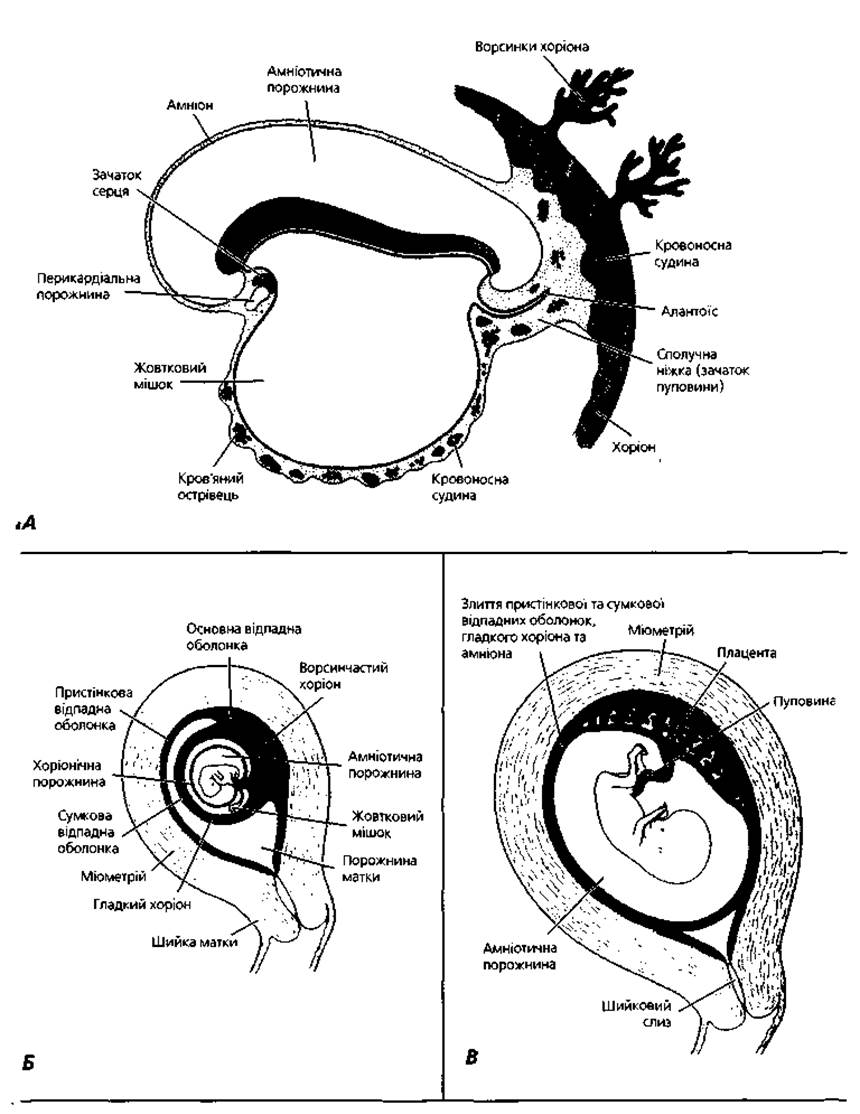
Fig. 2.13. Extraembryonic organs—placenta, umbilical cord, amnion, yolk sac, and allantois—at successive stages of prenatal ontogenesis: A - 19th day of development; B - end of the second month; C - end of the third month: fusion of the amnion and chorion, obliteration of the uterine cavity resulting from the fusion of the smooth chorion and the parietal decidua
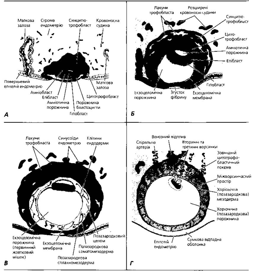
Fig. 2.14. Successive stages of placental development. A - Human blastocyst on the 7.5th day of development. B - Human blastocyst on the 9th day of development. C - Human blastocyst at about the 12th day of development. D - Human embryo at the beginning of the second month of development. At the embryonic pole, the chorionic villi are numerous and well-developed, whereas at the abembryonic pole, they are sparse and poorly developed
The fetal part of the placenta is formed by the villous chorion, a derivative of the trophoblast. A distinction is made between the villous (or stem/frondosum) chorion, whose villi invade the endometrium in the area of the decidua basalis, and the smooth chorion, which represents the site of contact between the trophoblast and the decidua capsularis. Chorionic villi are treelike, branching outgrowths of the trophoblast in the area of its contact with the uterine mucosa (Fig. 2.14, D).
Initially, the trophoblast forms primary villi, which consist solely of trophoblast elements. During the 2nd–3rd weeks of embryogenesis, as the extraembryonic mesoderm invades the trophoblast, secondary epithelio-mesenchymal villi are formed. Finally, with the invasion of microcirculatory blood vessels into the mesenchyme of the secondary chorionic villi, tertiary villi are formed. Thus, the core of each tertiary chorionic villus consists of embryonic connective tissue penetrated by blood vessels, containing a significant number of Collagen and elastic fibers. Cellular elements include macrophages known as Kashchor–Hofbauer cells, as well as fibroblasts, which produce collagen and elastic fibers. The mesenchyme of the tertiary villi is separated from the surface epithelium by a basement membrane. The villous epithelium—simple cuboidal—is called cytotrophoblast. The surface of the cytotrophoblast is covered by syncytiotrophoblast, which is formed by the fusion of surface epithelial cells of the villi into a continuous, multinucleated cytoplasmic mass (symplast) (Fig. 2.15, C; 2.16, B).
The formation of the human placenta occurs most intensively from the third to the sixth week of embryogenesis. This period is called placentation and represents a critical stage in the embryo's life. Placental formation is completed by the third month of prenatal ontogenesis. Notably, throughout pregnancy, the chorionic villi undergo continuous remodeling. For instance, primary villi are initially replaced by secondary villi, which in turn are replaced by tertiary villi. Starting from the 2nd month of pregnancy, the cytotrophoblast gradually thins and is replaced by the syncytiotrophoblast. Microvilli appear on the surface of the latter. In the second half of pregnancy, the trophoblast of some villi regresses, and their surface becomes covered by a fibrinoid, eosinophilic layer known as Langhans fibrinoid.
Morphologically, the human placenta (Fig. 2.15) is classified as a discoid, hemochorial villous placenta. This means that the placenta is disk-shaped, and the chorionic villi are bathed directly in maternal blood within intervillous spaces (lacunae) (Figs. 2.16, 2.17). During placentation, as the embryo embeds deeper into the uterine mucosa, the trophoblastic epithelium first comes into contact with the epithelium of the endometrium and uterine glands. Under the influence of Proteolytic Enzymes secreted by the trophoblast, the epithelium dissolves, allowing the chorionic villi to invade the connective tissue stroma of the uterine mucosa. Continued growth of the chorion leads to contact between the chorionic villi and the endothelium of microvessels, followed by the destruction of their walls. Blood leaking from the damaged vessels fills the intervillous spaces.
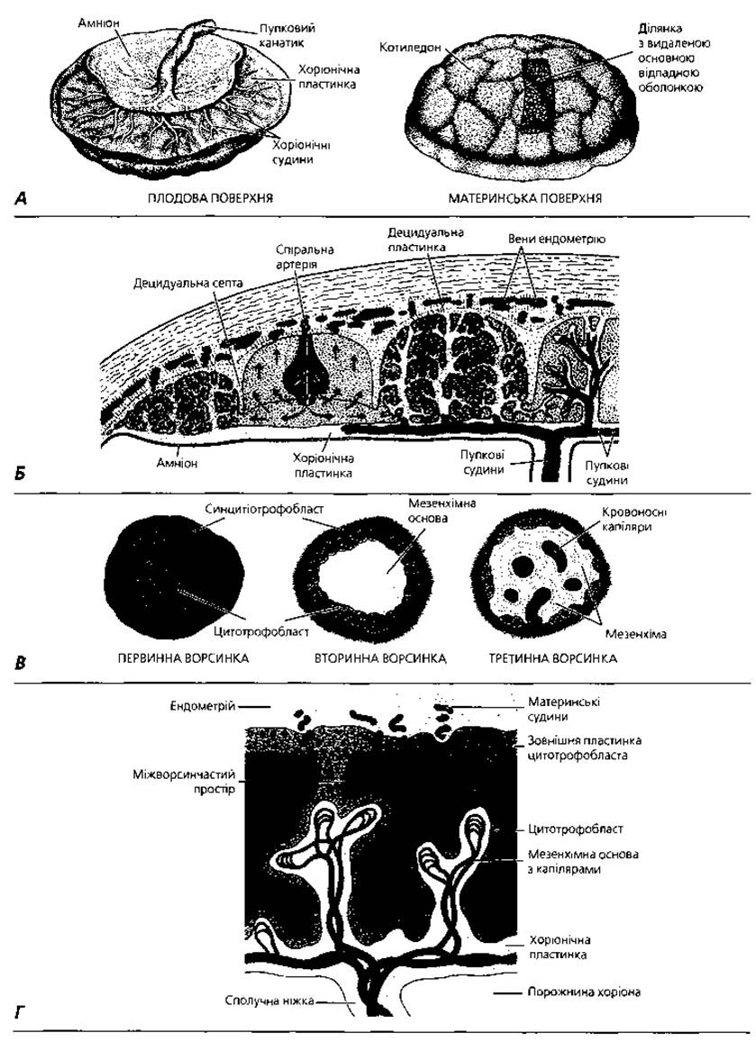
Fig. 2.15. Placenta: A - external appearance of the placenta and umbilical cord at the end of pregnancy; B - schematic representation of the structure and Blood Circulation in the placenta during the second half of pregnancy; C - diagram of the development of placental villi, cross-section; D - diagram of The structure of placental villi at the end of the fourth week of development, longitudinal section
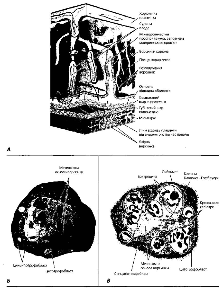
Fig. 2.16. Placenta. A - three-dimensional reconstruction. Arrows indicate the direction of blood flow from the endometrial vessels into the intervillous space and back, driven by pressure differences between the arterial and venous systems; B - semi-schematic representation of the structure of a chorionic villus in early pregnancy, x 550; C - semi-schematic representation of the ultrastructure of a chorionic villus in the second half of pregnancy
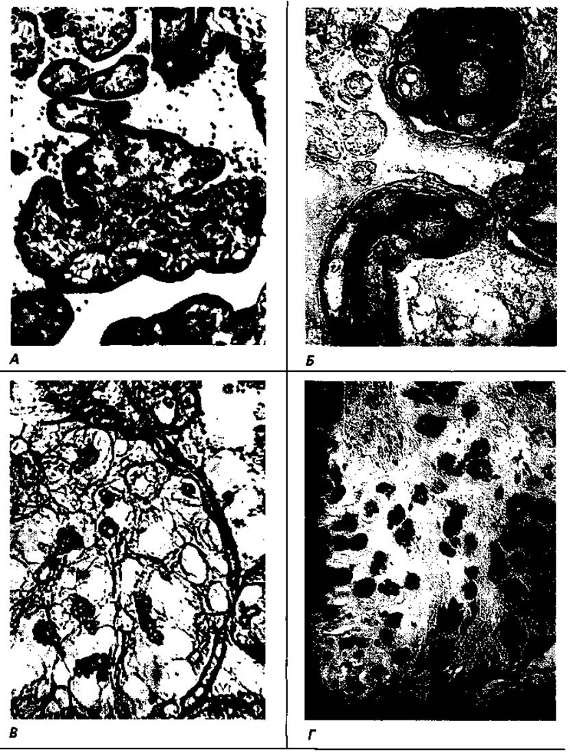
Fig. 2.17. Details of placental microstructure at term: A - light Microscopy of cross-sectioned chorionic villi, x 100; B - connective tissue stroma of chorionic villi, histochemical reaction with soybean lectin, x 100; C - Kashchor–Hofbauer cells in the stroma of a chorionic villus, selective detection with peanut lectin, x 300; D - decidual cells of the maternal part of the placenta, selective detection with soybean lectin, x 100
The Structural and functional unit of the placenta is the cotyledon. Each cotyledon corresponds to the branching territory of a single stem villus bathed in maternal blood. A stem (or anchoring) villus is a large outgrowth of the chorionic plate, firmly fused with the decidua basalis, from the lateral surfaces of which numerous branching twigs of smaller chorionic villi extend. The human placenta contains about 200 cotyledons. Adjacent cotyledons are separated by connective tissue partitions known as septa, which carry arterial branches delivering oxygen- and nutrient-rich blood to the placental lacunae. Wide openings of lacunar Veins allow maternal blood to drain out of the placenta. The walls of the lacunae are formed by endometrial connective tissue covered with layers of an amorphous substance known as Rohr's fibrinoid. A portion of the decidua basalis located between the villous and smooth chorion at the edge of the placental disk fuses tightly with the chorion, forming the marginal plate (closing ring), which prevents blood from leaking into the uterine cavity.
The placenta features a hemochorial (placental) barrier that separates the maternal blood from the fetal blood. The hemochorial barrier comprises the endothelial cells and basement membrane of the chorionic villi blood capillaries, connective tissue surrounding the microvasculature enriched with macrophages and fibroblasts, the basement membrane of the chorionic villi, a layer of syncytiotrophoblast, and Langhans fibrinoid located on the surface of the latter.
The placenta performs numerous vital Functions. Through circulation within the placental blood system, fetal blood is enriched with essential substances—Amino Acids, glucose, Lipids, electrolytes, Vitamins, hormones, Antibodies, and oxygen—while being cleared of carbon dioxide and other harmful metabolic waste products. By producing progesterone, estrogens, placental lactogen, chorionic gonadotropin, and other BIOLOGICALLY ACTIVE SUBSTANCES necessary for maintaining a normal pregnancy, the placenta acts as a temporary endocrine gland. Among these hormones, chorionic gonadotropin is secreted in significant amounts and can be detected in maternal blood as early as the third to fourth day following implantation, a marker utilized in clinical practice for the early Diagnosis of pregnancy. The hemoplacental barrier protects the embryonic organism from many environmental hazards that could otherwise pass from the maternal blood to the fetus. It is important to remember, however, that the hemoplacental barrier is permeable to alcohol, nicotine, drugs, and many medications; therefore, a pregnant woman should strictly limit—and ideally completely eliminate—the consumption of these substances.
The umbilical cord (umbilicus) is a connective-tissue cord enclosing major blood vessels (two Arteries and one vein) that ensure circulation between the fetus and the placenta (Fig. 2.18). In addition to blood vessels, histological preparations of the umbilical cord may reveal remnants of the vitelline duct, which connects the yolk sac to the embryonic body, as well as the allantois. The core of the umbilical cord consists of mucous connective tissue known as Wharton's jelly, which contains a high concentration of hyaluronic acid. The latter provides turgor (firmness) to the umbilical cord and prevents compression of its vessels, thereby maintaining an uninterrupted connection between mother and fetus. Cellular elements found within the umbilical cord include tissue basophils, which help regulate BLOOD FLOW IN the umbilical vessels, and Hofbauer cells, which protect the fetus against intrauterine infection.
The umbilical vein carries oxygen- and nutrient-rich blood from the placenta to the fetus. The umbilical arteries transport deoxygenated blood from the fetus back to the placenta. The surface of the umbilical cord is covered by a simple cuboidal (amniotic) epithelium, which transitions on one end into the amniotic epithelium and merges on the other with the embryonic ectodermal skin covering in the region of the umbilical ring.
The amnion is a continuous membrane surrounding the fetus that, starting from the seventh week of embryogenesis, participates in The production of Amniotic Fluid (Fig. 2.19). The amniotic membrane consists of two parts: an inner epithelial layer and an outer connective-tissue layer. In the Early stages of embryogenesis, the amniotic epithelium is simple squamous. Beginning in the third month of prenatal ontogenesis, the epithelium becomes columnar where the amnion adheres to the placenta, while retaining a cuboidal shape in other areas. The columnar epithelium of the placental disk region is involved in the production of amniotic fluid, whereas the extraplacental cuboidal epithelium participates in its resorption. The epithelium is separated from the underlying connective-tissue Base of the amnion by a basement membrane.
The connective tissue of the amniotic wall is divided into two layers: a deep compact layer and a superficial spongy layer. The compact connective tissue layer includes an acellular zone located close to the basement membrane and a more superficial cellular zone. The latter is rich in fibroblast-like cells, along with bundles of collagen and reticular fibers. The spongy Superficial layer of the amniotic wall is formed by mucous connective tissue traversed in various directions by bundles of collagen fibers. The spongy layer of the amnion has a high content of hydrated proteoglycan complexes.
The presence of the amniotic membrane ensures that the fetus develops in an aqueous environment with an optimal balance of electrolytes, Proteins, and CARBOHYDRATES. Amniotic fluid contains antibodies, which play a crucial role in protecting the embryo from pathogens. The aqueous medium also serves a vital cushioning function, absorbing various shocks and impacts to prevent mechanical injury to the fetus.
The yolk sac (saccus vitellinus) is a vesicle connected to the gut tube, whose wall is lined internally with epithelium and formed externally by connective tissue. Following the formation of the body folds, the yolk sac remains linked to the primitive gut via the vitelline duct. In the early stages of embryogenesis, the yolk sac functions as a hematopoietic organ. Primordial Germ Cells—gonocytoblasts—migrate from the wall of the yolk sac into the developing gonads. From the 7th to 8th week of embryogenesis onward, the yolk sac undergoes regression. Remnants of it can be observed within the umbilical cord as a narrow epithelial tube.
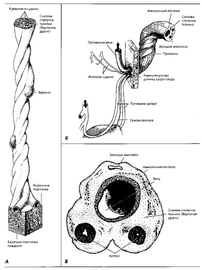
Fig. 2.18. Schematic representation of the umbilical cord of a human term fetus: A - transition zone where the umbilical cord meets the placenta; B - uteroplacental/feto-umbilical junction and The Relationship of the cord with adjacent fetal structures, with arrows indicating the direction of fetal blood flow; C - semi-schematic cross-section of the umbilical cord, x6
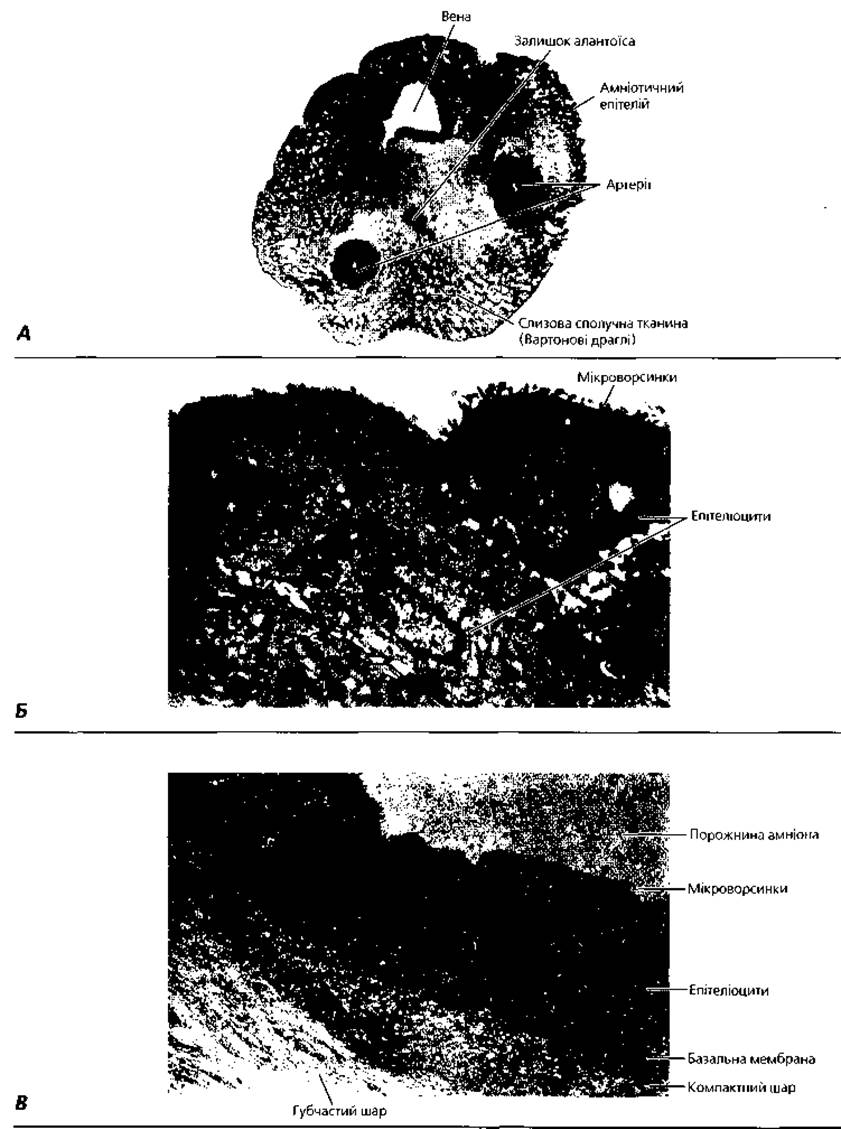
Fig. 2.19. Structural details of the umbilical cord and amniotic membrane of a human term fetus: A - photomicrograph of a cross-section of the umbilical cord, x6; B - electron micrograph of the amniotic epithelium on the outer surface of the cord, x3000; C - electron micrograph of the amniotic wall, x4000
The allantois is a finger-like outgrowth of the ventral wall of the caudal primitive gut that grows into the connecting stalk (amniotic stalk). In early embryogenesis, the allantois functions in nutrition, gas exchange, and excretion. Blood vessels grow from the embryo to the chorion through the allantois, with their terminal branches terminating within the stroma of the chorionic villi. Recent findings indicate that in early human ontogenesis, the allantois acts as an analog of the avian bursa of Fabricius, serving as a central organ for B-lymphocytopoiesis. Beginning in the second month of embryogenesis, the allantois undergoes regression.
Critical Periods of development. Throughout ontogenesis, there are phases characterized by heightened sensitivity of the organism to the damaging EFFECTS OF ENVIRONMENTAL factors. These phases are known as critical periods of development. The Concept of Critical periods was first formulated by the Australian physician Norman Gregg in 1944. The Russian embryologist P. G. Svetlov made a significant contribution to the further development of this theory.
The foundation for critical periods lies in the transition of the embryonic organism from one morphofunctional stage to the next, qualitatively distinct one. This qualitative restructuring of the organism is accompanied by cell proliferation, determination, and differentiation among its constituent cells. Such periods of heightened vulnerability during progenesis include Meiosis (the maturation stage of germ cells) and Fertilization. In prenatal ontogenesis, critical periods include implantation (days 6–8), placentation and the development of axial organ primordia (weeks 3–8), the period of rapid brain development (weeks 15–20), the formation of major functional physiological systems (weeks 20–24), and the birth process itself. In Postnatal ontogenesis, critical developmental periods include the neonatal period (the child's first year of life) and Puberty (ages 11–16).
Damaging effects on the organism—particularly during critical developmental windows—can be caused by chemical agents (including medications), ionizing radiation (including diagnostic X-rays), Hypoxia, malnutrition, psychoactive substances (including nicotine and alcohol), Viruses, and Bacteria. Chemical substances capable of crossing the hemoplacental barrier are especially dangerous During the first months of pregnancy because they tend to accumulate in embryonic tissues and organs, thereby significantly increasing the likelihood of teratogenic or toxic damage.
Early human embryogenesis is schematically illustrated in Figure 2.20.
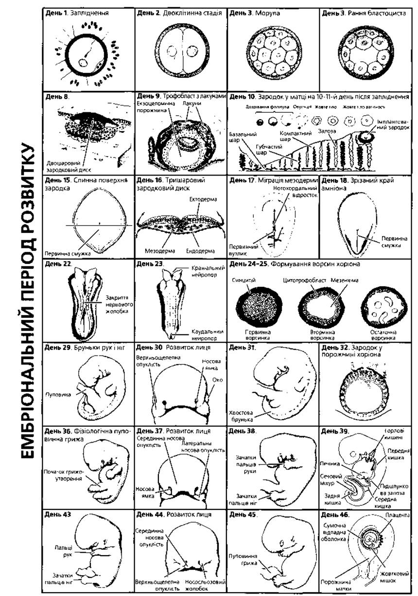
Fig. 2.20. Schematic representation of major embryological events and human embryo Morphology by weeks and days, from week 1 to week 49
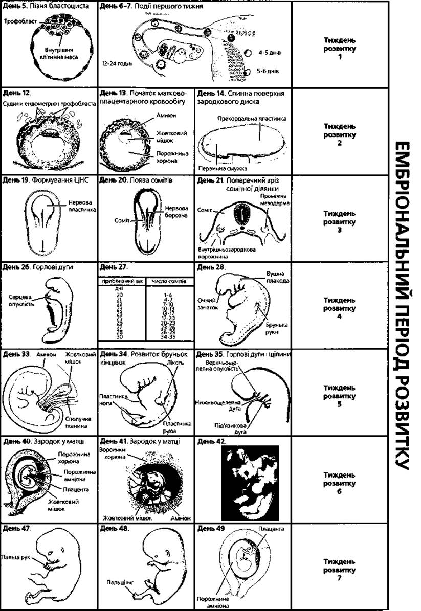
Fig. 2.20. (continued)
Key terms to remember
1. Gastrulation. 2. Ectoderm. 3. Endoderm. 4. Mesoderm. 5. Mesenchyme. 6. Germ disc. 7. Amniotic cavity. 8. Epiblast. 9. Primary and secondary yolk sac. 10. Hypoblast. 11. Chorion. 12. Connecting stalk. 13. Primitive streak. 14. Primitive node. 15. Allantois. 16. Notochord. 17. Body folds. 18. Somites. 19. Dermatome. 20. Myotome. 21. Sclerotome. 22. Splanchnotome. 23. Nephrotome (intermediate mesoderm). 24. Neural tube. 25. Neurulation. 26. Brain vesicles. 27. Ganglionic plate. 28. Neural crest. 29. Placodes. 30. Prechordal plate. 31. Surface ectoderm. 32. Extraembryonic organs. 33. Placenta. 34. Maternal part of the placenta. 35. Decidua basalis. 36. Fetal part of the placenta. 37. Chorionic villus. 38. Syncytiotrophoblast. 39. Cytotrophoblast. 40. Hofbauer cells. 41. Langhans fibrinoid. 42. Placentation. 43. Hemochorial discoid villous placenta. 44. Cotyledon. 45. Rohr's fibrinoid. 46. Hemochorial barrier. 47. Umbilical cord. 48. Amnion. 49. Yolk sac. 50. Allantois. 51. Critical periods of development.
Last update: 09/08/2026
Editorial and Educational Adaptation: This material has been compiled based on the primary/original source text. The project team performed an editorial review, corrected technical inaccuracies, structured sections, and adapted the content for an educational format.
What was processed:
- elimination of formatting defects (OCR errors, structural breaks, corrupted characters);
- editorial organization of content;
- standardization of terminology in accordance with academic sources;
- verification of factual statements against the original source text.
All mentions of the author, publication year, and origin of the primary text have been preserved in accordance with the source.