Human Physiology - William F. Ganong 2002
Blood Circulation
Dynamics of Blood and Lymph Circulation
Blood Circulation in Capillaries
At any given time, only 5% of the circulating Blood is contained within the capillaries; however, this 5% is arguably the most crucial part of the blood volume, as it is precisely across the capillary walls that oxygen and nutrients enter the interstitial fluid, while carbon dioxide and Metabolic waste products return to the Circulation. The exchange that occurs across the Capillary Wall is vital for tissue survival.
Class="center">
Fig. 30-20. Blood pressure in men (dark circles) and women (light circles) of various ages. Clinical measurements were compared with daytime and night-time values obtained using continuous ambulatory monitoring devices. Note that values are lower in women than in men, but increase after menopause. Furthermore, pressures measured at night are lower, and There is a general upward trend in blood pressure throughout life (data from O’Brien E et al: Twenty-four hour ambulatory blood pressure in men and women aged 17 to 80 years: The Allied Irish Bank Study. J Hypertens 1991;9:335).
Methods of Investigation
Obtaining precise measurements of blood pressure and capillary blood flow is challenging. Capillaries in the mesentery of experimental animals, as well as in the human nailbed, can be examined under a dissecting Microscope. Using these and other methods, patterns of blood flow under various conditions have been observed in these and other Tissues. Capillary pressure has been determined by measuring the external pressure required to occlude the capillaries, or the pressure needed to initiate the flow of a physiological saline solution through a micropipette inserted so that its tip enters the arteriolar end of a capillary.
Capillary Pressure and Blood Flow
Capillary pressure can vary significantly; nevertheless, in the capillaries of the nailbed, it averages 32 mm Hg at the arteriolar end and 15 mm Hg at the venular end. The pulse pressure is approximately 5 mm Hg at the arteriolar end and drops to zero at the venular end. Capillaries are short, yet blood moves slowly (about 0.07 cm/s) because the total cross-sectional area of the capillary bed is large. The transit time of blood from the arteriolar to the venular end in an average-sized capillary is 1–2 s.
Equilibrium with Interstitial Fluid
As noted above, the capillary wall is a thin membrane composed of endothelial Cells. Substances pass through the gaps between endothelial cells and through fenestrae, where present. Some substances cross the cells via Vesicular Transport or, in the case of lipid-soluble compounds, directly through the Cytoplasm.
In addition, transport across capillaries can occur via diffusion and filtration (see Chapter 1). Quantitatively, diffusion is by far the more important mechanism for the timely exchange of nutrients and metabolites between blood and tissue. Because the concentrations of oxygen and glucose are higher in the blood than in the interstitial fluid, these substances diffuse into the interstitial fluid, whereas carbon dioxide moves in the opposite direction.
The rate of filtration at any point along a capillary depends on the balance of forces sometimes referred to as Starling forces, named after the physiologist who first detailed this mechanism. One of these forces is the hydrostatic gradient (the difference between the hydrostatic pressure in the capillary and that in the interstitial fluid) at that site. Interstitial fluid pressure varies among Organs; evidence indicates that in subcutaneous tissue it is subatmospheric—about -2 mm Hg. Pressure is positive in The Liver and Kidneys, and equals 6 mm Hg in the Brain. The other force is the osmotic pressure gradient across the capillary wall (the difference between the colloid Osmotic Pressure of the plasma and the colloid osmotic pressure of the interstitial fluid). This component is directed inward.
Thus,

where k is the capillary filtration coefficient; Pc is the capillary hydrostatic pressure; Pi is the interstitial hydrostatic pressure; пс is the capillary osmotic pressure; п1 is the interstitial osmotic pressure.
Normally, п1 is negligible, so the osmotic pressure gradient (пс - п1) is essentially equal to the oncotic pressure. The capillary filtration coefficient is taken into account and is proportional to the permeability of the capillary wall and the available filtration surface area. The balance of Starling forces along a typical Muscle capillary is illustrated in Fig. 30-21. Fluid enters the interstitial space at the arteriolar end of the capillary, where the filtration pressure across the wall exceeds the oncotic pressure, and is reabsorbed into the vessel at the venular end, where the oncotic pressure exceeds the filtration pressure. In other capillary beds, the balance of Starling forces may differ. For instance, fluid is filtered out of the vessels along virtually the entire length of renal glomerular capillaries, whereas fluid is absorbed into capillaries along nearly their entire length in the intestine.
It is important to note that the concentrations of small molecules in the lumen and tissue are often equilibrated at the arteriolar end of the capillary. Under such conditions, net diffusion can be augmented by an increase in blood flow; that is, the exchange is flow-limited (Fig. 30-22). Conversely, the Transport of substances that do not reach equilibrium between the capillary and the interstitium is termed diffusion-limited.
It is estimated that capillaries filter about 24 L of fluid each day. This accounts for roughly 0.3% of the Cardiac Output. Nearly 85% of the filtered fluid is reabsorbed into the capillaries, with the remainder returning to the bloodstream via The Lymphatic system.
Active and Inactive Capillaries
In resting tissues, most capillaries are collapsed, and blood predominantly flows through thoroughfare channels from arterioles into venules. In active tissues, metarterioles and precapillary sphincters dilate. Intracapillary pressure rises above the critical closing pressure of the vessels, and blood flows through all capillaries. The relaxation of the smooth muscle in metarterioles and precapillary sphincters is mediated by vasodilator metabolites released in active tissues (see Chapter 31). Additionally, a reduction in The activity of sympathetic vasoconstrictor nerves innervating the vascular smooth muscle may also play a role.
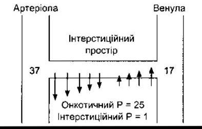
Fig. 30-21. Schematic representation of the pressure gradient across the wall of a muscle capillary. Numbers at the arteriolar and venular ends of the capillary represent the hydrostatic pressure (mm Hg) at those locations. Arrows indicate the approximate volume and direction of fluid movement. In this example, the net pressure gradient at the arteriolar end of the capillary is 11 mm Hg ([37-1]-25) outward; at the opposite end, it is 9 mm Hg (25-[17-1]) inward.

Fig. 30-22. Capillary exchange under conditions of altered flow and diffusion. A and V denote the arteriolar and venular ends of the capillary, respectively. Substance X in the blood reaches equilibrium with its concentration in the tissue (efflux into the tissue equals influx in the opposite direction) before blood leaves the capillary, whereas substance Y does not reach equilibrium. With other factors held constant, The amount of substance X entering the tissue can increase only if blood flow increases; thus, it is flow-dependent. The movement of substance Y is diffusion-dependent.
Following pathological stimulation via an axon reflex, substance P is released (see Chapter 32), which increases capillary permeability. Bradykinin and histamine also increase the permeability of these vessels. When capillaries are subjected to mechanical irritation, they empty of blood (white reaction; see Chapter 32), though this is likely due to the constriction of precapillary sphincters.
Last update: 10/08/2026
Editorial and Educational Adaptation: This material has been compiled based on the primary/original source text. The project team performed an editorial review, corrected technical inaccuracies, structured sections, and adapted the content for an educational format.
What was processed:
- elimination of formatting defects (OCR errors, structural breaks, corrupted characters);
- editorial organization of content;
- standardization of terminology in accordance with academic sources;
- verification of factual statements against the original source text.
All mentions of the author, publication year, and origin of the primary text have been preserved in accordance with the source.