Human Histology - O.D. Lutsyk 2003
Embryology
Germ cells. Fertilization. Cleavage. Implantation
Embryology is a general biological science that investigates the laws of embryonic formation and developmental processes. In contemporary times, it is a rapidly evolving field. Understanding the principles of embryology makes it possible to successfully address such conditions as male and Female Infertility—which are becoming increasingly prevalent in developed nations—determine the sex of an unborn child, and enable early prenatal prediction of potential developmental anomalies, among other Applications. Embryology is closely linked with clinical medicine, particularly obstetrics and gynecology, medical genetics, endocrinology, as well as molecular biology and biochemistry.
The individual development of an Organism—known as ontogenesis—comprises two main stages: prenatal and Postnatal ontogenesis. Ontogenesis is preceded by progenesis, The process of forming male and female Gametes (sex Cells) required to generate a new organism; hence, progenesis is also termed gametogenesis. The fusion of male and female sex cells produces a single-celled embryo, the zygote. This marks the beginning of Prenatal ontogenesis, or The Development of the organism before birth. In humans, normal prenatal ontogenesis lasts approximately 280 days (40 weeks). The initial period of prenatal ontogenesis spans days 1 through 7 of development. During this time, successive mitotic Divisions of the zygote yield a multicellular organism—the blastocyst—which implants into the uterine wall at the end of this initial period (the process of implantation).
The Embryonic period of development extends from the second to the eighth week, during which the rudiments of Tissues, Organs, and Organ Systems appear within the embryo. The fetal period of prenatal ontogenesis lasts from the third to the ninth month and culminates in childbirth. This period involves the further Structural and functional maturation of fetal tissues and organs, as well as the differentiation of their constituent cells. Progenesis, along with the initial and embryonic periods, constitutes the proper Subject Matter of embryology. In a broader sense, embryology also encompasses the fetal and early postnatal (perinatal) periods of organismal development.
Postnatal ontogenesis begins at birth and concludes with the death of the organism. Concluding this General Overview of ontogenesis, it should be noted that the late 19th century saw the formulation of the fundamental biogenetic law (the Haeckel-Müller law), which states that prenatal ontogenesis is a brief recapitulation of phylogenesis—meaning that the individual Development of the embryo concisely repeats the evolutionary history of its species.
Spermatozoa (Fig. 2.1). The process by which male sex cells are formed is called Spermatogenesis. It takes place in the male gonad—the Testis—specifically within its convoluted seminiferous tubules. A detailed examination of spermatogenesis is provided in the chapter "Male Reproductive System".
A mature human spermatozoon is approximately 60 µm long. Its anterior oval region is called the HEAD (measuring 4.5 µm in length, 3 µm in width, and 1 µm in thickness), while the posterior, elongated thread-like region is the tail. Between them lies the connecting piece (neck). The tail comprises the intermediate, principal, and terminal pieces. The sperm head contains a Nucleus with a haploid set of Chromosomes. The human sperm chromosomal Complement includes twenty-two autosomes and one gonosome (sex chromosome), which may be either X or Y. Based on the presence of the X or Y chromosome, spermatozoa are divided into two types: those carrying the Y chromosome (androsperms), which give rise to a male organism, and those carrying the X chromosome (gynosperms), which initiate a female organism upon fusing with an ovum.
The anterior part of the sperm head is capped by a modified Golgi complex derivative known as the acrosome. This is a vesicle filled with hydrolytic Enzymes—such as Trypsin and hyaluronidase—which are essential for digesting the egg coats and successfully executing Fertilization. The connecting piece (neck) of the spermatozoon, a narrow region of Cytoplasm situated behind The Nucleus, contains a proximal centriole (located in a nuclear fossa) and a distal centriole, from which the axonemal complex of the tail originates. The axoneme is composed of 9 peripheral pairs of microtubules and a central pair. In addition, specialized cytoskeletal elements are located in the neck and tail. Surrounding the centrioles in the neck are 9 segmented columns distally continuous with 9 dense fibers that run parallel to the axonemal microtubules in the midpiece. In this region, the dense fibers are encircled by spiral Cell/35.html">Mitochondria, forming the so-called mitochondrial sheath. The midpiece is approximately 7 µm long. The mitochondria provide the energy required for sperm motility.
In the principal piece of the tail (approximately 40 µm in length), the axoneme is surrounded by a fibrous sheath consisting of longitudinal columns interconnected by Ribs. The terminal piece of the sperm tail is about 5 µm long and contains only the axonemal complex.
Class="center">
Fig. 2.1. Spermatozoon: A – Morphology of a mature human spermatozoon; B – scanning electron micrograph of a spermatozoon within the uterine cavity, x 2000
Spermatozoa possess A number of functional characteristics that enable them to fertilize the ovum. For instance, thanks to a motile tail, male sex cells can travel in a directed manner at speeds of up to 50 µm/s. The direction of travel is determined by the sperm's ability to navigate toward the oocyte, which releases specific sperm attractants known as gynogamones (these substances are widespread in the animal kingdom, though not yet identified in humans). The ability of spermatozoa to respond to chemical stimuli is termed chemotaxis. Spermatozoa are also capable of swimming against a fluid current—the secretory products of the epithelial cells lining the uterine tubes and uterine glands. This property is called rheotaxis. A mildly alkaline environment is optimal for sperm viability, whereas acidic pH values cause male sex cells to lose motility, agglutinate, and rapidly perish.
In humans, normal ejaculation yields 3–5 mL of semen containing approximately 350 million spermatozoa. For successful fertilization, the total sperm count in the ejaculate must be at least 150 million, with a concentration of no less than 60 million per 1 mL of semen. Under optimal conditions (Temperature and pH), spermatozoa can reach the uterine cavity within 0.5–1 hour after sexual intercourse, and the ampulla of the Uterine tube—where contact with the oocyte occurs—within 1.5–2 hours. Spermatozoa retain their fertilizing capacity for 36–88 hours post-ejaculation.
The Ovum (Fig. 2.2). Female sex cells are produced in the female gonad—the Ovary—in a process called oogenesis. Oogenesis comprises three phases: multiplication, growth, and maturation; the final phase is completed in the uterine tube following ovulation (the release of the oocyte from the ovary). A detailed description of oogenesis is presented in the chapter "FEMALE REPRODUCTIVE SYSTEM".
A post-ovulatory human oocyte is spherical, measures approximately 130 µm in diameter, and is surrounded by the zona pellucida and follicular epithelial cells (corona radiata / granulosa layer). Upon interacting with spermatozoa, the oocyte completes the second meiotic division, transforming from a secondary oocyte into a mature ovum.
The oocyte nucleus contains a haploid set of chromosomes comprising 22 autosomes and a single X sex chromosome. The oocyte cytoplasm is rich in nutrient inclusions—yolk (deutoplasm). Yolk consists of high-energy lipophosphoprotein complexes accumulated within Golgi vesicles. Based on its yolk content, the human ovum is classified as secondary oligolecithal (oligosolecithal), and according to yolk distribution (cytotopography), as isolecithal (evenly distributed throughout the cytoplasm). The secondary evolutionary emergence of oligolecithal eggs in humans and placental mammals is due to the transition of the embryo to maternal Nutrition, which eliminates the need to accumulate massive yolk reserves. The oocyte cytoplasm contains a well-developed rough Endoplasmic reticulum, abundant Ribosomes, various RNA species, and tubulins. Concentrated in the peripheral cytoplasmic zones (beneath the oolemma) are numerous cortical granules. These represent complexes of Proteoglycans and Glycoproteins assembled within Golgi vesicles. Cortical granules ensure The formation of the fertilization membrane, which is impermeable to sperm and protects the oocyte against polyspermy (the penetration of more than one spermatozoon into the cytoplasm).
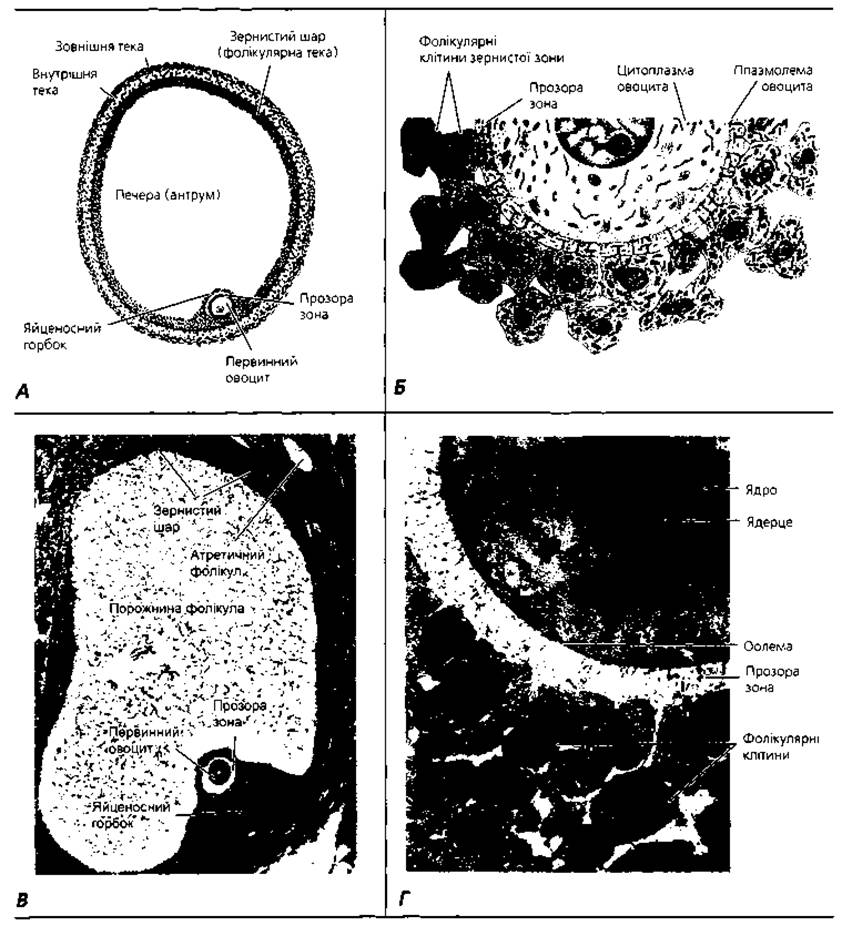
Fig. 2.2. Pre-ovulatory oocyte within a tertiary (mature) ovarian follicle. Schematic drawings based on light (A) and electron (B) Microscopy; C – photomicrograph of a histological section of a pre-ovulatory follicle 10 mm in diameter, x 60; D – electron micrograph of a primary oocyte fragment with adjacent follicular cells, x 1800
Throughout a woman's reproductive lifespan (roughly from ages 15 to 50), approximately 400–500 oocytes mature and are capable of being fertilized, though the actual number of fertilized eggs is typically much lower. The oocyte utilizes its nutrient reserve within 12–24 hours post-ovulation, after which an unfertilized egg perishes.
Fertilization (Fig. 2.3) is the process of fusion between male and female sex cells, resulting in a single-celled embryo—the zygote. In humans, fertilization takes place in the ampullary region of the uterine tube. The oocyte reaches this site passively, driven by contractions of the fimbriae, the beating of epithelial cilia, and peristaltic contractions of the muscular wall of the uterine tube.
Spermatozoa reach the ampulla of the uterine tubes owing to their physiological adaptations—motility, chemotaxis, and rheotaxis. It should be noted that sperm preparation for fertilization begins during their transit through the Ductus deferens. At this stage, modifications occur in the surface glycopolymers of the sperm Plasma Membrane (glycocalyx), protecting the male sex cells from destruction within the female reproductive tract. Following ejaculation, spermatozoa can remain quiescent for several days in the distal portion of the uterine tube isthmus. After ovulation, these "depot" spermatozoa become activated and begin migrating toward the oocyte in the ampulla of the uterine tube.
Spermatozoa freshly deposited in the female reproductive tract are incapable of fertilizing an egg. To acquire this capacity, they must undergo capacitation and the acrosome reaction. The core process of capacitation—which occurs in the uterine tube and lasts approximately 7 hours in humans—involves interactions between the spermatozoon and the mucosal surface of the uterine tube. During this process, glycoprotein coats and seminal fluid Proteins are stripped from The Plasma Membrane overlying the acrosomal region of the sperm. Only capacitated spermatozoa can undergo the acrosome reaction and penetrate the corona radiata of the oocyte.
The acrosome reaction occurs after the spermatozoon binds to the zona pellucida and is triggered by proteins residing within it. The reaction involves the release of enzymes required for zona penetration—such as acrosin and trypsin-like substances. Of the roughly 350 million spermatozoa entering the female reproductive tract during ejaculation, only 300–500 reach the site of fertilization, and only a single sperm fertilizes the ovum, while the others merely assist in breaching the barriers surrounding the oocyte. During fertilization, a spermatozoon must successively overcome three barriers: the corona radiata, the zona pellucida, and the oocyte plasma membrane. Capacitated spermatozoa readily traverse the Cells of the corona radiata.

Fig. 2.3. Fertilization: A – a primary oocyte is expelled during ovulation from a mature ovarian follicle and, through fimbrial contractions and ciliary beating of the uterine tube, enters its ampullary region; B – successive phases of sperm penetration through the egg coats: phase 1 – adhesion; phase 2 – penetration through the zona pellucida; phase 3 – penetration of the sperm nucleus and proximal centriole through the oolemma
The zona pellucida is a critical barrier along the sperm's path. It is composed primarily of glycoproteins—ZP1, ZP2, and ZP3. The zona pellucida mediates sperm binding and induces the acrosome reaction. Binding is facilitated by the ZP3 protein, to which the spermatozoon possesses specific receptors. Simultaneously, this process triggers the acrosome reaction, which can be viewed as a specialized form of exocytosis. The outer acrosomal membrane and the sperm cell membrane fuse, releasing acrosomal enzymes that digest the molecules of the zona pellucida, carving out a narrow tunnel through which the spermatozoon passes. Primary adhesion between the spermatozoon and the oocyte is mediated in part by interactions between oocyte surface Integrins and their ligands on the sperm surface. Following adhesion, the Plasma Membranes of the sperm and egg fuse, and the sperm head and tail enter the oocyte cytoplasm, whereas the sperm plasma membrane remains fixed to the oocyte surface.
Following sperm penetration into the oocyte cytoplasm, the cortical reaction is initiated. This reaction involves the exocytosis of cortical granule contents beyond the oocyte plasma membrane. High-molecular-weight Biopolymers of the cortical granules interact with the oocyte's plasma membrane glycocalyx to form the fertilization envelope, which is impermeable to spermatozoa. The zona pellucida alters its Structure and composition to prevent the binding and penetration of additional spermatozoa, thereby ensuring monospermic fertilization.
A series of sequential changes takes place within the cytoplasm of the fertilized oocyte. Specifically, the second maturation division is completed, resulting in the formation of a mature ovum and one polar body from the secondary oocyte. Once inside the oocyte, the sperm nucleus migrates forward and transforms into the male pronucleus, while the female pronucleus forms from the egg nucleus. Each pronucleus replicates its DNA, after which their nuclear envelopes dissolve, establishing a common zygotic metaphase plate with a mitotic spindle. This guarantees the diploid chromosome number in the cells of the future embryo. The interaction of the pronucleuses marks the completion of fertilization proper. The centriole, essential for zygotic division, is delivered into the oocyte via the sperm neck. In addition to paternal chromosomes, the spermatozoon contributes its Mitochondrial Genome along with mitochondria, as well as a Cleavage-triggering signal protein, to the new organism.
The zygote undergoes an active redistribution of cytoplasmic material known as ooplasmic segregation. This process forms the so-called presumptive zones—specific Regions of the zygote cytoplasm that are destined to develop into particular parts or structures of the embryonic organism.
Cleavage (Fig. 2.4) is the post-fertilization stage of embryonic development comprising a series of successive mitotic divisions that transform the zygote into a multicellular organism. Cleavage spans the period from the first to the end of the sixth day. Its hallmark feature is an extremely short interphase between successive mitoses, causing the newly formed embryonic cells to progressively decrease in size. The cells constituting the embryo during the cleavage period are called blastomeres. Due to this successive reduction in blastomere size, the overall dimensions of the embryo on the sixth day of development do not exceed those of the zygote, allowing the blastocyst to fit entirely within the space bounded by the fertilization envelope. Cleavage concludes when the size of the blastomeres approaches that of the somatic cells of the organism.

Fig. 2.4. Cleavage: A - diagram of successive stages of fertilization and cleavage; B - phase-contrast image of a human zygote and two-cell embryo; C - scanning electron micrograph of an eight-cell embryo
Human zygotes are characterized by total (holoblastic), subequal, and asynchronous cleavage. These terms indicate that the cleavage furrow passes completely through the zygote or embryo; the resulting blastomeres are nearly identical in size (subequal); and different blastomeres enter mitotic division at varying times (asynchronously).
At the eight-cell stage, blastomeres form a loosely arranged cluster of cells. However, following the third cleavage division—approximately 3 days post-fertilization—they maximize their intercellular contact area, forming a compact cellular mass held together by tight junctions. Known as compaction, this process results in the segregation of outer cells from inner cells, which communicate via Gap Junctions (nexuses). At this stage, the embryo is termed a morula (resembling a mulberry). The central cells of the morula are interconnected by nexuses, through which Intercellular signaling takes place, forming the inner cell mass. The peripheral cells, linked by tight junctions, create a barrier that restricts the internal microenvironment of the morula, thereby forming the outer cell mass (trophoblast).
On the 4th day following fertilization, the embryo enters the Uterus. At this time, a fluid-filled cavity called the blastocoele appears within it, formed by fluid transported into the embryo from its microenvironment by trophoblast cells. The embryo is now referred to as a blastocyst. It resembles a vesicle whose wall is formed by elongated trophoblast cells. Inside lies the blastocoele, with the inner cell mass (or embryoblast) localized at one pole along the inner surface of the trophoblast. The embryo proper and certain extraembryonic structures develop from the embryoblast (partial or complete splitting of the inner cell mass leads to the formation of twins). The trophoblast gives rise to the chorion, the embryonic portion of the Placenta. By the end of the 4th day of development, enzymes produced by trophoblast cells dissolve the fertilization envelope (the modified zona pellucida), thereby creating the conditions necessary for subsequent implantation.
Implantation (Fig. 2.5) is the process by which the embryo embeds into the uterine mucosa, taking place on the 7th-8th days of development. Prior to this, driven by peristaltic contractions, ciliary beating, and the movement of epithelial secretions within the uterine tube, the embryo travels from the ampullary region to the uterine cavity. This constitutes the so-called tubal period of embryonic life (days one through four). From the fifth to the seventh day, the embryo resides in the uterine cavity as a free blastocyst (not attached to any specific region of the endometrium). During these periods, embryonic nutrition relies partly on the oocyte's stored nutrients and partly on secretions from the uterine tubes and endometrial glands. Days seven and eight, when the blastocyst makes contact with the uterine mucosa, are considered a critical developmental period due to the risk of implantation failure and subsequent embryonic demise. Implantation comprises two distinct phases: adhesion and invasion.

Fig. 2.5. Implantation: A - diagram of a human embryo (blastocyst) on days 4-5 of development; B - photomicrograph of a 107-cell human embryo; C - invasion of the trophoblast at the embryonic pole of the blastocyst into the uterine mucosa on days 5-6 post-fertilization; D - diagram of a human embryo upon completion of implantation (day 12 of development)
Adhesion (attachment) involves the binding of the blastocyst to the endometrial surface. Typically, the blastocyst attaches between the excretory ducts of two adjacent uterine glands, highlighting the crucial role of their secretory products in mediating adhesion. In turn, uterine glandular secretion is regulated by progesterone produced by the corpus luteum of the ovary. Implantation cannot succeed without establishing a specific hormonal Background and the corresponding hormone-dependent remodeling of the uterine mucosa. Evidence suggests that this initial phase of implantation is driven by molecular interactions between high-molecular-weight glycoproteins from uterine secretions and the endometrial cell glycocalyx with specific lectin-like proteins on the blastocyst surface.
Invasion (penetration) of the blastocyst into the uterine mucosa is mediated by trophoblast enzymatic systems. Driven by these enzymes, epithelial cells are destroyed first, followed by the Connective Tissue and, ultimately, the walls of endometrial Blood Vessels. Consequently, the blastocyst sinks into the implantation crypt and becomes enveloped by connective tissue. The epithelial defect heals through the proliferation of endometrial epithelial cells. The histiotrophic period of Embryogenesis persists until the trophoblast erodes the vessel walls and establishes contact with maternal blood, spanning the first four weeks of development. During this time, the embryo is nourished by absorbing nutrients from uterine gland secretions and the products of endometrial tissue breakdown by the trophoblast. The haematotrophic (hemo-trophic) period extends from the moment of contact with maternal blood until birth (from the second to the ninth month). Throughout this period, the supply of nutrients and gas exchange for the embryo and fetus are maintained entirely by the maternal Circulatory system.
In certain cases (such as strictures of the uterine tube or aberrant blastocyst migration), the embryo may fail to reach the uterine cavity. This most frequently results in Tubal Pregnancy, wherein the embryonic trophoblast implants into the mucosa of the uterine tubes. Such anomalous implantation can cause rupture of the uterine tube as the embryo enlarges, leading to severe Hemorrhage and maternal mortality. Instances of embryonic attachment to ovarian tissues, the Peritoneum, and even the hepatic parenchyma have also been documented. With the advent of modern reproductive technologies, a new medical and legal concept has emerged—surrogacy. In this Procedure, women who have undergone Hysterectomy or have medical contraindications to carrying a pregnancy obtain oocytes that are fertilized extracorporeally using sperm from the partner or a donor; the resulting embryo at the 18-32 blastomere stage is then transferred into the uterus of another woman who carries the fetus to term. During In vitro fertilization, it is possible to pre-select the sex of the future child: a single blastomere is biopsied from several resulting blastosomes; cytogenetic analysis of these cells identifies the presence of X or Y sex chromosomes; and a blastocyst with the desired chromosomal profile is transferred to the uterus. The transfer of embryos previously cryopreserved in liquid nitrogen has also been successfully implemented, and the cryopreservation of oocytes and sperm has likewise proven feasible.
In recent years, gamete intrafallopian transfer has gained widespread use. In this procedure, oocytes retrieved from a woman's Ovaries are combined with paternal spermatozoa and placed directly into the uterine tube, where fertilization takes place. Advanced sperm-sorting Methods have been developed to remove defective or abnormal spermatozoa from seminal fluid. Procedures are also practiced to wash spermatozoa free of antisperm Antibodies that may cause infertility. Techniques for pre-selecting the sex of the offspring have been established: differential centrifugation or Electrophoresis yields fractions of X- or Y-chromosome-bearing spermatozoa (androsperms or gynosperms); introducing these purified sperm fractions into the female reproductive tract allows the sex of the future child to be determined prior to embryonic development. Microsurgical techniques on the oocyte have been developed to facilitate fertilization. Notably, intracytoplasmic sperm injection (ICSI) is employed, wherein a single spermatozoon is microinjected directly into the oocyte in a laboratory Setting for artificial fertilization. The early-stage embryo is subsequently transferred to the uterus for further development. These Examples demonstrate that medical embryology now enables the correction of numerous reproductive pathologies that were until recently considered incurable.
Advancements in medical embryology drive the development of novel therapeutic strategies for various diseases. For instance, recent years have seen the development of embryo cloning for cell therapy. In this approach, the nucleus is removed from an oocyte and replaced with the nucleus of a cumulus cell, fibroblast, or another somatic cell microinjected into the ooplasm. The oocyte, now harboring a diploid set of chromosomes, is incubated in a solution of bioactive substances that trigger cleavage. By days 4-5 of cloning, a blastocyst comprising approximately 100 blastomeres is formed. The inner cell mass is isolated from the blastocyst and further cultured to generate stem cell populations. These cells can differentiate into diverse mature stem cell types suitable for transplantation back into the patient from whom the genetic material was originally derived. For example, pancreatic islet-like cells generated in this manner can be utilized to treat diabetes, while neural cells can be applied to repair Spinal Cord injuries or treat Parkinson's disease.
Key Terms to Remember
1. Embryology. 2. Ontogeny. 3. Progenesis. 4. Biogenetic law. 5. Spermatozoon. 6. Sperm head. 7. Sperm tail. 8. Acrosome. 9. Chemotaxis. 10. Rheotaxis. 11. Ejaculation. 12. Ovum (Egg cell). 13. Yolk (Deutoplasm). 14. Secondary oligolecithal egg cell. 15. Cortical granules. 16. Fertilization. 17. Zygote. 18. Capacitation. 19. Sperm penetration into the ovum. 20. Cortical reaction. 21. Fertilization envelope. 22. Monospermy. 23. Pronuclei. 24. Ooplasmic segregation. 25. Presumptive Zones of the zygote. 26. Cleavage. 27. Morula. 28. Blastomeres. 29. Blastocoele. 30. Total subequal asynchronous cleavage. 31. Compaction. 32. Embryoblast. 33. Trophoblast. 34. Blastocyst. 35. Implantation. 36. Adhesion phase. 37. Invasion phase. 38. Histiotrophic nutrition period. 39. Haematotrophic nutrition period. 40. In vitro fertilization.
Last update: 09/08/2026
Editorial and Educational Adaptation: This material has been compiled based on the primary/original source text. The project team performed an editorial review, corrected technical inaccuracies, structured sections, and adapted the content for an educational format.
What was processed:
- elimination of formatting defects (OCR errors, structural breaks, corrupted characters);
- editorial organization of content;
- standardization of terminology in accordance with academic sources;
- verification of factual statements against the original source text.
All mentions of the author, publication year, and origin of the primary text have been preserved in accordance with the source.